Anti-PSD95 Antibody (56452)
$457.00
| Host | Quantity | Applications | Species Reactivity | Data Sheet | |
|---|---|---|---|---|---|
| Mouse | 100ug | WB,IHC,ICC/IF,AM | Mouse, Rat, Bovine |  |
SKU: 56452
Categories: Antibody Products, Neuroscience and Signal Transduction Antibodies, Products
Overview
Product Name Anti-PSD95 Antibody (56452)
Description Anti-PSD95 Mouse Monoclonal Antibody
Target PSD95
Species Reactivity Mouse, Rat, Bovine
Applications WB,IHC,ICC/IF,AM
Host Mouse
Clonality Monoclonal
Clone ID 6G6-1C9
Isotype IgG2a
Immunogen Recombinant rat PSD95
Properties
Form Liquid
Concentration Lot Specific
Formulation PBS, pH 7.4.
Buffer Formulation Phosphate Buffered Saline
Buffer pH pH 7.4
Format Purified
Purification Purified by Protein G affinity chromatography
Specificity Information
Specificity This antibody recognizes mouse, rat, and bovine PSD95.
Target Name Disks large homolog 4
Target ID PSD95
Uniprot ID P31016
Alternative Names Postsynaptic density protein 95, PSD-95, Synapse-associated protein 90, SAP-90, SAP90
Gene Name Dlg4
Sequence Location Cell membrane, Cell junction, synapse, postsynaptic density, Cell junction, synapse, Cytoplasm, Cell projection, axon, Cell projection, dendritic spine, Cell projection, dendrite, Cell junction, synapse, presynapse
Biological Function Postsynaptic scaffolding protein that plays a critical role in synaptogenesis and synaptic plasticity by providing a platform for the postsynaptic clustering of crucial synaptic proteins (PubMed:15317815, PubMed:15358863, PubMed:19596852, PubMed:23300088, PubMed:26679993). Interacts with the cytoplasmic tail of NMDA receptor subunits and shaker-type potassium channels. Required for synaptic plasticity associated with NMDA receptor signaling. Overexpression or depletion of DLG4 changes the ratio of excitatory to inhibitory synapses in hippocampal neurons. May reduce the amplitude of ASIC3 acid-evoked currents by retaining the channel intracellularly. May regulate the intracellular trafficking of ADR1B (By similarity). Also regulates AMPA-type glutamate receptor (AMPAR) immobilization at postsynaptic density keeping the channels in an activated state in the presence of glutamate and preventing synaptic depression (PubMed:19596852).Under basal conditions, cooperates with FYN to stabilize palmitoyltransferase ZDHHC5 at the synaptic membrane through FYN-mediated phosphorylation of ZDHHC5 and its subsequent inhibition of association with endocytic proteins (By similarity). {UniProtKB:P78352, UniProtKB:Q62108, PubMed:15317815, PubMed:15358863, PubMed:19596852, PubMed:23300088, PubMed:26679993}.
Research Areas Neuroscience
Background Postsynaptic Density Protein 95 (PSD95), also known as Synapse Associated Protein 90kDa, is a member of the membrane- associated guanylate kinase family of proteins. PSD95 is a scaffolding protein involved in the assembly and function of the postsynaptic density complex. This family of proteins consists of N-terminal variable segments followed by three amino-terminal PDZ domains, an upstream SH3 domain, and an inactive carboxy-terminal guanylate kinase domain. The first and second PDZ domains localize NMDA receptors and K+ channels to synapses, and the third binds to neuroligins, neuronal cell adhesion molecules. PSD95 also binds to neuronal nitric oxide synthase. PSD95 is implicated in experience-dependent plasticity and plays an indispensable role in learning. Mutations in PSD95 are associated with autism.
Application Images


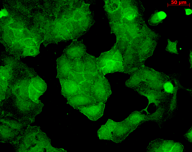
Description Immunocytochemistry/Immunofluorescence analysis using Mouse Anti-PSD95 Monoclonal Antibody, Clone 6G6 (56452). Tissue: HaCaT cells. Species: Human. Fixation: Cold 100% methanol for 10 minutes at -20°C. Primary Antibody: Mouse Anti-PSD95 Monoclonal Antibody (56452) at 1:100 for 1 hour at RT. Secondary Antibody: FITC Goat Anti-Mouse (green) at 1:50 for 1 hour at RT. Localization: Junction staining.

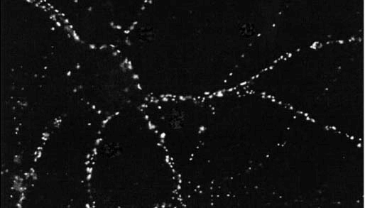
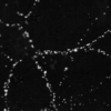
Description Immunocytochemistry/Immunofluorescence analysis using Mouse Anti-PSD95 Monoclonal Antibody, Clone 6G6 (56452). Tissue: dissociated hippocampal neurons. Species: Rat. Fixation: Cold 4% paraformaldehyde/0.2% glutaraldehyde in 0.1M sodium phosphate buffer. Primary Antibody: Mouse Anti-PSD95 Monoclonal Antibody (56452) at 1:1000 for 12 hours at 4°C. Secondary Antibody: FITC Goat Anti-Mouse IgG (green) at 1:50 for 30 minutes at RT. Magnification: 10X. Courtesy of: Mary Kennedy, Caltech.

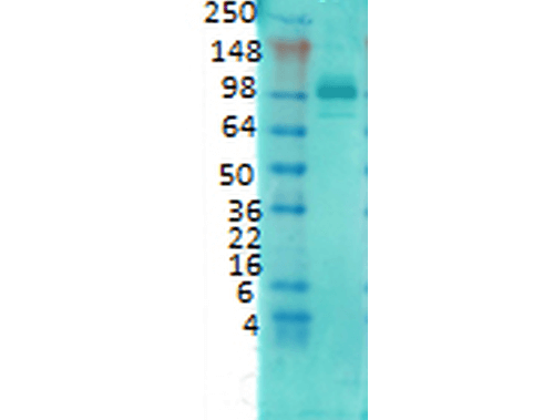
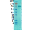
Description Western Blot analysis of Rat brain membrane lysate showing detection of PSD95 protein using Mouse Anti-PSD95 Monoclonal Antibody, Clone 6G6 (56452). Primary Antibody: Mouse Anti-PSD95 Monoclonal Antibody (56452) at 1:1000.
Handling
Storage This antibody is stable for at least one (1) year at -20°C.
Dilution Instructions Dilute in PBS or medium that is identical to that used in the assay system.
Application Instructions Immunoblotting: use at 4ug/mL. A band of ~100 kDa is detected.
Immunocytochemistry: use at 3-15ug/mL
These are recommended concentrations. User should determine optimal concentrations for their application.
Positive control: Rat brain tissue
Immunocytochemistry: use at 3-15ug/mL
These are recommended concentrations. User should determine optimal concentrations for their application.
Positive control: Rat brain tissue
References & Data Sheet
Data Sheet  Download PDF Data Sheet
 Download PDF Data Sheet

