Anti-PD-L1 Antibody (34068)
$233.00
Overview
Product Name Anti-PD-L1 Antibody (34068)
Description Anti-PD-L1 Rabbit Polyclonal Antibody
Target PD-L1
Species Reactivity Human
Applications IHC
Host Rabbit
Clonality Polyclonal
Clone ID G411.1
Isotype IgG
Immunogen Recombinant human PD-L1.
Properties
Form Liquid
Concentration Lot Specific
Formulation Tris buffer, pH 7.3-7.7, 1% BSA, 0.1% sodium azide.
Buffer Formulation Tris
Buffer pH pH 7.3-7.7
Buffer Anti-Microbial 0.1% Sodium Azide
Buffer Protein Stabilizer 1% Bovine Serum Albumin
Format Purified
Purification Purified by immunoaffinity chromatography
Specificity Information
Specificity Human PD-L1. Reactivity with other species has not been investigated.
Target Name Programmed cell death 1 ligand 1
Target ID PD-L1
Uniprot ID Q9NZQ7
Alternative Names PD-L1, PDCD1 ligand 1, Programmed death ligand 1, hPD-L1, B7 homolog 1, B7-H1, CD antigen CD274
Gene Name CD274
Sequence Location Cell membrane, Early endosome membrane, Recycling endosome membrane
Biological Function Plays a critical role in induction and maintenance of immune tolerance to self (PubMed:11015443, PubMed:28813417, PubMed:28813410). As a ligand for the inhibitory receptor PDCD1/PD-1, modulates the activation threshold of T-cells and limits T-cell effector response (PubMed:11015443, PubMed:28813417, PubMed:28813410). Through a yet unknown activating receptor, may costimulate T-cell subsets that predominantly produce interleukin-10 (IL10) (PubMed:10581077). {PubMed:10581077, PubMed:11015443, PubMed:28813410, PubMed:28813417}.; The PDCD1-mediated inhibitory pathway is exploited by tumors to attenuate anti-tumor immunity and escape destruction by the immune system, thereby facilitating tumor survival (PubMed:28813417, PubMed:28813410). The interaction with PDCD1/PD-1 inhibits cytotoxic T lymphocytes (CTLs) effector function (By similarity). The blockage of the PDCD1-mediated pathway results in the reversal of the exhausted T-cell phenotype and the normalization of the anti-tumor response, providing a rationale for cancer immunotherapy (By similarity). {UniProtKB:Q9EP73, PubMed:28813410, PubMed:28813417}.
Research Areas Cancer research
Background Programmed Death-Ligand 1 (PD-L1), also known as CD274 or B7 Homolog 1 (B7-H1), is a transmembrane protein involved in suppressing the immune system and rendering tumor cells resistant to CD8+ T cell-mediated lysis through binding of the Programmed Death-1 (PD-1) receptor. Overexpression of PD-L1 may allow cancer cells to evade the actions of the host immune system. In renal cell carcinoma, upregulation of PD-L1 has been linked to increased tumor aggressiveness and risk of death, and, in ovarian cancer, higher expression of this protein has led to significantly poorer prognosis. PD-L1 has also been linked to systemic lupus erythematosus and cutaneous melanoma. When considered in adjunct with CD8+ tumor-infi- ltrating lymphocyte density, expression levels of PD-L1 may be a useful predictor of multiple cancer types, including stage III non-small cell lung cancer, hormone receptor negative breast cancer, and sentinel lymph node melanoma.
Application Images


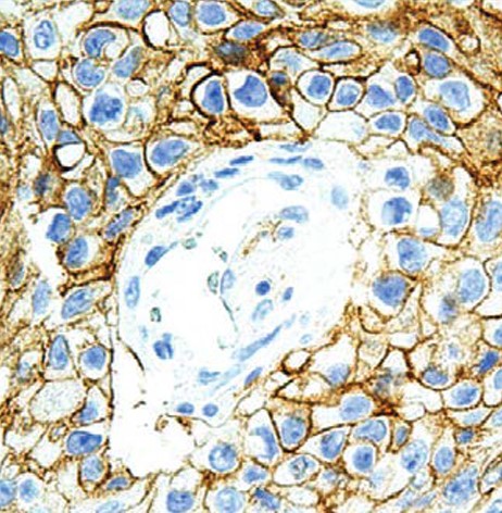
Description Immunohistochemistry: use at a dilution of 1:100-1:200 on formalin-fixed, paraffin-embedded samples after heat-induced epitope retrieval at pH 9 for 10-30 minutes. Detection of PD-L1 in human lung tumor with #34068 diluted 1:100-1:200.
Handling
Storage Store at 2-8°C. Do not freeze.
Dilution Instructions Dilute in PBS or medium that is identical to that used in the assay system.
Application Instructions
Immunohistochemistry: use at a dilution of 1:100-1:200 on formalin-fixed, paraffin-embedded samples after heat-induced epitope retrieval at pH 9 for 10-30 minutes.
Immunohistochemistry: use at a dilution of 1:100-1:200 on formalin-fixed, paraffin-embedded samples after heat-induced epitope retrieval at pH 9 for 10-30 minutes.
References & Data Sheet
Data Sheet  Download PDF Data Sheet
 Download PDF Data Sheet


