Anti-p53 Antibody (34065)
$225.00
Overview
Product Name Anti-p53 Antibody (34065)
Description Anti-p53 Mouse Monoclonal Antibody
Target p53
Species Reactivity Human
Applications IHC
Host Mouse
Clonality Monoclonal
Clone ID G053.1
Isotype IgG2b
Immunogen Recombinant human p53.
Properties
Form Liquid
Concentration Lot Specific
Formulation Tris buffer, pH 7.3-7.7, 1% BSA, 0.1% sodium azide.
Buffer Formulation Tris
Buffer pH pH 7.3-7.7
Buffer Anti-Microbial 0.1% Sodium Azide
Buffer Protein Stabilizer 1% Bovine Serum Albumin
Format Purified
Purification Purified by immunoaffinity chromatography
Specificity Information
Specificity Human p53. Reactivity with other species has not been investigated.
Target Name Cellular tumor antigen p53
Target ID p53
Uniprot ID P04637
Alternative Names Antigen NY-CO-13, Phosphoprotein p53, Tumor suppressor p53
Gene Name TP53
Sequence Location Cytoplasm, Nucleus, Nucleus, PML body, Endoplasmic reticulum, Mitochondrion matrix, Cytoplasm, cytoskeleton, microtubule organizing center, centrosome
Biological Function Acts as a tumor suppressor in many tumor types; induces growth arrest or apoptosis depending on the physiological circumstances and cell type. Involved in cell cycle regulation as a trans-activator that acts to negatively regulate cell division by controlling a set of genes required for this process. One of the activated genes is an inhibitor of cyclin-dependent kinases. Apoptosis induction seems to be mediated either by stimulation of BAX and FAS antigen expression, or by repression of Bcl-2 expression. Its pro-apoptotic activity is activated via its interaction with PPP1R13B/ASPP1 or TP53BP2/ASPP2 (PubMed:12524540). However, this activity is inhibited when the interaction with PPP1R13B/ASPP1 or TP53BP2/ASPP2 is displaced by PPP1R13L/iASPP (PubMed:12524540). In cooperation with mitochondrial PPIF is involved in activating oxidative stress-induced necrosis; the function is largely independent of transcription. Induces the transcription of long intergenic non-coding RNA p21 (lincRNA-p21) and lincRNA-Mkln1. LincRNA-p21 participates in TP53-dependent transcriptional repression leading to apoptosis and seems to have an effect on cell-cycle regulation. Implicated in Notch signaling cross-over. Prevents CDK7 kinase activity when associated to CAK complex in response to DNA damage, thus stopping cell cycle progression. Isoform 2 enhances the transactivation activity of isoform 1 from some but not all TP53-inducible promoters. Isoform 4 suppresses transactivation activity and impairs growth suppression mediated by isoform 1. Isoform 7 inhibits isoform 1-mediated apoptosis. Regulates the circadian clock by repressing CLOCK-ARNTL/BMAL1-mediated transcriptional activation of PER2 (PubMed:24051492). {PubMed:11025664, PubMed:12524540, PubMed:12810724, PubMed:15186775, PubMed:15340061, PubMed:17317671, PubMed:17349958, PubMed:19556538, PubMed:20673990, PubMed:20959462, PubMed:22726440, PubMed:24051492, PubMed:9840937}.
Research Areas Cancer research
Background p53, like Retinoblastoma protein, is a tumor suppressor protein. Mutations in p53 are found in most tumor types. p53 protein binds DNA which in turn stimulates another gene to produce a protein called p21 that interacts with a cell division-stimulating protein (cdk2). When p21 is complexed with cdk2 the cell cannot pass through to the next stage of cell division. Mutant p53 can no longer bind DNA in an effective way, and, as a result, the p21 protein is not made available to act as the ‘stop signal' for cell division. Thus cells divide uncontrollably, and form tumors.
Application Images


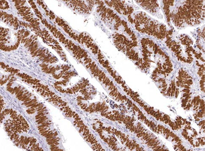
Description Immunohistochemistry: use at a dilution of 1:100-1:200 on formalin-fixed, paraffin-embedded samples after heat-induced epitope retrieval at pH 9 for 10-30 minutes. Detection of p53 in human colon carcinoma with #34065 diluted 1:100-1:200.
Handling
Storage Store at 2-8°C. Do not freeze.
Dilution Instructions Dilute in PBS or medium that is identical to that used in the assay system.
Application Instructions
Immunohistochemistry: use at a dilution of 1:100-1:200 on formalin-fixed, paraffin-embedded samples after heat-induced epitope retrieval at pH 9 for 10-30 minutes.
Immunohistochemistry: use at a dilution of 1:100-1:200 on formalin-fixed, paraffin-embedded samples after heat-induced epitope retrieval at pH 9 for 10-30 minutes.
References & Data Sheet
Data Sheet  Download PDF Data Sheet
 Download PDF Data Sheet


