Anti-Malondialdehyde Mouse Monoclonal Antibody (56615)
$441.00
| Host | Quantity | Applications | Species Reactivity | Data Sheet | |
|---|---|---|---|---|---|
| Mouse | 100 ug | WB,ICC/IF,FACS,FCM,ELISA | Species Independent |  |
SKU: 56615
Categories: Antibody Products, Heat Shock and Stress Protein Antibodies, Products
Overview
Product Name Anti-Malondialdehyde Mouse Monoclonal Antibody (56615)
Description Anti-Malondialdehyde Mouse Monoclonal Antibody
Target Malondialdehyde
Species Reactivity Species Independent
Applications WB,ICC/IF,FACS,FCM,ELISA
Host Mouse
Clonality Monoclonal
Clone ID 6H6
Isotype IgG1
Immunogen Synthetic Malondialdehyde modified Keyhole Limpet Hemocyanin (KLH).
Properties
Form Liquid
Concentration 1 mg/mL
Formulation PBS pH 7.4, 50% glycerol, 0.09% Sodium azide
Buffer Formulation Phosphate Buffered Saline
Buffer pH pH 7.4
Buffer Anti-Microbial 0.09% Sodium azide
Buffer Cryopreservative 50% glycerol
Format Purified
Purification Protein G Purified
Background Malondialdehyde (MDA) is the biomarker in greatest diagnostic use, due to its molecular stability. This three-carbon, low-molecular weight aldehyde has a strong affinity for amino acids, which results in adduct formation to both free amino acids and proteins. Increased MDA levels have been found at correlating levels in breast cancer, and lung cancer patients. Other diseased states with elevated MDA levels include diabetes and Alzheimer’s disease. Multiple laboratory techniques exist for quantification of MDA levels, including the thiobarbituric acid reactive substances (TBARS) assay. In addition to use as a biomarker, MDA has been shown to have mutagenic effects on tissues themselves as adduct formation can result in DNA cross-linking.
Specificity Information
Specificity Specific for Malondialdehyde conjugated proteins. Does not detect free Malondialdehyde. Does not cross-react with Acrolein, Crotonaldehyde, Hexanoyl Lysine, 4-Hydroxy-2-hexenal, 4-Hydroxy nonenal, or Methylglyoxal modified proteins.
Target Name Malondialdehyde
Target ID Malondialdehyde
Alternative Names Malondialdehyde, MDA, Malondialdehyde (MDA), Malonic aldehyde Propanedial, 1,3-Propanedial, Malonaldehyde
CAS Number 542-78-9
PubChem ID 10964
Research Areas Cancer | Oxidative Stress | Lipid peroxidation | Neuroscience | Neurodegeneration | Alzheimer's Disease
Application Images


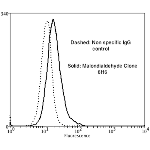
Description Flow Cytometry analysis using Mouse Anti-Malondialdehyde Monoclonal Antibody, Clone 6H6 . Tissue: Neuroblastoma cells (SH-SY5Y). Species: Human. Fixation: 90% Methanol. Primary Antibody: Mouse Anti-Malondialdehyde Monoclonal Antibody at 1:50 for 30 min on ice. Secondary Antibody: Goat Anti-Mouse: PE at 1:100 for 20 min at RT. Isotype Control: Non Specific IgG. Cells were subject to oxidative stress by treating with 250 µM H2O2 for 24 hours.

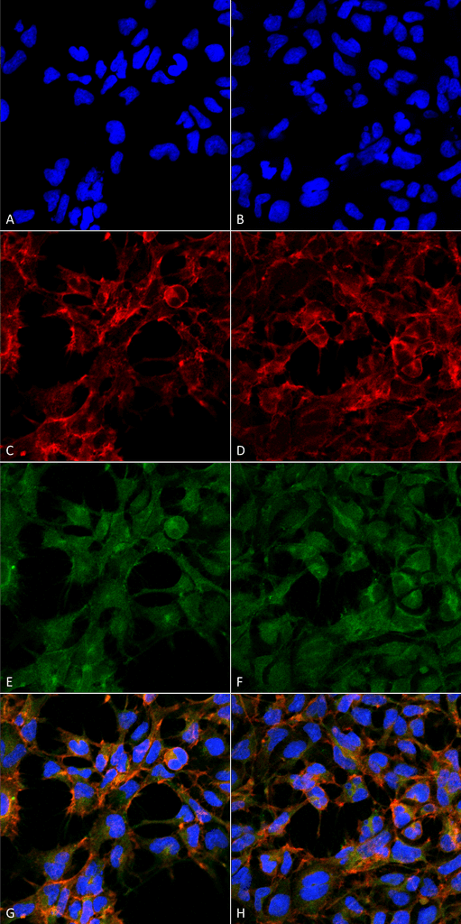
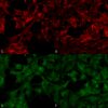
Description Immunocytochemistry/Immunofluorescence analysis using Mouse Anti-Malondialdehyde Monoclonal Antibody, Clone 6H6 . Tissue: Embryonic kidney epithelial cell line (HEK293). Species: Human. Fixation: 5% Formaldehyde for 5 min. Primary Antibody: Mouse Anti-Malondialdehyde Monoclonal Antibody at 1:50 for 30-60 min at RT. Secondary Antibody: Goat Anti-Mouse Alexa Fluor 488 at 1:1500 for 30-60 min at RT. Counterstain: Phalloidin Alexa Fluor 633 F-Actin stain; DAPI (blue) nuclear stain at 1:250, 1:50000 for 30-60 min at RT. Magnification: 20X (2X Zoom). (A,C,E,G) - Untreated. (B,D,F,H) - Cells cultured overnight with 50 µM H2O2. (A,B) DAPI (blue) nuclear stain. (C,D) Phalloidin Alexa Fluor 633 F-Actin stain. (E,F) Malondialdehyde Antibody. (G,H) Composite. Courtesy of: Dr. Robert Burke, University of Victoria.

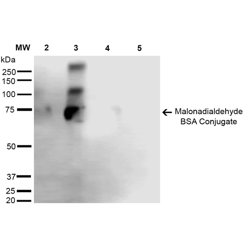
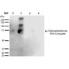
Description Western Blot analysis of Malondialdehyde-BSA Conjugate showing detection of 67 kDa Malondialdehyde protein using Mouse Anti-Malondialdehyde Monoclonal Antibody, Clone 6H6 . Lane 1: Molecular Weight Ladder (MW). Lane 2: Malondialdehyde-BSA (0.5 µg). Lane 3: Malondialdehyde-BSA (2.0 µg). Lane 4: BSA (0.5 µg). Lane 5: BSA (2.0 µg) . Block: 5% Skim Milk in TBST. Primary Antibody: Mouse Anti-Malondialdehyde Monoclonal Antibody at 1:1000 for 2 hours at RT. Secondary Antibody: Goat Anti-Mouse IgG: HRP at 1:2000 for 60 min at RT. Color Development: ECL solution for 5 min in RT. Predicted/Observed Size: 67 kDa.
Handling
Storage This antibody is stable for at least one (1) year at -20°C. Avoid multiple freeze-thaw cycles.
Dilution Instructions Dilute in PBS or medium which is identical to that used in the assay system.
Application Instructions WB (1:1000); ICC/IF (1:50); FACS (1:50); FCM (1:50); ELISA (1:1000); optimal dilutions for assays should be determined by the user.
References & Data Sheet
Data Sheet  Download PDF Data Sheet
 Download PDF Data Sheet

