Anti-Hsp70 Antibody (11117)
$447.00
| Host | Quantity | Applications | Species Reactivity | Data Sheet | |
|---|---|---|---|---|---|
| Mouse | 100ug | WB,IHC,ICC/IF,IP,AM | Human, Mouse, Rat, Fish, Avian, Amphibian, Drosophila, Yeast |  |
SKU: 11117
Categories: Antibody Products, Heat Shock and Stress Protein Antibodies, Products
Overview
Product Name Anti-Hsp70 Antibody (11117)
Description Anti-Hsp70 clone 3A3 Mouse Monoclonal Antibody
Target Hsp70
Species Reactivity Human, Mouse, Rat, Fish, Avian, Amphibian, Drosophila, Yeast
Applications WB,IHC,ICC/IF,IP,AM
Host Mouse
Clonality Monoclonal
Clone ID 3A3
Isotype IgG1
Immunogen Recombinant human Hsp70 expressed in E. coli.
Properties
Form Liquid
Concentration Lot Specific
Formulation PBS, pH 7.4 and 50% glycerol.
Buffer Formulation Phosphate Buffered Saline
Buffer pH pH 7.4
Buffer Cryopreservative 50% Glycerol
Format Purified
Purification Purified by Protein G affinity chromatography
Specificity Information
Specificity This antibody recognizes human, mouse, rat, fish, avian, amphibian, Drosophila, and yeast Hsp70, Hsc70, p75, and, following heat shock, Hsp72.
Target ID Hsp70
Uniprot ID P08107
Gene ID 3303
Accession Number NP_005336.3
Research Areas Heat Shock& Stress Proteins
Background Hsp70 binds ATP with high affinity and possesses a weak ATPase activity which can be stimulated by binding to unfolded proteins and synthetic peptides. Hsp70 recognizes and binds to nascent polypeptide chains as well as partially folded intermediates of proteins preventing their aggregation and misfolding. The binding of ATP triggers a critical conformational change leading to the release of the bound protein. The ability of Hsp70 to undergo cycles of binding and release from hydrophobic stretches of partially unfolded proteins is the basis for their role in a variety of intracellular functions such as protein synthesis, protein folding and oligomerization and protein transport.
Application Images
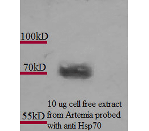


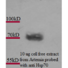
Description Western Blot analysis of Artemia franciscanna (brine shrimp) cell lysates showing detection of Hsp70 protein using Mouse Anti-Hsp70 Monoclonal Antibody, Clone 3A3 (11117). Primary Antibody: Mouse Anti-Hsp70 Monoclonal Antibody (11117) at 1:1000. Courtesy of: Alison King.

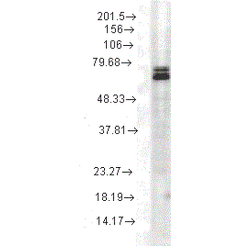
Description Western Blot analysis of Rat cell lysates showing detection of Hsp70 protein using Mouse Anti-Hsp70 Monoclonal Antibody, Clone 3A3 (11117). Load: 15 µg. Block: 1.5% BSA for 30 minutes at RT. Primary Antibody: Mouse Anti-Hsp70 Monoclonal Antibody (11117) at 1:1000 for 2 hours at RT. Secondary Antibody: Sheep Anti-Mouse IgG: HRP for 1 hour at RT.

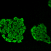
Description Immunocytochemistry/Immunofluorescence analysis using Mouse Anti-HSP70 Monoclonal Antibody, Clone 3A3 (11117). Tissue: Cervical cancer cell line (HeLa). Species: Human. Fixation: 4% Formaldehyde for 15 min at RT. Primary Antibody: Mouse Anti-HSP70 Monoclonal Antibody (11117) at 1:100 for 60 min at RT. Secondary Antibody: Goat Anti-Mouse ATTO 488 at 1:100 for 60 min at RT. Counterstain: DAPI (blue) nuclear stain at 1:5000 for 5 min RT. Localization: Cytoplasm. Magnification: 40X.
Handling
Storage This antibody is stable for at least one (1) year at -20°C.
Dilution Instructions Dilute in PBS or medium which is identical to that used in the assay system.
Application Instructions Immunoblotting: use at 0.2ug/mL (ECL). A band of ~70 kDa is detected.
Immunoprecipitation: use at 1-2ug/mLImmunohistochemistry/Immunocyto- chemistry: use at 2 ug/mLUser should determine optimal concentrations for their applications.
Positive control: Heat-shocked HeLa cell lysate.
Immunoprecipitation: use at 1-2ug/mLImmunohistochemistry/Immunocyto- chemistry: use at 2 ug/mLUser should determine optimal concentrations for their applications.
Positive control: Heat-shocked HeLa cell lysate.
References & Data Sheet
Data Sheet  Download PDF Data Sheet
 Download PDF Data Sheet


