New Products, News from QED Bioscience
IHC Troubleshooting
Related Antibodies from QED Bioscience
Name – Catalog No.
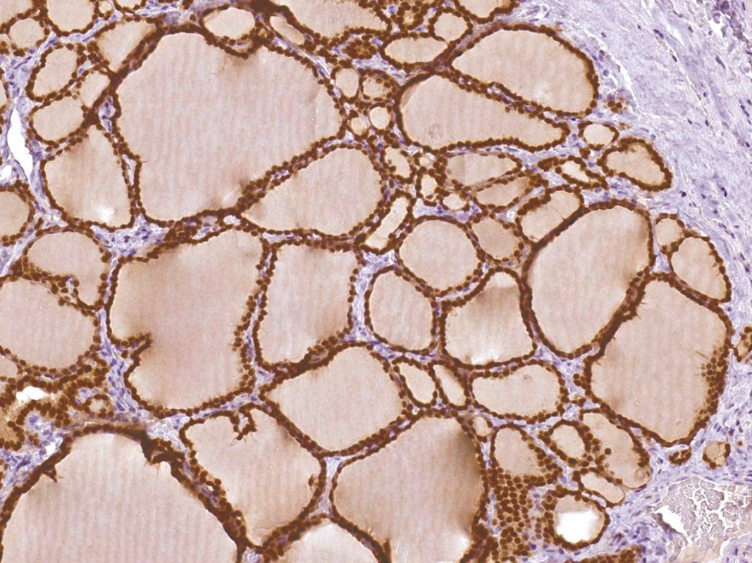
Immunohistochemistry (IHC) is an effective microscopy-based technique for visualizing cellular components in tissue samples, and although IHC images are fascinating and tremendously informative, the art of creating useful IHC images requires attention to several important factors and awareness of potential problems. Here are some troubleshooting tips to help with achieving crisp, clean IHC images free of background staining and non-specific antibody binding.
Problem: High background staining
Cause #1: Endogenous peroxidase
Peroxidase-labeled secondary antibodies with chromogenic substrates are commonly used reagents for detecting binding of primary antibodies in IHC, but endogenous peroxidase activity in cells, such as erythrocytes and granulocytes, will react with chromogenic peroxidase substrates. This can lead to background staining of tissues that have a high blood content or heavy granulocyte infiltrate and make interpretation of IHC samples difficult.
Solution: Block endogenous peroxidase activity by applying a weak solution of H 2 O 2 to tissue sections before applying antibody reagents. A 3% solution of H 2 O 2 in distilled water is commonly used and is highly effective, although some antigens may be sensitive to H 2 O 2 , in which case a lower concentration of H 2 O 2 (perhaps as low as 0.5%) should be used.
Cause #2: Endogenous phosphatase
Many types of alkaline phosphatases (AP) are present in human tissues, and these can produce background staining when a chromogenic AP substrate is used to detect binding of AP-conjugated secondary antibody. Endogenous alkaline phosphates are usually more of a problem in frozen tissues, but, in some cases, this enzyme activity can survive formalin fixation.
Solution: Most endogenous alkaline phosphatase activity can be blocked by applying 1mM levamisole to tissue sections between the primary and secondary antibody steps.
Cause #3: Insufficient washing
Antibodies might remain on the surface of slides, even where there are no relevant areas of tissue sections, due to ionic charge or because some antibodies are “sticky”.
Solution: Be sure to wash slides gently with PBS a minimum of 3 times. For some antibodies, 5 washes might be necessary.
Problem: No staining
Cause #1: Primary antibody is not suitable for IHC
Although many antibodies can recognize their target antigen in ELISA or Western blot, they might not perform well in IHC either because the antigen is not present in tissue sections at detectable levels, or the conformation of the antigen in tissues is different than the conformation of the antigen the antibody was made against. The latter is particularly true if an antibody is made against a linear peptide whose sequence is derived from a protein of interest; that peptide sequence might not be accessible to antibody binding in the native, three-dimensional conformation of the protein.
Solution: Increasing antibody concentration and/or extending incubation times can improve the detection of low-level antigens. Unfortunately, it isn’t possible to change an antibody that isn’t suitable for IHC into one that is.
Cause #2: Primary antibody is too dilute
Antibody-antigen reactions are governed by the ratio of antibody:antigen, so it’s important to use antibodies at optimal concentrations relative to the amount of antigen present in samples.
Solution: Test a range of antibody dilutions on a positive control sample in which you know the antigen of interest is present in order to determine the optimal dilution of primary antibody.
Cause #3: Target antigen is a nuclear protein
Intact nuclear membranes don’t allow penetration of large molecules like antibodies.
Solution: A detergent such as Triton X-100 can dissolve lipid in nuclear membranes which facilitates antibody penetration. A 0.5-1.0% solution of Triton X-100 in PBS works well on tissue sections when applied before the primary antibody; for immunocytochemistry, use a 0.05% solution of Triton X-100 in PBS.
Cause #4: Fixation alters the antigen
Fixatives like formalin cross-link proteins in tissues which is good for preserving tissue integrity but can mask epitopes and, as a result, block primary antibody from binding to its target. Fortunately, epitope masking is usually reversible.
Solution: Antigen retrieval methods are used to unmask epitopes, and there are two types of antigen retrieval methods, enzymatic and heating.
Enzymatic Antigen Retrieval is believed to work by cleaving proteins that have masked epitopes.
Enzymes commonly used for this purpose are:
Proteinase K (20 g/ml in Tris-EDTA buffer, pH 8)
Trypsin (0.5% in dH 2 0)
Pepsin (0.1% in 10mM HCl)
Incubate slides with the enzyme of choice 10-20 minutes at 37 o C after deparaffinizing and before blocking.
Heating Antigen Retrieval probably works by hydrolyzing fixative-induced cross-links. Methods which require 10-40 minutes in a steamer or microwave (in five minute increments) or a water bath at 95-100 o C followed by 20 minutes cooling at room temperature include:
Citrate buffer (10 mM Sodium Citrate, 0.05% Tween 20, pH 6)
Citrate-EDTA buffer (10 mM Sodium Citrate, 2 mM EDTA, 0.05% Tween 20, pH 6.2)
EDTA (1 mM EDTA, 0.05% Tween 20, pH 8)
Treat slides with these methods after deparaffinizing and before blocking.
Cause #5: Secondary antibody doesn’t recognize primary antibody
Solution: This is an easy one. As an example, if your primary antibody is a mouse antibody, the secondary antibody must be anti-mouse. The species of the secondary antibody doesn’t matter – it can be produced in rabbit or goat or another species – as long as it is made against the species of your primary antibody.
Problem: Non-specific staining
Cause #1: Insufficient blocking
Many types of mammalian cells can bind primary and/or secondary antibodies non-specifically via Fc receptors present on cell membranes.
Solution: This type of non-specific antibody binding can be eliminated by blocking tissue sections or cell preparations with a 10% solution of normal serum for 30-60 minutes before applying primary antibodies.
Immunoglobulin present in normal serum will occupy Fc receptors. The normal serum should be of a species that’s different than the species of primary antibody. For example, if your primary antibody is a mouse or rabbit antibody, block with 10% normal goat serum.
Cause #2: Concentration of primary or secondary antibody is too high
Excess antibody precipitates on tissue sections.
Solution: Try decreasing antibody concentrations and test on a negative control tissue to confirm specificity of staining. Also, make sure that tissue sections aren’t allowed to dry during the IHC procedure.
Cause #3: Secondary antibody reacts with tissue samples.
The majority of commercial secondary antibodies are enzyme-conjugated polyclonal antibodies, such as HRP-conjugated rabbit anti-mouse IgG or HRP-conjugated goat anti-mouse IgG. However, some of these secondary antibodies might cross-react with other species IgG. For example, an anti-mouse IgG secondary antibody might also recognize human IgG due to similarities between the two species’ IgGs. If human IgG is present and preserved in a human tissue sample, a cross-reactive secondary antibody will bind to it as well as to a primary mouse antibody. In some cases this type of staining can be recognized as artifactual because staining occurs in areas of tissue sections where the target antigen shouldn’t be located. In other cases, though, it isn’t possible to distinguish specific vs. non-specific staining, and that can lead to incorrect interpretations of IHC results.
Solution: Use a secondary antibody that is cross-absorbed on other species immunoglobulins to remove unwanted antibody cross-reactivities. These are available from many manufacturers who offer secondary antibodies.
Summary
There are many variables to consider when performing IHC analyses – different tissues, different antigen targets, different antibody reagents – so optimizing conditions and reagent concentrations for each application is key to achieving picture-perfect IHC results.


