Anti-GABARAP Antibody (56569)
$466.00
SKU: 56569
Categories: Antibody Products, Neuroscience and Signal Transduction Antibodies, Products
Overview
Product Name Anti-GABARAP Antibody (56569)
Description Anti-GABARAP Rabbit Polyclonal Antibody
Target GABARAP
Species Reactivity Human, Mouse
Applications WB,ICC/IF
Host Rabbit
Clonality Polyclonal
Immunogen Synthetic peptide corresponding to the C-terminus of human GABARAP
Properties
Form Liquid
Concentration 1.0 mg/mL
Formulation PBS, pH 7.4, 50% glycerol, 0.09% sodium azide.
Buffer Formulation Phosphate Buffered Saline
Buffer pH pH 7.4
Buffer Anti-Microbial 0.09% Sodium Azide
Buffer Cryopreservative 50% Glycerol
Format Purified
Purification Purified by peptide immuno-affinity chromatography
Specificity Information
Specificity This antibody recognizes human and mouse GABARAP.
Target Name γ-aminobutyric acid receptor-associated protein
Target ID GABARAP
Uniprot ID O95166
Alternative Names GABA(A receptor-associated protein, MM46
Gene Name GABARAP
Sequence Location Cytoplasmic vesicle, autophagosome membrane, Endomembrane system, Cytoplasm, cytoskeleton, Golgi apparatus membrane, Cytoplasmic vesicle
Biological Function Ubiquitin-like modifier that plays a role in intracellular transport of GABA(A) receptors and its interaction with the cytoskeleton (PubMed:9892355). Involved in autophagy: while LC3s are involved in elongation of the phagophore membrane, the GABARAP/GATE-16 subfamily is essential for a later stage in autophagosome maturation (PubMed:15169837, PubMed:20562859, PubMed:22948227). Through its interaction with the reticulophagy receptor TEX264, participates in the remodeling of subdomains of the endoplasmic reticulum into autophagosomes upon nutrient stress, which then fuse with lysosomes for endoplasmic reticulum turnover (PubMed:31006538). Also required for the local activation of the CUL3(KBTBD6/7) E3 ubiquitin ligase complex, regulating ubiquitination and degradation of TIAM1, a guanyl-nucleotide exchange factor (GEF) that activates RAC1 and downstream signal transduction (PubMed:25684205). Thereby, regulates different biological processes including the organization of the cytoskeleton, cell migration and proliferation (PubMed:25684205). Involved in apoptosis (PubMed:15977068). {PubMed:15169837, PubMed:15977068, PubMed:20562859, PubMed:22948227, PubMed:25684205, PubMed:31006538, PubMed:9892355}.
Research Areas Neuroscience
Background Gamma-aminobutyric acid A receptors (GABA(A) receptors) are ligand-gated chloride channels that mediate inhibitory neurotransmission. This protein clusters neurotransmitter receptors by mediating interaction with the cytoskeleton. In addition, GABARAP has an important role in autophagy; it is crucial for autophagosome formation and sequestering cytosolic cargo into double-membrane vesicles which is then followed by degradation after fusion with lysosomes.
Application Images


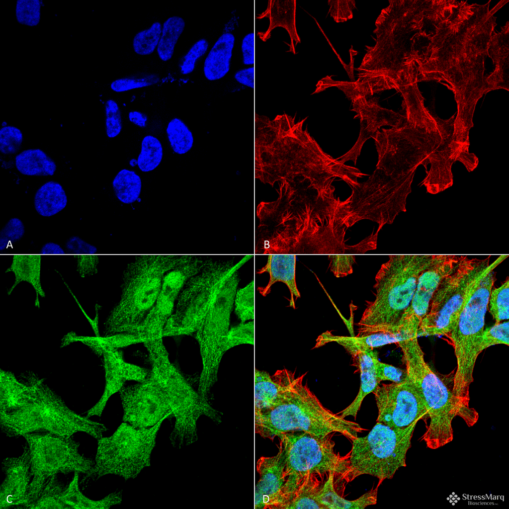
Description Immunocytochemistry/Immunofluorescence analysis using Rabbit Anti-GABARAP Polyclonal Antibody (56569). Tissue: Neuroblastoma cell line (SK-N-BE). Species: Human. Fixation: 4% Formaldehyde for 15 min at RT. Primary Antibody: Rabbit Anti-GABARAP Polyclonal Antibody (56569) at 1:100 for 60 min at RT. Secondary Antibody: Goat Anti-Rabbit ATTO 488 at 1:100 for 60 min at RT. Counterstain: Phalloidin Texas Red F-Actin stain; DAPI (blue) nuclear stain at 1:1000, 1:5000 for 60min RT, 5min RT. Localization: Endomembrane System, Cytoplasm, Cytoskeleton, Golgi Apparatus Membrane, Cytoplasmic Vesicle. Magnification: 60X. (A) DAPI (blue) nuclear stain (B) Phalloidin Texas Red F-Actin stain (C) GABARAP Antibody (D) Composite.
Handling
Storage This antibody is stable for at least one (1) year at -20°C.
Dilution Instructions Dilute in PBS or medium that is identical to that used in the assay system.
Application Instructions Immunoblotting: use at dilution of 1:1,000. A band of ~14-16kDa is detected. Other bands observed at ~40kDa (complex) and ~26kDa (dimer).
Immunofluorescence: use at dilution of 1:100
These are recommended working dilutions.
Endusers should determine optimal dilutions for their applications.
Immunofluorescence: use at dilution of 1:100
These are recommended working dilutions.
Endusers should determine optimal dilutions for their applications.
References & Data Sheet
Data Sheet  Download PDF Data Sheet
 Download PDF Data Sheet


