Anti-GABA(B)R2 Antibody (56523)
$466.00
| Host | Quantity | Applications | Species Reactivity | Data Sheet | |
|---|---|---|---|---|---|
| Mouse | 100ug | WB,IHC,ICC/IF | Human, Mouse, Rat |  |
SKU: 56523
Categories: Antibody Products, Neuroscience and Signal Transduction Antibodies, Products
Overview
Product Name Anti-GABA(B)R2 Antibody (56523)
Description Anti-GABA (B)R2 Mouse Monoclonal Antibody
Target GABA(B)R2
Species Reactivity Human, Mouse, Rat
Applications WB,IHC,ICC/IF
Host Mouse
Clonality Monoclonal
Clone ID S81-2
Isotype IgG1
Immunogen Fusion protein corresponding to aa 861-912 of rat GABA(B)R2 (accession no. NP_113990.1).
Properties
Form Liquid
Concentration 1.0 mg/mL
Formulation PBS, pH 7.4, 50% glycerol, 0.09% sodium azide.Purified by Protein G affinity chromatography.
Buffer Formulation Phosphate Buffered Saline
Buffer pH pH 7.4
Buffer Anti-Microbial 0.09% Sodium Azide
Buffer Cryopreservative 50% Glycerol
Format Purified
Purification Purified by Protein G affinity chromatography
Specificity Information
Specificity This antibody recognizes human, mouse, and rat GABA(B)R2. It does not cross- react with GABA(B)R1.
Target Name γ-aminobutyric acid type B receptor subunit 2
Target ID GABA(B)R2
Uniprot ID O88871
Alternative Names GABA-B receptor 2, GABA-B-R2, GABA-BR2, GABABR2, Gb2, G-protein coupled receptor 51
Gene Name Gabbr2
Sequence Location Cell membrane, Cell junction, synapse, postsynaptic cell membrane, Perikaryon, Cell projection, dendrite
Biological Function Component of a heterodimeric G-protein coupled receptor for GABA, formed by GABBR1 and GABBR2 (PubMed:9872315, PubMed:9872317, PubMed:9872744). Within the heterodimeric GABA receptor, only GABBR1 seems to bind agonists, while GABBR2 mediates coupling to G proteins (PubMed:9872317, PubMed:10658574). Ligand binding causes a conformation change that triggers signaling via guanine nucleotide-binding proteins (G proteins) and modulates the activity of down-stream effectors, such as adenylate cyclase (PubMed:9872315, PubMed:9872317, Ref.4, PubMed:10075644, PubMed:9872744, PubMed:10924501). Signaling inhibits adenylate cyclase, stimulates phospholipase A2, activates potassium channels, inactivates voltage-dependent calcium-channels and modulates inositol phospholipid hydrolysis (PubMed:9872315, PubMed:9872317, PubMed:10457184, PubMed:9872744, PubMed:10924501). Plays a critical role in the fine-tuning of inhibitory synaptic transmission (PubMed:9872317, PubMed:10457184, PubMed:9872744). Pre-synaptic GABA receptor inhibits neurotransmitter release by down-regulating high-voltage activated calcium channels, whereas postsynaptic GABA receptor decreases neuronal excitability by activating a prominent inwardly rectifying potassium (Kir) conductance that underlies the late inhibitory postsynaptic potentials (PubMed:9872744, PubMed:10924501). Not only implicated in synaptic inhibition but also in hippocampal long-term potentiation, slow wave sleep, muscle relaxation and antinociception (By similarity). {UniProtKB:O75899, PubMed:10075644, PubMed:10457184, PubMed:10658574, PubMed:10924501, PubMed:9872315, PubMed:9872317, PubMed:9872744, Ref.4}.
Research Areas Neuroscience
Background GABA (g-aminobutyric acid) is the primary inhibitory neurotransmitter in the central nervous system and interacts with three different receptors: GABA(A), GABA(B), and GABA(C). GABA(B) receptor is coupled to G proteins that modulate slow inhibitory synaptic transmission. Functional GABA(B) receptors form heterodimers of GABA(B)R1 and GABA(B)R2 in which GABA(B)R1 binds a ligand and GABA(B)R2 is the primary G protein contact site.
Application Images


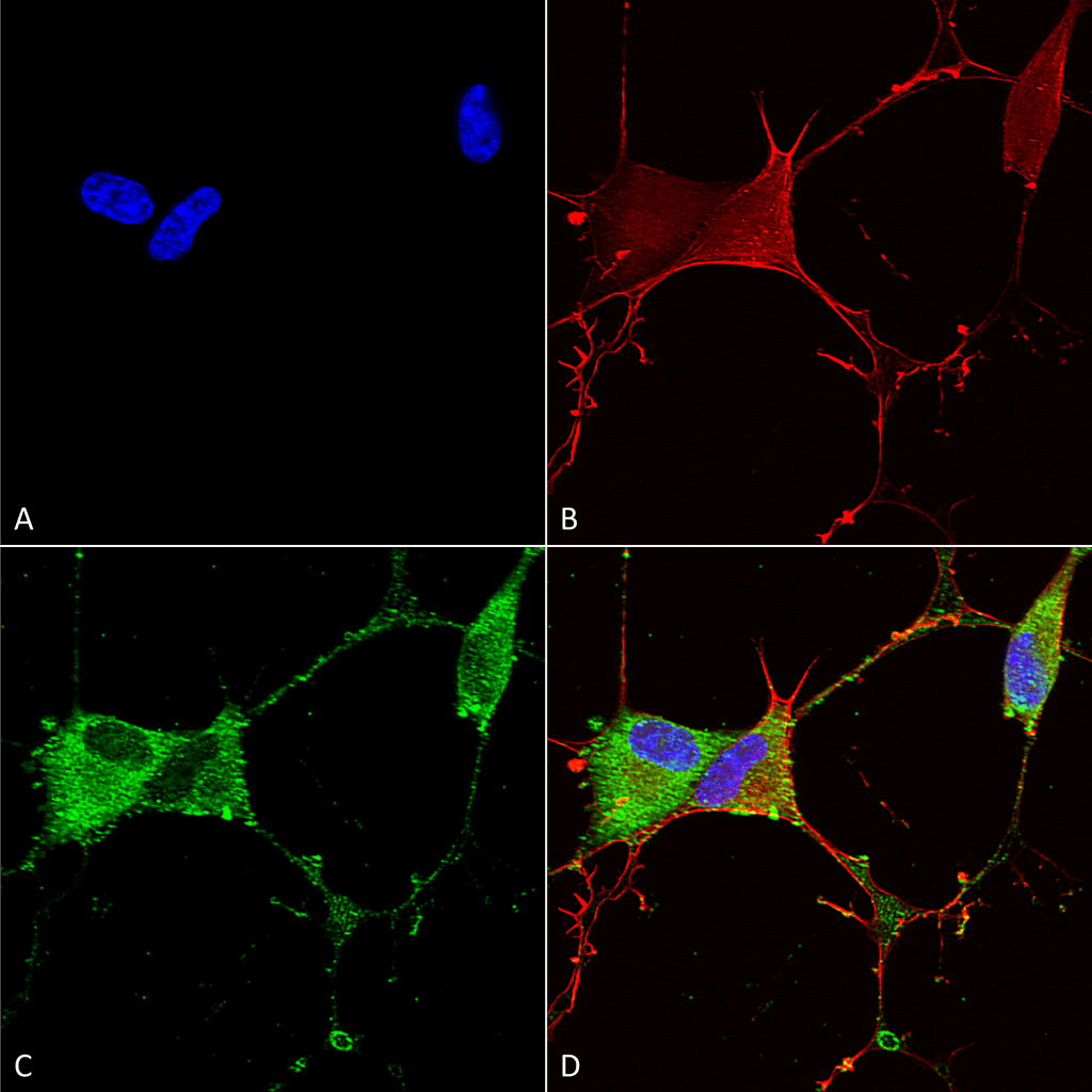
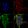
Description Immunocytochemistry/Immunofluorescence analysis using Mouse Anti-GABA-B Receptor 2 Monoclonal Antibody, Clone S81-2 (56523). Tissue: Neuroblastoma cells (SH-SY5Y). Species: Human. Fixation: 4% PFA for 15 min. Primary Antibody: Mouse Anti-GABA-B Receptor 2 Monoclonal Antibody (56523) at 1:100 for overnight at 4°C with slow rocking. Secondary Antibody: AlexaFluor 488 at 1:1000 for 1 hour at RT. Counterstain: Phalloidin-iFluor 647 (red) F-Actin stain; Hoechst (blue) nuclear stain at 1:800, 1.6mM for 20 min at RT. (A) Hoechst (blue) nuclear stain. (B) Phalloidin-iFluor 647 (red) F-Actin stain. (C) GABA-B Receptor 2 Antibody (D) Composite.

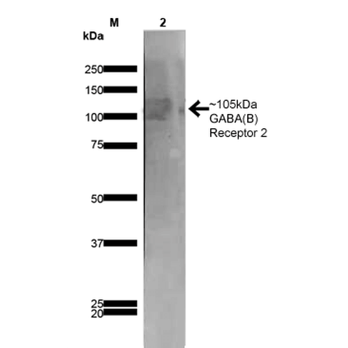
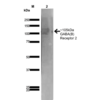
Description Western Blot analysis of Rat Brain Membrane showing detection of ~105 kDa GABA B Receptor 2 protein using Mouse Anti-GABA B Receptor 2 Monoclonal Antibody, Clone S81-2 (56523). Lane 1: MW Ladder. Lane 2: Rat Brain Membrane (10 µg). . Load: 10 µg. Block: 5% milk. Primary Antibody: Mouse Anti-GABA B Receptor 2 Monoclonal Antibody (56523) at 1:1000 for 1 hour at RT. Secondary Antibody: Goat Anti-Mouse IgG: HRP at 1:200 for 1 hour at RT. Color Development: TMB solution for 10 min at RT. Predicted/Observed Size: ~105 kDa.

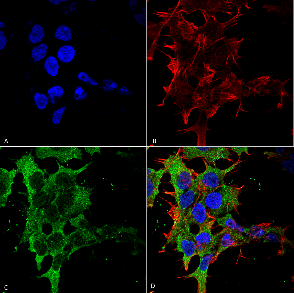
Description Immunocytochemistry/Immunofluorescence analysis using Mouse Anti-GABA-B Receptor 2 Monoclonal Antibody, Clone S81-2 (56523). Tissue: Neuroblastoma cell line (SK-N-BE). Species: Human. Fixation: 4% Formaldehyde for 15 min at RT. Primary Antibody: Mouse Anti-GABA-B Receptor 2 Monoclonal Antibody (56523) at 1:100 for 60 min at RT. Secondary Antibody: Goat Anti-Mouse ATTO 488 at 1:100 for 60 min at RT. Counterstain: Phalloidin Texas Red F-Actin stain; DAPI (blue) nuclear stain at 1:1000, 1:5000 for 60min RT, 5min RT. Localization: Cell Membrane. Magnification: 60X. (A) DAPI (blue) nuclear stain. (B) Phalloidin Texas Red F-Actin stain. (C) GABA-B Receptor 2 Antibody. (D) Composite.
Handling
Storage This product is stable for at least 1 year at -20°C. Freeze in multiple aliquots to avoid repeated freeze-thaw cycles.
Dilution Instructions Dilute in PBS or medium that is identical to that used in the assay system.
Application Instructions Immunoblotting: use at 1-2ug/mL. A band of ~105kDa is detected.
Immunohistochemistry: use at 1-5ug/mL.
These are recommended concentrations.
Enduser should determine optimal concentrations for their applications.
Positive control: Rat brain membranes.
Immunohistochemistry: use at 1-5ug/mL.
These are recommended concentrations.
Enduser should determine optimal concentrations for their applications.
Positive control: Rat brain membranes.
References & Data Sheet
Data Sheet  Download PDF Data Sheet
 Download PDF Data Sheet

