Anti-Phosphotyrosine Antibody (56494)
$466.00
SKU: 56494
Categories: Antibody Products, Neuroscience and Signal Transduction Antibodies, Products
Overview
Product Name Anti-Phosphotyrosine Antibody (56494)
Description Anti-Phosphotyrosine Mouse Monoclonal Antibody
Target Phosphotyrosine
Species Reactivity All
Applications WB,IHC,ICC/IF,IP
Host Mouse
Clonality Monoclonal
Clone ID G104
Isotype IgG1
Immunogen Phosphotyrosine, alanine and glycine in a 1:1:1 ratio polymerized in the presence of KLH with 1-ethyl-3-(3'- dimethylaminopropyl) carbodiimide.
Properties
Form Liquid
Concentration 1.0 mg/mL
Formulation PBS, pH 7.4
Buffer Formulation Phosphate Buffered Saline
Buffer pH pH 7.4
Format Purified
Purification Purified by Protein G affinity chromatography
Specificity Information
Specificity This antibody reacts with phosphotyrosine and detects phosphotyrosine in proteins of unstimulated and stimulated cell lysates. It does not cross-react with phosphoserine or phosphothreonine.
Target ID Phosphotyrosine
CAS Number 60-18-4
PubChem ID 1153
Research Areas Neuroscience
Background Protein phosphorylation is an important posttranslational modification that serves many key functions in regulation of protein activity, localization, and protein-protein interactions. Phosphorylation is catalyzed by specific protein kinases that remove a phosphate group from ATP and attach it covalently to a recipient protein. Some kinases act on serine and threonine, others act on tyrosine, and many act on all three. Phosphorylation of tyrosine is considered one of the key steps in signal transduction and regulation of enzymatic activity. Antibodies to phosphotyrosine are useful in identifying protein substrates of tyrosine kinase.
Application Images


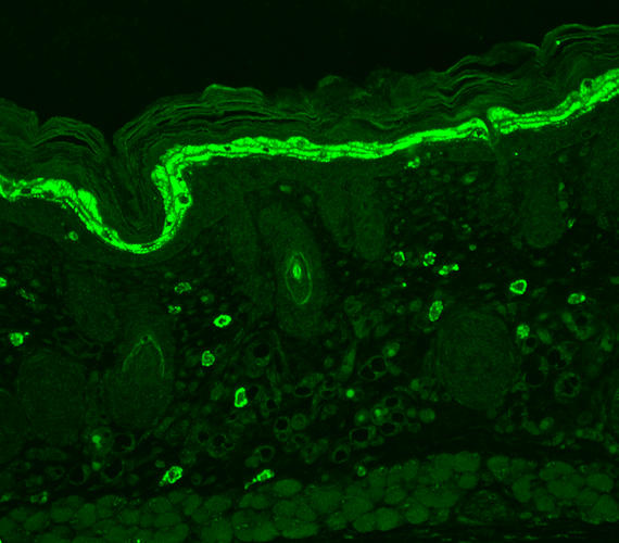
Description Immunohistochemistry analysis using Mouse Anti-Phosphotyrosine Monoclonal Antibody, Clone G104 (56494). Tissue: backskin. Species: Mouse. Fixation: Bouin's Fixative and paraffin-embedded. Primary Antibody: Mouse Anti-Phosphotyrosine Monoclonal Antibody (56494) at 1:100 for 1 hour at RT. Secondary Antibody: FITC Goat Anti-Mouse (green) at 1:50 for 1 hour at RT. Localization: Stratum granulosum staining in the epidermis. Some dermal staining.

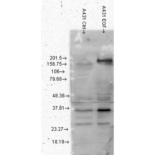
Description Western Blot analysis of Human A431 cell lysates showing detection of Phosphotyrosine protein using Mouse Anti-Phosphotyrosine Monoclonal Antibody, Clone G104 (56494). Load: 15 µg. Block: 1.5% BSA for 30 minutes at RT. Primary Antibody: Mouse Anti-Phosphotyrosine Monoclonal Antibody (56494) at 1:1000 for 2 hours at RT. Secondary Antibody: Sheep Anti-Mouse IgG: HRP for 1 hour at RT. Left: normal, right: EGF treated.
Handling
Storage This antibody is stable for at least one (1) year at -20°C.
Dilution Instructions Dilute in PBS or medium that is identical to that used in the assay system.
Application Instructions Immunoblotting: use at 1ug/mL.
Immunohistochemistry: use at 1-10ug/mL.
Immunofluorescence: use at 1-10ug/mL.
Positive control: Rat tissue lysate
These are recommended concentrations;
Enduser should determine optimal concentrations for their applications.See specific product references below for protocols and more information.
Immunohistochemistry: use at 1-10ug/mL.
Immunofluorescence: use at 1-10ug/mL.
Positive control: Rat tissue lysate
These are recommended concentrations;
Enduser should determine optimal concentrations for their applications.See specific product references below for protocols and more information.
References & Data Sheet
References Garton AJ et al. 1996 Mol Cell Biol 16: 6408-6418.
PMID 8887669
Data Sheet  Download PDF Data Sheet
 Download PDF Data Sheet


