Anti-p95/Nibrin Antibody (56249)
$503.00
SKU: 56249
Categories: Antibody Products, Cancer Research Antibodies, Products
Overview
Product Name Anti-p95/Nibrin Antibody (56249)
Description Anti-p95 Mouse Monoclonal Antibody
Target p95/Nibrin
Species Reactivity Human
Applications WB,ICC/IF,IP
Host Mouse
Clonality Monoclonal
Clone ID 1D7
Isotype IgG1
Immunogen Fusion protein containing the complete coding region of human p95/nibrin expressed in E. coli.
Properties
Form Liquid
Concentration Lot Specific
Formulation PBS, pH 7.4.
Buffer Formulation Phosphate Buffered Saline
Buffer pH pH 7.4
Format Purified
Purification Purified by Protein G affinity chromatography
Specificity Information
Specificity This antibody recognizes the 95 kD nibrin protein which contains a forkhead-associated domain that is adjacent to BRCT, a breast cancer C- terminal domain involved in protein- protein interactions. p95/nibrin is a member of the Mre11/Rad50 double- strand break repair complex.
Target Name Nibrin
Target ID p95/Nibrin
Uniprot ID O60934
Alternative Names Cell cycle regulatory protein p95, Nijmegen breakage syndrome protein 1
Gene Name NBN
Sequence Location Nucleus, Nucleus, PML body, Chromosome, telomere, Chromosome
Biological Function Component of the MRE11-RAD50-NBN (MRN complex) which plays a critical role in the cellular response to DNA damage and the maintenance of chromosome integrity. The complex is involved in double-strand break (DSB) repair, DNA rPubMed:10888888, PubMed:15616588, PubMed:19759395, PubMed:23762398, PubMed:26438602, PubMed:9705271}.
Research Areas Cancer research
Application Images


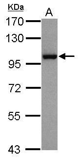
Description Sample (30 ug of whole cell lysate)
A: HepG2
7.5% SDS PAGE
56249 diluted at 1:1000
The HRP-conjugated anti-mouse IgG antibody was used to detect the primary antibody.
A: HepG2
7.5% SDS PAGE
56249 diluted at 1:1000
The HRP-conjugated anti-mouse IgG antibody was used to detect the primary antibody.

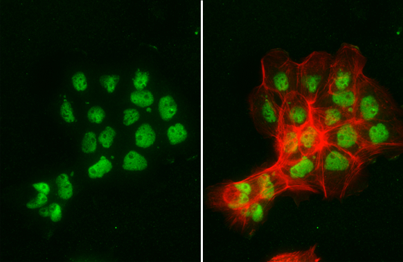
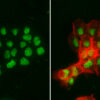
Description NBS1 antibody [1D7] detects NBS1 protein at nucleus by immunofluorescent analysis.Sample: A431 cells were fixed in 4% paraformaldehyde at RT for 15 min.Green: NBS1 stained by NBS1 antibody [1D7] (56249) diluted at 1:500.Red: phalloidin, a cytoskeleton marker, diluted at 1:200.

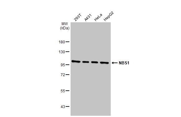
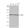
Description Various whole cell extracts (30 ug) were separated by 7.5% SDS-PAGE, and the membrane was blotted with NBS1 antibody [1D7] (56249) diluted at 1:500. The HRP-conjugated anti-mouse IgG antibody was used to detect the primary antibody.
Handling
Storage This antibody is stable for at least one (1) year at -70°C. Avoid multiple freeze- thaw cycles.
Dilution Instructions Dilute in PBS or medium which is identical to that used in the assay system.
Application Instructions Immunoblotting,
Immunoprecipitation:: use at 1-2 ug/mL. In immunoblots, a band of 95 kD is detected.
Positive controls: MCF-7, HeLa, or Raji cells.
Immunoprecipitation:: use at 1-2 ug/mL. In immunoblots, a band of 95 kD is detected.
Positive controls: MCF-7, HeLa, or Raji cells.
References & Data Sheet
Data Sheet  Download PDF Data Sheet
 Download PDF Data Sheet

