Anti-Interleukin-21 (IL-21) (IN) Antibody (41008)
$469.00
SKU: 41008
Categories: Antibody Products, Growth Factors/Cytokines/Receptor Antibodies, Products
Overview
Product Name Anti-Interleukin-21 (IL-21) (IN) Antibody (41008)
Description Anti-IL-21 (IN) Rabbit Polyclonal Antibody
Target Interleukin-21 (IL-21) (IN)
Species Reactivity Human
Applications ELISA,WB,ICC
Host Rabbit
Clonality Polyclonal
Immunogen Peptide corresponding to aa 121-135 of human IL-21 precursor.
Properties
Form Liquid
Concentration Lot Specific
Formulation PBS, pH 7.4.
Buffer Formulation Phosphate Buffered Saline
Buffer pH pH 7.4
Format Purified
Purification Purified by peptide immuno-affinity chromatography
Specificity Information
Specificity This antibody recognizes human IL-21, a cytokine that has significant homology to IL-2, IL-4, and IL-15. The receptor for IL-21, IL-21R, complexes with the common cytokine receptor g chain, gc, and mediates IL-21 signaling. IL-21 and its receptor activate the JAK-STAT signaling pathway. IL-21 is expressed in activated T cells and the HL-60 and THP-1 cell lines. IL-21 is associated with the proliferation and maturation of NK, B and T cell populations.
Target Name Interleukin-21
Target ID Interleukin-21 (IL-21) (IN)
Uniprot ID Q9HBE4
Alternative Names IL-21, Za11
Gene Name IL21
Sequence Location Secreted
Biological Function Cytokine with immunoregulatory activity. May promote the transition between innate and adaptive immunity. Induces the production of IgG(1) and IgG(3) in B-cells (By similarity). Implicated in the generation and maintenance of T follicular helper (Tfh) cells and the formation of germinal-centers. Together with IL6, control the early generation of Tfh cells and are critical for an effective antibody response to acute viral infection (By similarity). May play a role in proliferation and maturation of natural killer (NK) cells in synergy with IL15. May regulate proliferation of mature B- and T-cells in response to activating stimuli. In synergy with IL15 and IL18 stimulates interferon gamma production in T-cells and NK cells (PubMed:11081504, PubMed:15178704). During T-cell mediated immune response may inhibit dendritic cells (DC) activation and maturation (By similarity). {UniProtKB:Q9ES17, PubMed:11081504, PubMed:15178704}.
Research Areas Growth Factors, Cytokines, Receptors
Application Images
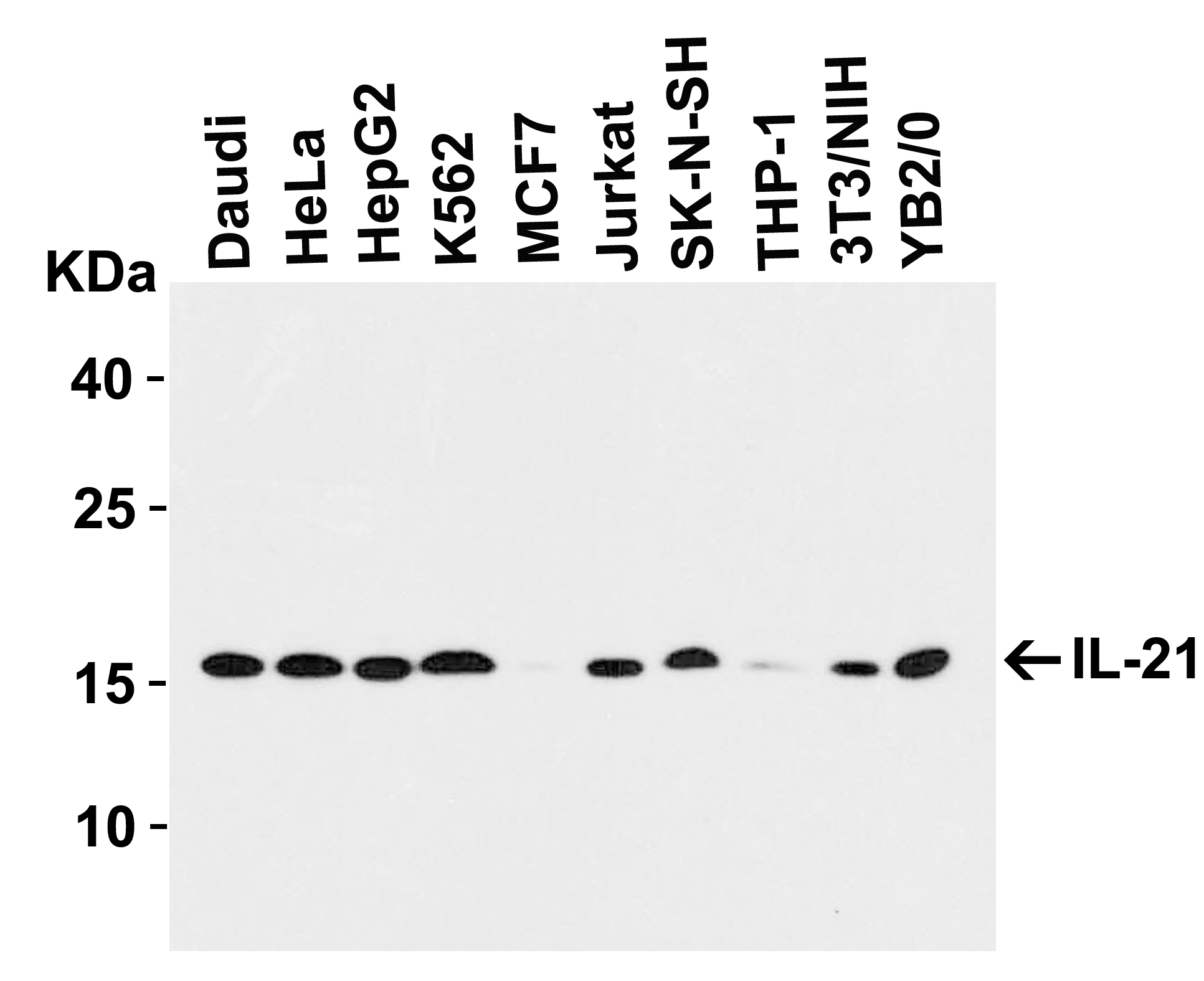

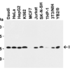
Description Western Blot Validation in Human, Mouse and Rat Cell Lines
Loading: 15 ug of lysates per lane. Antibodies: IL-21 41008, (1 ug/mL), 1h incubation at RT in 5% NFDM/TBST.Secondary: Goat anti-rabbit IgG HRP conjugate at 1:10000 dilution.
Loading: 15 ug of lysates per lane. Antibodies: IL-21 41008, (1 ug/mL), 1h incubation at RT in 5% NFDM/TBST.Secondary: Goat anti-rabbit IgG HRP conjugate at 1:10000 dilution.

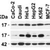
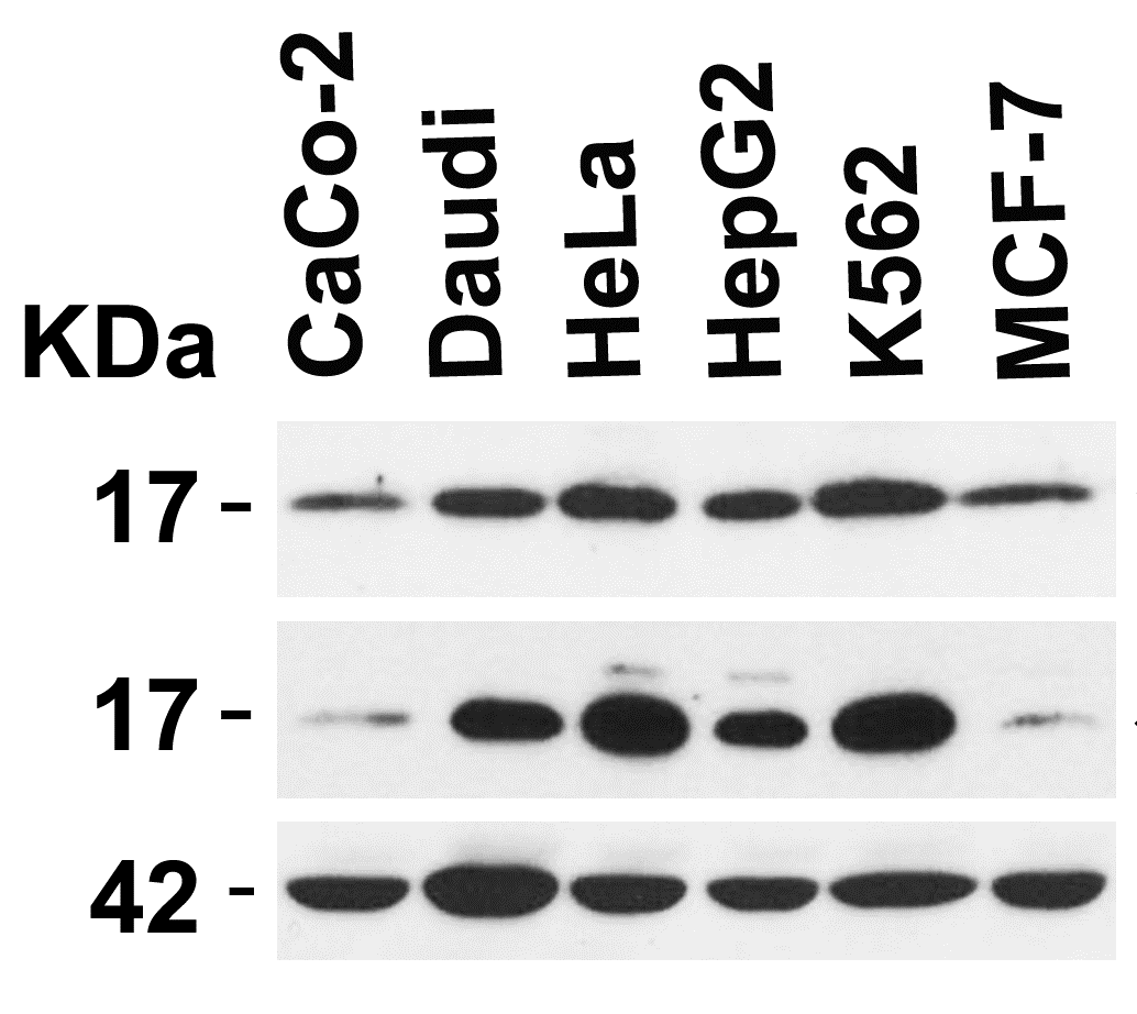
Description Independent Antibody Validation (IAV) via Protein Expression Profile in Cell Lines
Loading: 15 ug of lysates per lane. Antibodies: IL-21 41008 (1 ug/mL), IL-21 2463 (5 ug/mL), beta-actin (5 ug/mL) and beta-actin (1 ug/mL), 1h incubation at RT in 5% NFDM/TBST.Secondary: Goat anti-rabbit IgG HRP conjugate at 1:10000 dilution.
Loading: 15 ug of lysates per lane. Antibodies: IL-21 41008 (1 ug/mL), IL-21 2463 (5 ug/mL), beta-actin (5 ug/mL) and beta-actin (1 ug/mL), 1h incubation at RT in 5% NFDM/TBST.Secondary: Goat anti-rabbit IgG HRP conjugate at 1:10000 dilution.

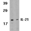
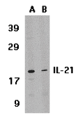
Description Western Blot Validation in Human HL-60 Cell Lysate
Loading: 15 ug of lysates per lane. Antibodies: IL-21 41008 (1 ug/mL), 1h incubation at RT in 5% NFDM/TBST.Secondary: Goat anti-rabbit IgG HRP conjugate at 1:10000 dilution.(A) Absence of blocking peptide(B) Presence of blocking peptide.
Loading: 15 ug of lysates per lane. Antibodies: IL-21 41008 (1 ug/mL), 1h incubation at RT in 5% NFDM/TBST.Secondary: Goat anti-rabbit IgG HRP conjugate at 1:10000 dilution.(A) Absence of blocking peptide(B) Presence of blocking peptide.

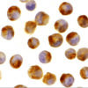
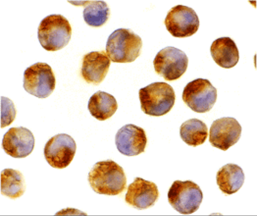
Description Immunocytochemistry Validation of IL-21 in K562 Cells
Immunocytochemical analysis of K562 cells using anti-IL-21 antibody (41008) at 2 ug/mL. Cells was fixed with formaldehyde and blocked with 10% serum for 1 h at RT; antigen retrieval was by heat mediation with a citrate buffer (pH6). Samples were incubated with primary antibody overnight at 4 C. A goat anti-rabbit IgG H&L (HRP) at 1/250 was used as secondary. Counter stained with Hematoxylin.
Immunocytochemical analysis of K562 cells using anti-IL-21 antibody (41008) at 2 ug/mL. Cells was fixed with formaldehyde and blocked with 10% serum for 1 h at RT; antigen retrieval was by heat mediation with a citrate buffer (pH6). Samples were incubated with primary antibody overnight at 4 C. A goat anti-rabbit IgG H&L (HRP) at 1/250 was used as secondary. Counter stained with Hematoxylin.

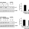
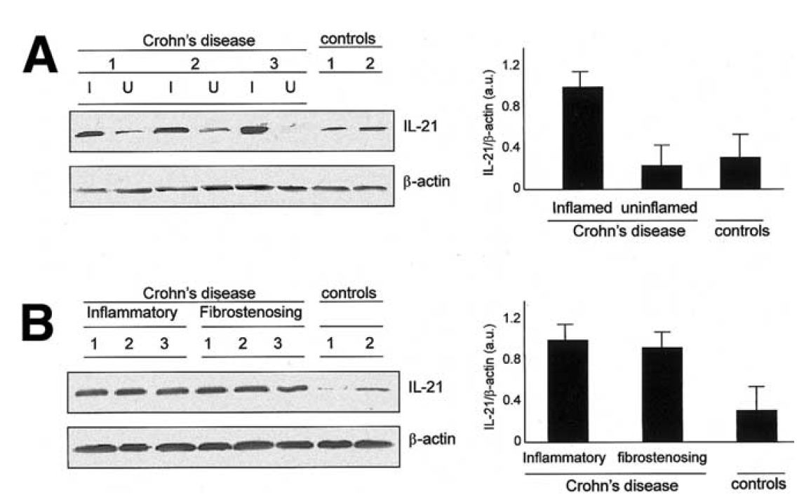
Description Induced Expression Validation of IL-21 expresssion in patients with Crohn’s Disease (Monteleone et al, 2416)
Enhanced IL-21 was observed in involved but not uninvolvedCD, and was not associated with any CD phenotype, such as fibrostenosing disease. IL-21 expression was detected by anti-IL-21 antibodies (41008).
Enhanced IL-21 was observed in involved but not uninvolvedCD, and was not associated with any CD phenotype, such as fibrostenosing disease. IL-21 expression was detected by anti-IL-21 antibodies (41008).

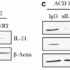
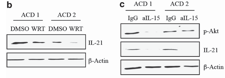
Description Regulation of IL-21 expresssion in duodenal biopsies of two patients with active Celiac disease (ACD) (Sarra et al., 2412)
(b) shows IL-21 expression levels treated with dimethyl sulfoxide (DMSO) or wortmannin (WRT). (c) shows IL-21 expression levels with the inhibition of IL-15 antibody (aIL-15). IgG isotype was used as a control.Both WB show IL-21 expression decreased with the treatment with WRT or the inhibition of IL-15 antibody.
(b) shows IL-21 expression levels treated with dimethyl sulfoxide (DMSO) or wortmannin (WRT). (c) shows IL-21 expression levels with the inhibition of IL-15 antibody (aIL-15). IgG isotype was used as a control.Both WB show IL-21 expression decreased with the treatment with WRT or the inhibition of IL-15 antibody.

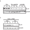
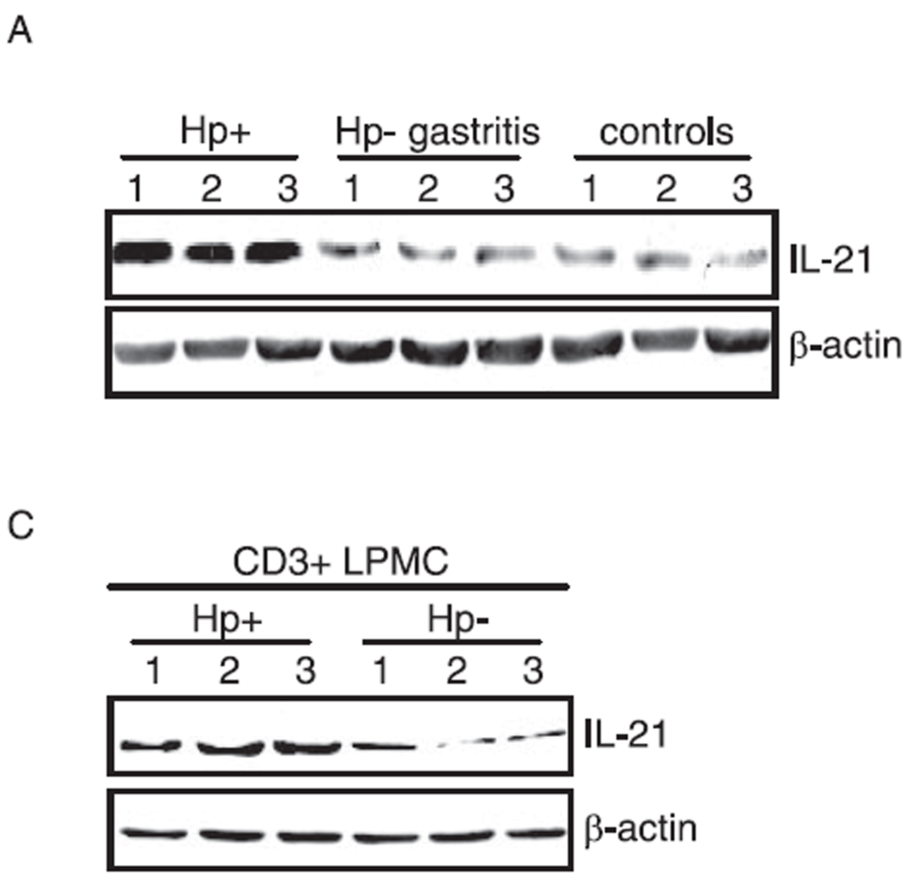
Description Induced Expression Validation of IL-21 expresssion in patients with Helicobacter Pylori (Hp) (Caruso et al., 2416)
(A) shows IL-21 expression levels from biopsies of patients with Hp infection and without Hp infection (n=3).(C) shows IL-21 expression levels in CD3+LPMC cells from Hp-positive and Hp-negative patients (n=3).Both WB show IL-21 expression increased in patients with Hp infection.
(A) shows IL-21 expression levels from biopsies of patients with Hp infection and without Hp infection (n=3).(C) shows IL-21 expression levels in CD3+LPMC cells from Hp-positive and Hp-negative patients (n=3).Both WB show IL-21 expression increased in patients with Hp infection.
Handling
Storage This antibody is stable for at least one (1) year at -20°C. Avoid multiple freeze- thaw cycles.
Dilution Instructions Dilute in PBS or medium that is identical to that used in the assay system.
Application Instructions Immunoblotting: use at 0.5-1 ug/mL. In immunoblots, a band of 18 kD is detected.
Positive control: HL60 or THP-1 cell lysates.
Positive control: HL60 or THP-1 cell lysates.
References & Data Sheet
Data Sheet  Download PDF Data Sheet
 Download PDF Data Sheet


