Anti-ZIP Kinase Antibody (2431)
$445.00
SKU: 2431
Categories: Antibody Products, Apoptosis Antibodies, Products
Overview
Product Name Anti-ZIP Kinase Antibody (2431)
Description Anti-ZIP Kinase Rabbit Polyclonal Antibody
Target ZIP Kinase
Species Reactivity Human
Applications ELISA,WB,ICC,IF
Host Rabbit
Clonality Polyclonal
Isotype IgG
Immunogen Peptide corresponding to aa 279- 298 of human ZIP kinase (accession no. BAA81746).
Properties
Form Liquid
Concentration Lot Specific
Formulation PBS, pH 7.4.
Buffer Formulation Phosphate Buffered Saline
Buffer pH pH 7.4
Format Purified
Purification Purified by peptide immuno-affinity chromatography
Specificity Information
Specificity This antibody recognizes full-length human, mouse, and rat ZIP kinase (52kDa).
Target Name Death-associated protein kinase 3
Target ID ZIP Kinase
Uniprot ID O43293
Alternative Names DAP kinase 3, EC 2.7.11.1, DAP-like kinase, Dlk, MYPT1 kinase, Zipper-interacting protein kinase, ZIP-kinase
Gene Name DAPK3
Gene ID 1613
Accession Number NP_001339
Sequence Location Nucleus, Cytoplasm
Biological Function Serine/threonine kinase which is involved in the regulation of apoptosis, autophagy, transcription, translation and actin cytoskeleton reorganization. Involved in the regulation of smooth muscle contraction. Regulates both type I (caspase-dependent) apoptotic and type II (caspase-independent) autophagic cell deaths signal, depending on the cellular setting. Involved in regulation of starvation-induced autophagy. Regulates myosin phosphorylation in both smooth muscle and non-muscle cells. In smooth muscle, regulates myosin either directly by phosphorylating MYL12B and MYL9 or through inhibition of smooth muscle myosin phosphatase (SMPP1M) via phosphorylation of PPP1R12A; the inhibition of SMPP1M functions to enhance muscle responsiveness to Ca(2+) and promote a contractile state. Phosphorylates MYL12B in non-muscle cells leading to reorganization of actin cytoskeleton. Isoform 2 can phosphorylate myosin, PPP1R12A and MYL12B. Overexpression leads to condensation of actin stress fibers into thick bundles. Involved in actin filament focal adhesion dynamics. The function in both reorganization of actin cytoskeleton and focal adhesion dissolution is modulated by RhoD. Positively regulates canonical Wnt/beta-catenin signaling through interaction with NLK and TCF7L2. Phosphorylates RPL13A on 'Ser-77' upon interferon-gamma activation which is causing RPL13A release from the ribosome, RPL13A association with the GAIT complex and its subsequent involvement in transcript-selective translation inhibition. Enhances transcription from AR-responsive promoters in a hormone- and kinase-dependent manner. Involved in regulation of cell cycle progression and cell proliferation. May be a tumor suppressor. {PubMed:10356987, PubMed:11384979, PubMed:11781833, PubMed:12917339, PubMed:15096528, PubMed:15367680, PubMed:16219639, PubMed:17126281, PubMed:17158456, PubMed:18084323, PubMed:18995835, PubMed:21169990, PubMed:21408167, PubMed:21454679, PubMed:21487036, PubMed:23454120}.
Research Areas Apoptosis
Background A novel serine/threonine kinase that mediates apoptosis has been identified and designated ZIP kinase. ZIP kinase contains an N-terminal kinase domain and a C- terminal leucine zipper structure and binds to ATF4, a member of ATF/CREB family. ZIP kinase has high sequence homology to DAP kinase (death-associated protein kinase), a mediator of apoptosis induced by gamma interferon. Overexpression of ZIP kinase induces apoptosis. ZIP and DAP kinases represent a novel kinase family which mediates apoptosis through their catalytic activities. Messenger RNA for ZIP kinase is widely expressed in a variety of human tissues.
Application Images


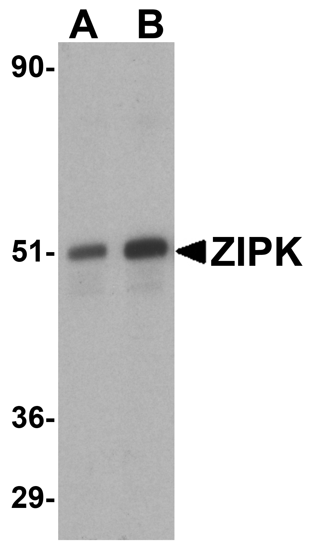
Description Western blot analysis of ZIP kinase in (A) HeLa and (B) Jurkat lysates with ZIP kinase antibody at 1 ug/mL.

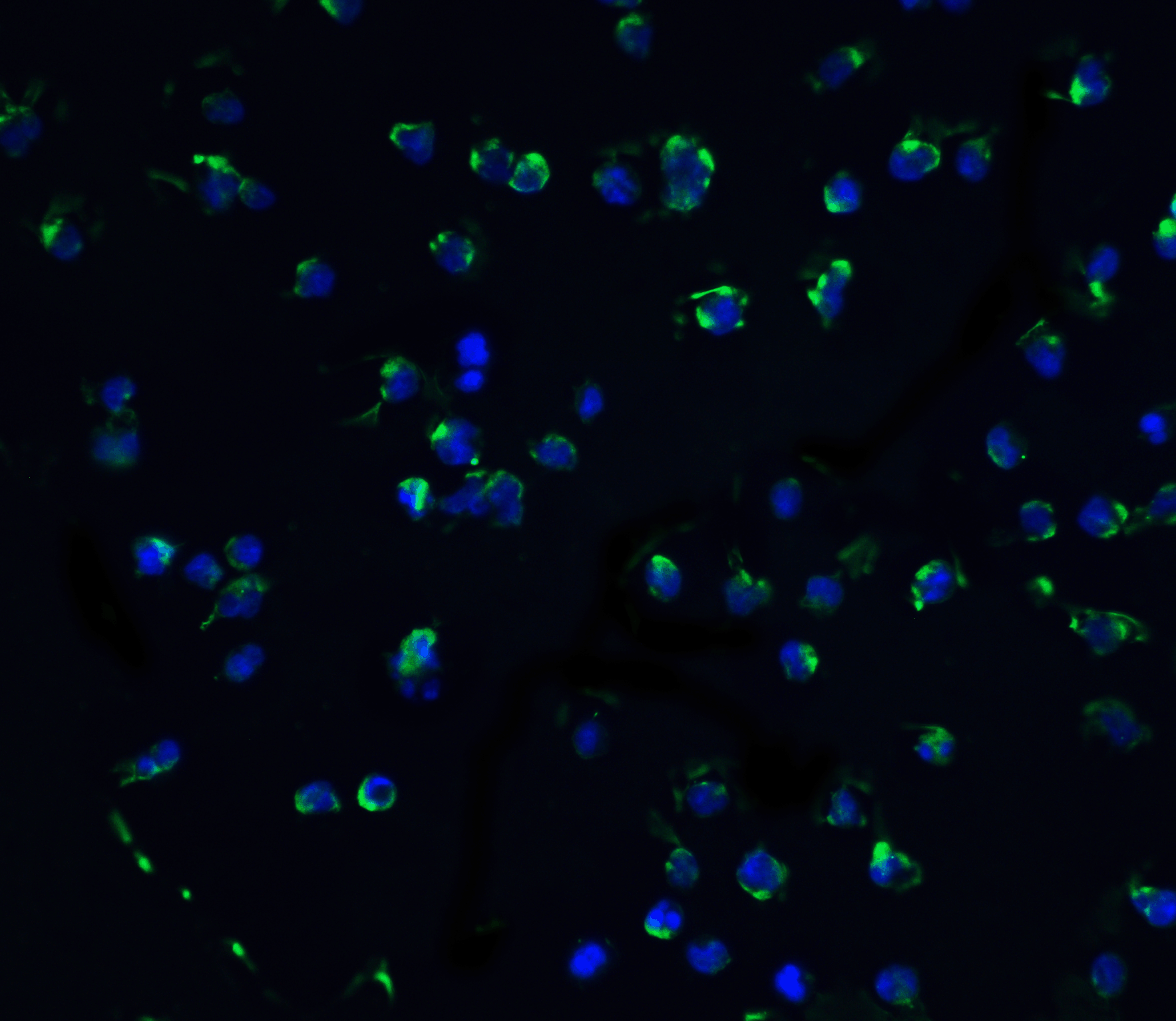
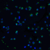
Description Immunofluorescence of ZIPK in Jurkat cells with ZIPK antibody at 10 ug/mL.

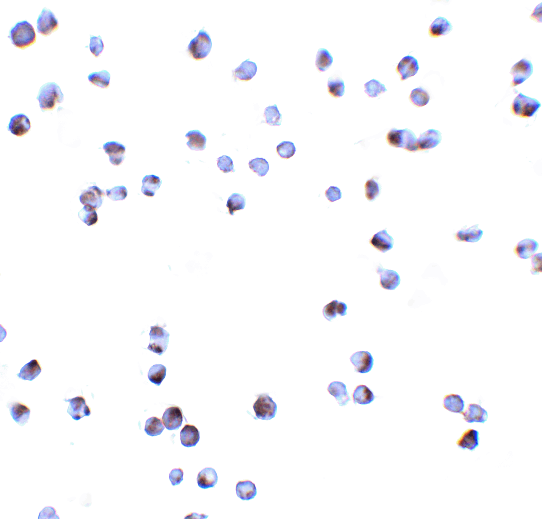
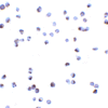
Description Immunocytochemistry of ZIPK in Jurkat cells with ZIPK antibody at 10 ug/mL.
Handling
Storage This antibody is stable for at least one (1) year at -20°C. Avoid multiple freeze-thaw cycles.
Dilution Instructions Dilute in PBS or medium which is identical to that used in the assay system.
Application Instructions Immunoblotting: use at 1ug/mL.
Positive control: HeLa cell lysate.
Immunocytochemistry: use at 10ug/mL.
These are recommended concentrations.
Enduser should determine optimal concentrations for their applications.
Positive control: HeLa cell lysate.
Immunocytochemistry: use at 10ug/mL.
These are recommended concentrations.
Enduser should determine optimal concentrations for their applications.
References & Data Sheet
Data Sheet  Download PDF Data Sheet
 Download PDF Data Sheet

