Anti-RICK (NT) Antibody (2427)
$445.00
SKU: 2427
Categories: Antibody Products, Apoptosis Antibodies, Products
Overview
Product Name Anti-RICK (NT) Antibody (2427)
Description Anti-RICK (NT) Rabbit Polyclonal Antibody
Target RICK (NT)
Species Reactivity Human
Applications ELISA,WB,ICC,IF
Host Rabbit
Clonality Polyclonal
Isotype IgG
Immunogen Synthetic peptide corresponding to aa 11-30 at the N-terminus of human RICK (accession no. O43353).
Properties
Form Liquid
Concentration Lot Specific
Formulation PBS, pH 7.4.
Buffer Formulation Phosphate Buffered Saline
Buffer pH pH 7.4
Format Purified
Purification Purified by peptide immuno-affinity chromatography
Specificity Information
Specificity This antibody recognizes full-length human, mouse, and rat RICK (60kDa).
Target Name Receptor-interacting serine/threonine-protein kinase 2
Target ID RICK (NT)
Uniprot ID O43353
Alternative Names EC 2.7.11.1, CARD-containing interleukin-1 β-converting enzyme-associated kinase, CARD-containing IL-1 β ICE-kinase, RIP-like-interacting CLARP kinase, Receptor-interacting protein 2, RIP-2, Tyrosine-protein kinase RIPK2, EC 2.7.10.2
Gene Name RIPK2
Gene ID 8767
Accession Number NP_003812
Sequence Location Cytoplasm
Biological Function Serine/threonine/tyrosine kinase that plays an essential role in modulation of innate and adaptive immune responses. Upon stimulation by bacterial peptidoglycans, NOD1 and NOD2 are activated, oligomerize and recruit RIPK2 through CARD-CARD domains. Contributes to the tyrosine phosphorylation of the guanine exchange factor ARHGEF2 through Src tyrosine kinase leading to NF-kappaB activation by NOD2. Once recruited, RIPK2 autophosphorylates and undergoes 'Lys-63'-linked polyubiquitination by E3 ubiquitin ligases XIAP, BIRC2 and BIRC3. The polyubiquitinated protein mediates the recruitment of MAP3K7/TAK1 to IKBKG/NEMO and induces 'Lys-63'-linked polyubiquitination of IKBKG/NEMO and subsequent activation of IKBKB/IKKB. In turn, NF-kappa-B is released from NF-kappa-B inhibitors and translocates into the nucleus where it activates the transcription of hundreds of genes involved in immune response, growth control, or protection against apoptosis. Plays also a role during engagement of the T-cell receptor (TCR) in promoting BCL10 phosphorylation and subsequent NF-kappa-B activation. Plays a role in the inactivation of RHOA in response to NGFR signaling (PubMed:26646181). {PubMed:14638696, PubMed:17054981, PubMed:18079694, PubMed:21123652, PubMed:21887730, PubMed:26646181}.
Research Areas Apoptosis
Background A CARD-containing serine/threonine kinase that regulates apoptosis has been identified and designated RICK (RIP-like interacting CLARP kinase). RICK contains an N-terminal kinase catalytic domain and a C-terminal CARD domain. RICK kinase domain has high sequence homology to that of RIP. Overexpression of RICK promotes the activation of caspase-8 and Fas-induced apoptosis. RICK represents a novel kinase that regulates Fas induced- apoptosis. The messenger RNA of RICK is expressed in a variety of human tissues.
Application Images


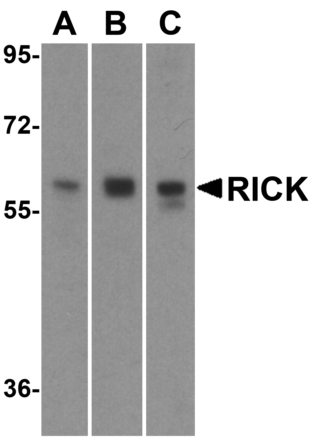
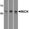
Description Western blot analysis of RICK in (A) HeLa, (B) Ramos and (C) EL4 cell lysate with RICK antibody at 1 ug/mL.

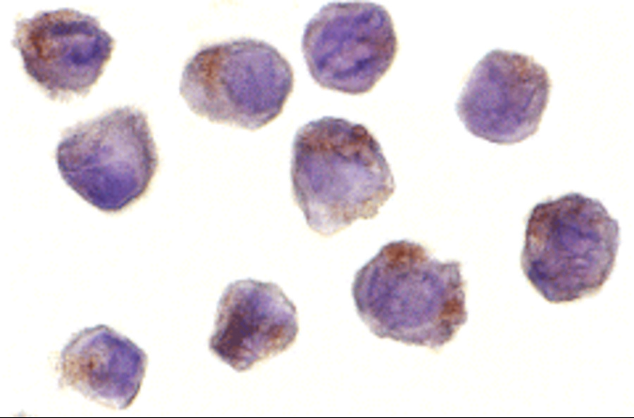
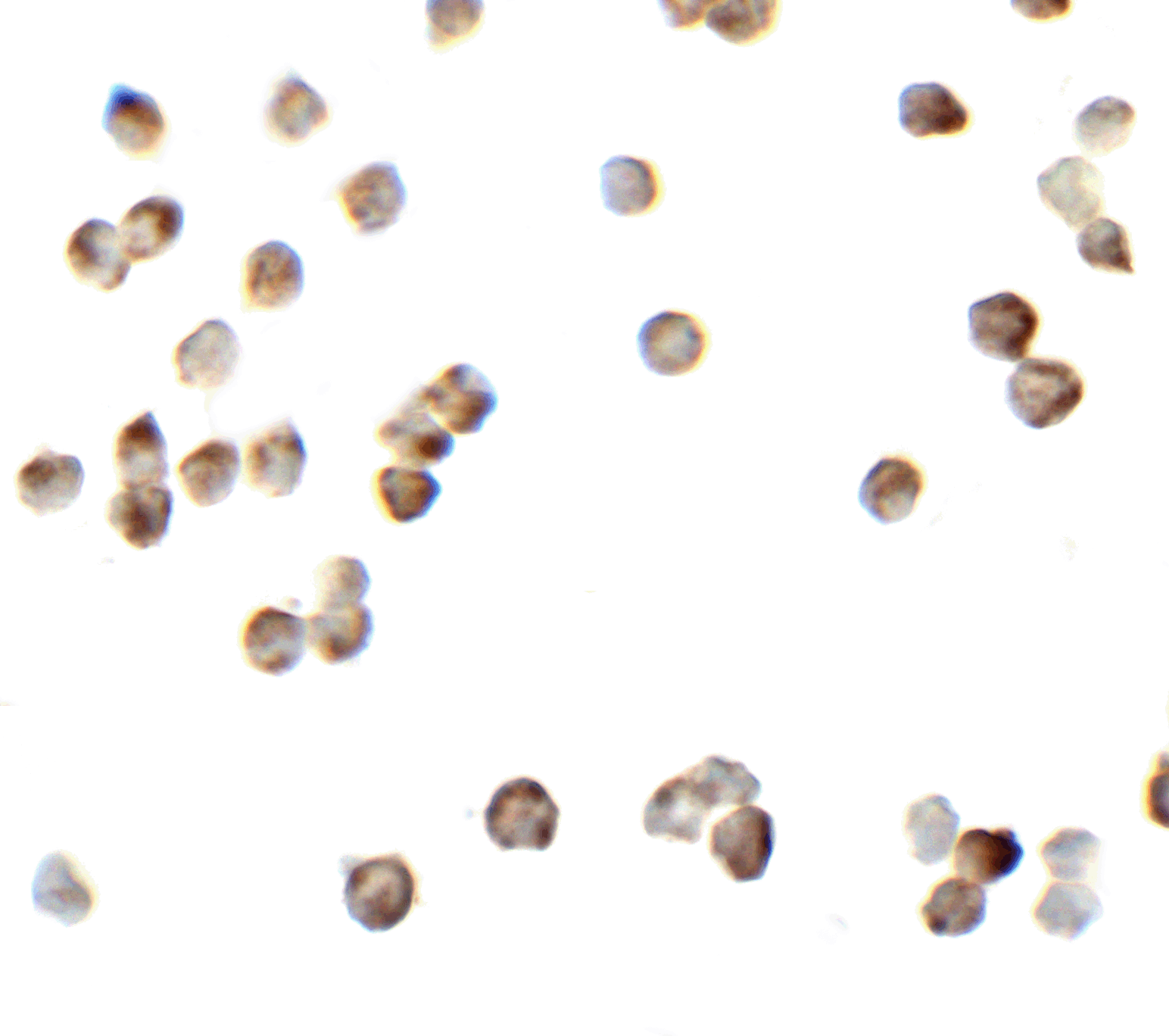
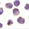
Description Immunocytochemistry of RICK in A431 cells with RICK antibody at 10 ug/mL.

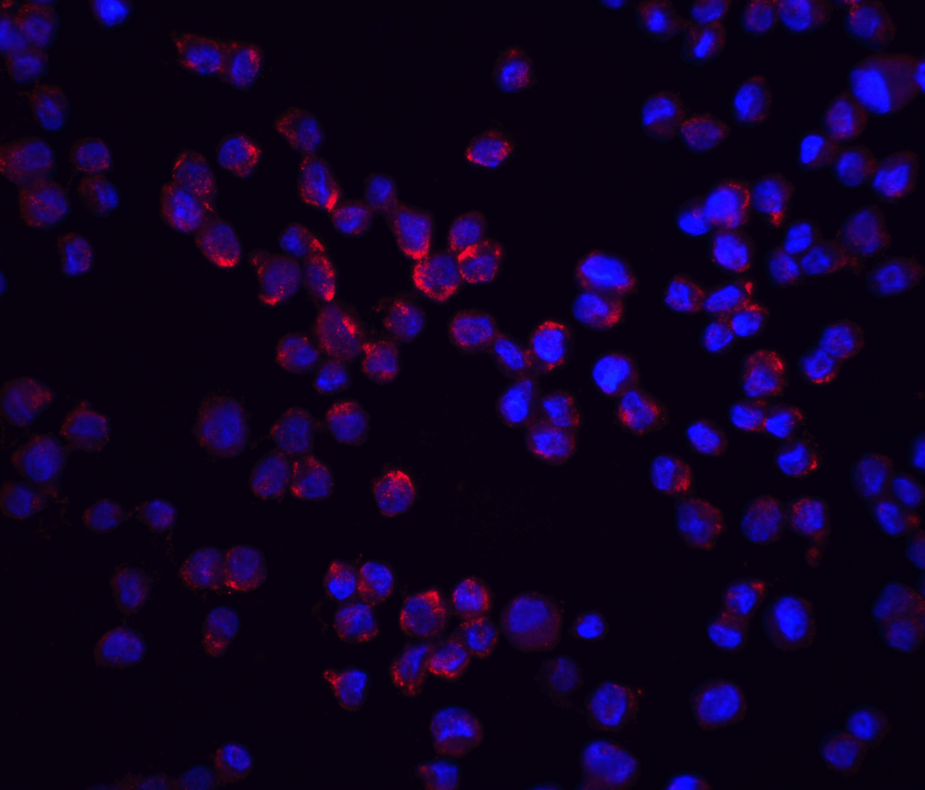
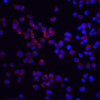
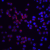
Description Immunofluorescence of RICK in K562 cells with RICK antibody at 20 ug/ml.

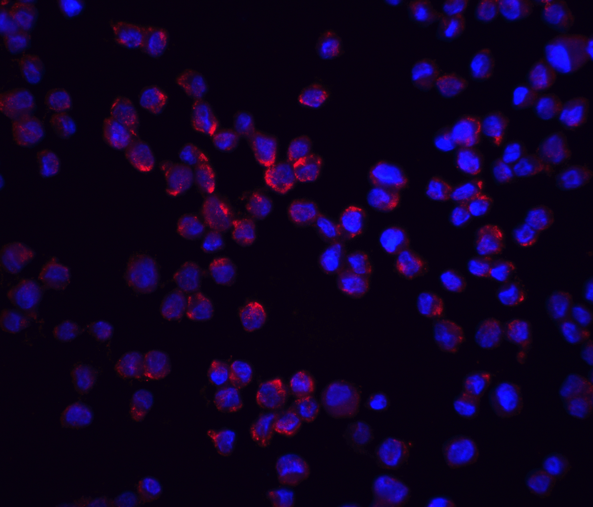
Description Immunofluorescence of RICK in K562 cells with RICK antibody at 20 ug/mL.
Red: RICK Antibody (2427)
Blue: DAPI staining
Blue: DAPI staining

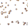
Description Immunocytochemistry of RICK in K562 cells with RICK antibody at 2.5 ug/mL.
Handling
Storage This antibody is stable for at least one (1) year at -20°C. Avoid multiple freeze-thaw cycles.
Dilution Instructions Dilute in PBS or medium which is identical to that used in the assay system.
Application Instructions Immunoblotting: use at 1ug/mL.
Positive control: A431 or K562 cell lysate.
Immunocytochemistry: use at 10ug/mL.
These are recommended concentrations.
Enduser should determine optimal concentrations for their applications.
Positive control: A431 or K562 cell lysate.
Immunocytochemistry: use at 10ug/mL.
These are recommended concentrations.
Enduser should determine optimal concentrations for their applications.
References & Data Sheet
Data Sheet  Download PDF Data Sheet
 Download PDF Data Sheet

