Anti-DR3 (CT) Antibody (2403)
$445.00
SKU: 2403
Categories: Antibody Products, Apoptosis Antibodies, Products
Overview
Product Name Anti-DR3 (CT) Antibody (2403)
Description Anti-DR3 (CT) Rabbit Polyclonal Antibody
Target DR3 (CT)
Species Reactivity Human
Applications ELISA,WB,IF
Host Rabbit
Clonality Polyclonal
Isotype IgG
Immunogen Peptide corresponding to aa 398-417 of human DR3 (accession no. AAQ88676).
Properties
Form Liquid
Concentration Lot Specific
Formulation PBS, pH 7.4.
Buffer Formulation Phosphate Buffered Saline
Buffer pH pH 7.4
Format Purified
Purification Purified by peptide immuno-affinity chromatography
Specificity Information
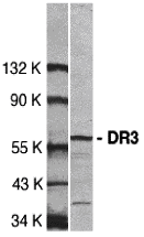
Specificity This antibody recognizes full-length human DR3 (59kDa).
Target Name Tumor necrosis factor receptor superfamily member 25
Target ID DR3 (CT)
Uniprot ID Q93038
Alternative Names Apo-3, Apoptosis-inducing receptor AIR, Apoptosis-mediating receptor DR3, Apoptosis-mediating receptor TRAMP, Death receptor 3, Lymphocyte-associated receptor of death, LARD, Protein WSL, Protein WSL-1
Gene Name TNFRSF25
Gene ID 8718
Accession Number NP_683868
Sequence Location [Isoform 1]: Cell membrane; Single-pass type I membrane protein.; [Isoform 2]: Cell membrane; Single-pass type I membrane protein.; [Isoform 9]: Cell membrane; Single-pass type I membrane protein.; [Isoform 11]: Cell membrane; Single-pass type I membrane protein.; [Isoform 3]: Secreted.; [Isoform 4]: Secreted.; [Isoform 5]: Secreted.; [Isoform 6]: Secreted.; [Isoform 7]: Secreted.; [Isoform 8]: Secreted.; [Isoform 10]: Secreted.; [Isoform 12]: Secreted.
Biological Function Receptor for TNFSF12/APO3L/TWEAK. Interacts directly with the adapter TRADD. Mediates activation of NF-kappa-B and induces apoptosis. May play a role in regulating lymphocyte homeostasis. {PubMed:8875942, PubMed:8994832, PubMed:9052839}.
Research Areas Apoptosis
Background Apoptosis is induced by certain cytokines including TNF and Fas ligand of the TNF family through their death domain-containing receptors, TNFR1 and Fas. Another cell death receptor has been identified and designated DR3, Wsl-1, Apo-3, TRAMP, and LARD. DR3 is widely expressed in tissues enriched in lymphocytes including peripheral blood, thymus, and spleen. Like TNFR1, DR3 induces apoptosis and NF- B activation.
Application Images


Description Western blot analysis of DR3 in Jurkat total cell lysate with DR3 antibody at 1:500 dilution.

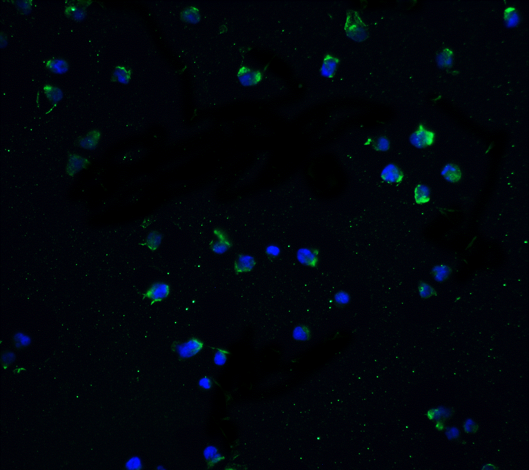
Description Immunofluorescence of DR3 in Jurkat cells with DR3 antibody at 20 ug/mL.
Green: DR3 Antibody (2403)
Blue: DAPI staining
Blue: DAPI staining
Handling
Storage This antibody is stable for at least one (1) year at -20°C. Avoid multiple freeze-thaw cycles.
Dilution Instructions Dilute in PBS or medium which is identical to that used in the assay system.
Application Instructions Immunoblotting: use at 2ug/mL.
Positive control: Whole cell lysate from Jurkat cells.
These are recommended concentrations.
Enduser should determine optimal concentrations for their applications.
Positive control: Whole cell lysate from Jurkat cells.
These are recommended concentrations.
Enduser should determine optimal concentrations for their applications.
References & Data Sheet
Data Sheet  Download PDF Data Sheet
 Download PDF Data Sheet


