Anti-BACE (CT) Antibody (2253)
$445.00
| Host | Quantity | Applications | Species Reactivity | Data Sheet | |
|---|---|---|---|---|---|
| Rabbit | 100ug | ELISA,WB,ICC,IHC-P,IF | Human, Mouse |  |
SKU: 2253
Categories: Antibody Products, Neuroscience and Signal Transduction Antibodies, Products
Overview
Product Name Anti-BACE (CT) Antibody (2253)
Description Anti-BACE (CT) Rabbit Polyclonal Antibody
Target BACE (CT)
Species Reactivity Human, Mouse
Applications ELISA,WB,ICC,IHC-P,IF
Host Rabbit
Clonality Polyclonal
Isotype IgG
Immunogen Peptide corresponding to aa 485-501 of human BACE. This sequence differs from those of mouse and rat by one amino acid.
Properties
Form Liquid
Concentration Lot Specific
Formulation PBS, pH 7.4.
Buffer Formulation Phosphate Buffered Saline
Buffer pH pH 7.4
Format Purified
Purification Purified by immunoaffinity chromatography
Specificity Information
Specificity This antibody recognizes human and mouse BACE (approx. 70 kD).
Target Name β-secretase 1
Target ID BACE (CT)
Uniprot ID P56817
Alternative Names EC 3.4.23.46, Aspartyl protease 2, ASP2, Asp 2, β-site amyloid precursor protein cleaving enzyme 1, β-site APP cleaving enzyme 1, Memapsin-2, Membrane-associated aspartic protease 2
Gene Name BACE1
Gene ID 23621
Accession Number NP_620428
Sequence Location Cell membrane, Golgi apparatus, trans-Golgi network, Endoplasmic reticulum, Endosome, Cell surface, Cytoplasmic vesicle membrane, Membrane raft, Lysosome, Late endosome, Early endosome, Recycling endosome, Cell projection, axon, Cell projection, dendrite
Biological Function Responsible for the proteolytic processing of the amyloid precursor protein (APP). Cleaves at the N-terminus of the A-beta peptide sequence, between residues 671 and 672 of APP, leads to the generation and extracellular release of beta-cleaved soluble APP, and a corresponding cell-associated C-terminal fragment which is later released by gamma-secretase (PubMed:10656250, PubMed:10677483, PubMed:20354142). Cleaves CHL1 (By similarity). {UniProtKB:P56818, PubMed:10656250, PubMed:10677483, PubMed:20354142}.
Research Areas Neuroscience
Background Accumulation of the amyloid-beta peptide (Abeta) in the cerebral cortex is a critical event in the pathogenesis of Alzheimer's disease. Abeta peptide is generated as a result of proteolytic cleavage of the beta-amyloid protein precursor (APP) at beta- and g-sites by two proteases. APP is first cleaved by beta- secretase to produce a soluble derivative of the protein and a membrane-anchored 99- amino acid carboxy-terminal fragment (C99). The C99 fragment serves as substrate for g-secretase to generate the 4 kD amyloid-beta peptide which is found in brains of patients with Alzheimer's disease. beta-secretase was recently identified and designated beta-site APP cleaving enzyme (BACE) and aspartyl protease 2 (Asp2). BACE/Asp2 is a transmembrane aspartic protease and co- localizes with APP.
Application Images


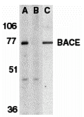
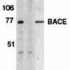
Description Western Blot Validation of BACE
Loading: 15 µg of lysates per lane. Antibodies: BACE (1 ug/mL), 1h incubation at RT in 5% NFDM/TBST.Secondary: Goat anti-rabbit IgG HRP conjugate at 1:10000 dilution.Lane A-C: human brain tissue lysate in the absence (A) or presence (B) of blocking peptide and mouse 3T3/NIH cell lysate (C).
Loading: 15 µg of lysates per lane. Antibodies: BACE (1 ug/mL), 1h incubation at RT in 5% NFDM/TBST.Secondary: Goat anti-rabbit IgG HRP conjugate at 1:10000 dilution.Lane A-C: human brain tissue lysate in the absence (A) or presence (B) of blocking peptide and mouse 3T3/NIH cell lysate (C).

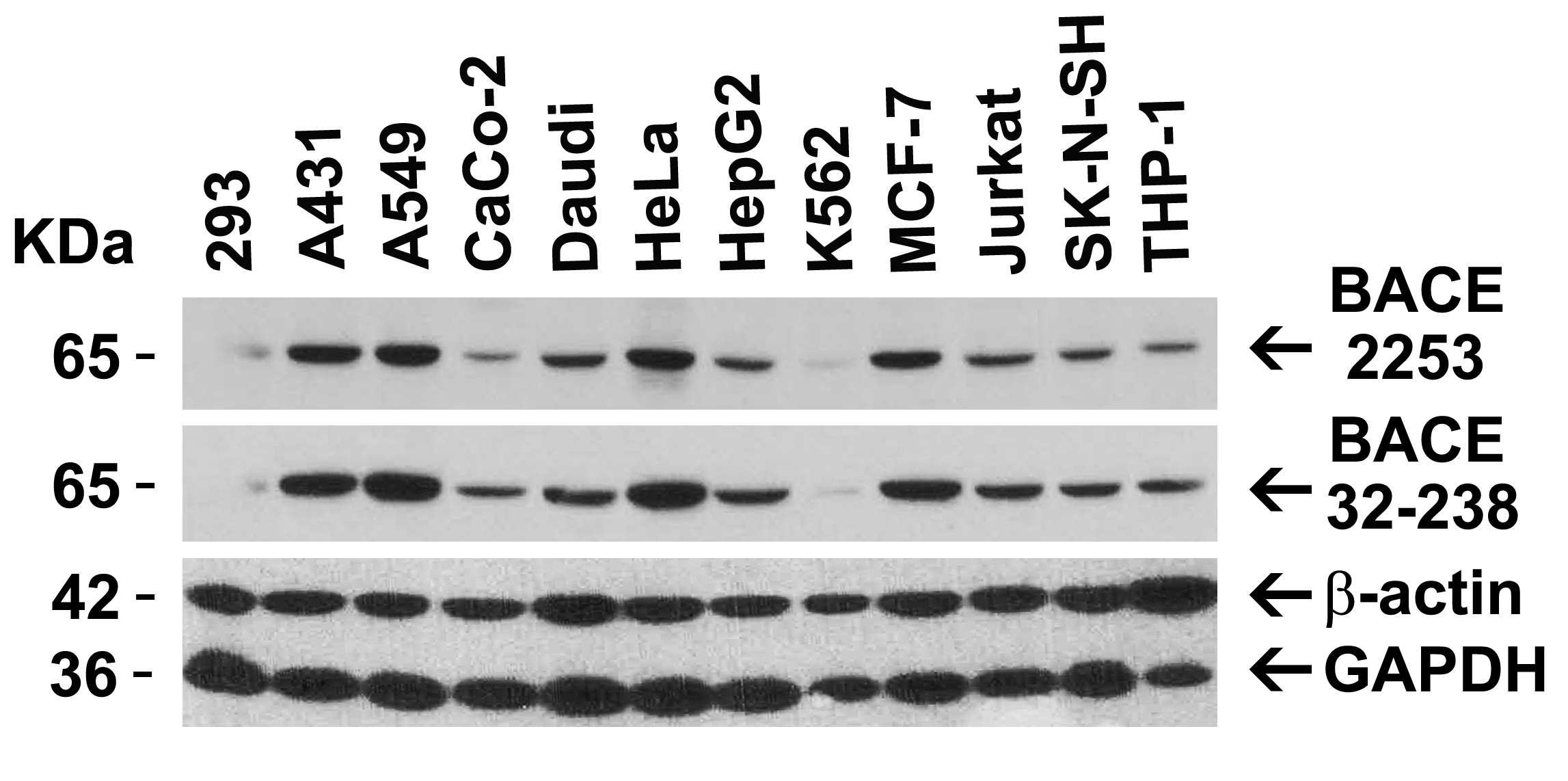
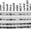
Description Independent Antibody Validation (IAV) via Protein Expression Profile in Cell Lines
Loading: 15 ug of lysates per lane. Antibodies: BACE 2253 (1 ug/mL), BACE 32-238 (1 ug/mL), beta-actin (1 ug/mL), and GAPDH (0.02 ug/mL), 1h incubation at RT in 5% NFDM/TBST.Secondary: Goat anti-rabbit IgG HRP conjugate at 1:10000 dilution.
Loading: 15 ug of lysates per lane. Antibodies: BACE 2253 (1 ug/mL), BACE 32-238 (1 ug/mL), beta-actin (1 ug/mL), and GAPDH (0.02 ug/mL), 1h incubation at RT in 5% NFDM/TBST.Secondary: Goat anti-rabbit IgG HRP conjugate at 1:10000 dilution.

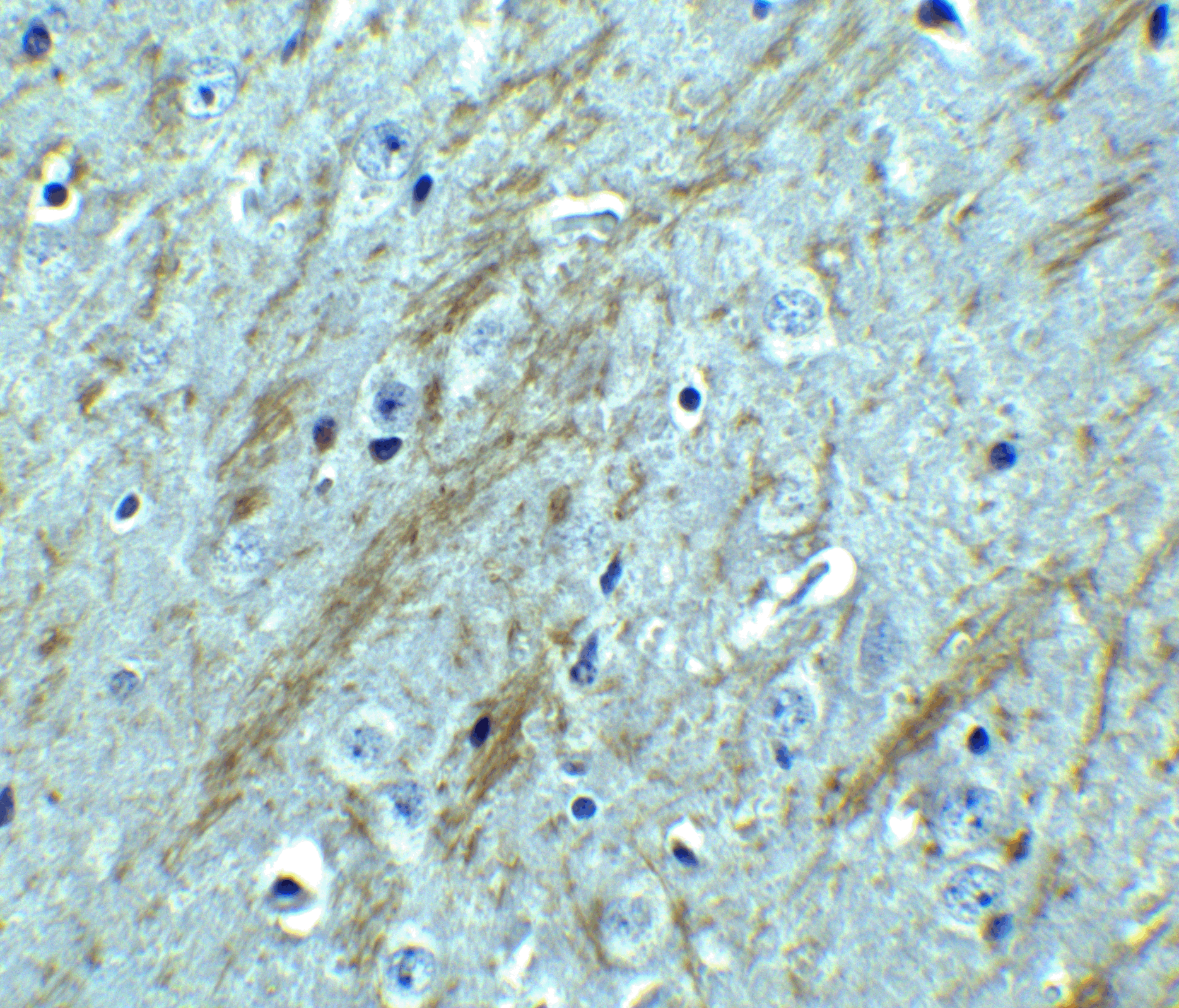
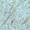
Description Immunohistochemistry Validation of BACE in Mouse Brain
Immunohistochemical analysis of paraffin-embedded mouse brain tissue using anti-BACE antibody (2253) at 2.5 ug/ml. Tissue was fixed with formaldehyde and blocked with 10% serum for 1 h at RT; antigen retrieval was by heat mediation with a citrate buffer (pH6). Samples were incubated with primary antibody overnight at 4C. A goat anti-rabbit IgG H&L (HRP) at 1/250 was used as secondary. Counter stained with Hematoxylin.
Immunohistochemical analysis of paraffin-embedded mouse brain tissue using anti-BACE antibody (2253) at 2.5 ug/ml. Tissue was fixed with formaldehyde and blocked with 10% serum for 1 h at RT; antigen retrieval was by heat mediation with a citrate buffer (pH6). Samples were incubated with primary antibody overnight at 4C. A goat anti-rabbit IgG H&L (HRP) at 1/250 was used as secondary. Counter stained with Hematoxylin.

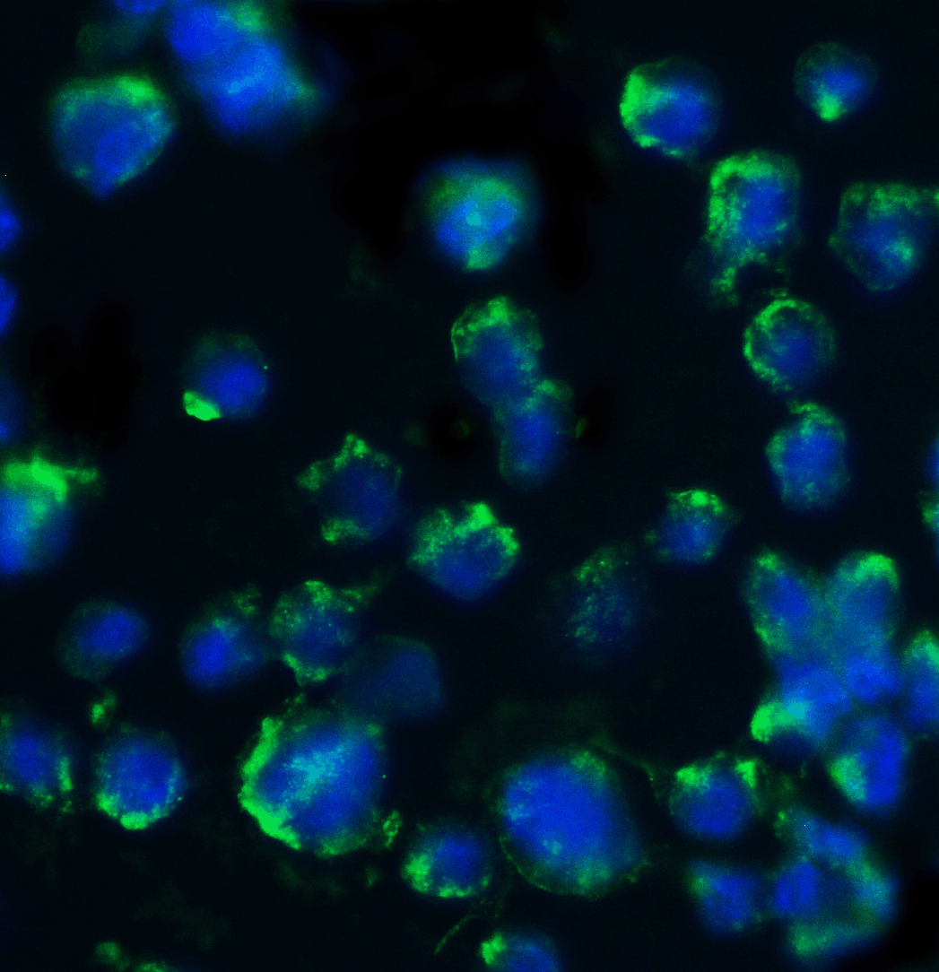
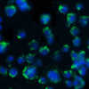
Description Immunofluorescence Validation of BACE in 3T3/NIH Cells
Immunofluorescent analysis of 4% paraformaldehyde-fixed mouse 3T3/NIH cells labeling BACE with 2253 at 20 ug/mL, followed by goat anti-rabbit IgG secondary antibody at 1/500 dilution (green) and DAPI staining (blue). Image showing both membrane and cytosol staining on 3T3/NIH cells.
Immunofluorescent analysis of 4% paraformaldehyde-fixed mouse 3T3/NIH cells labeling BACE with 2253 at 20 ug/mL, followed by goat anti-rabbit IgG secondary antibody at 1/500 dilution (green) and DAPI staining (blue). Image showing both membrane and cytosol staining on 3T3/NIH cells.

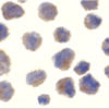
Description Immunocytochemistry Validation of BACE in 3T3/NIH Cells
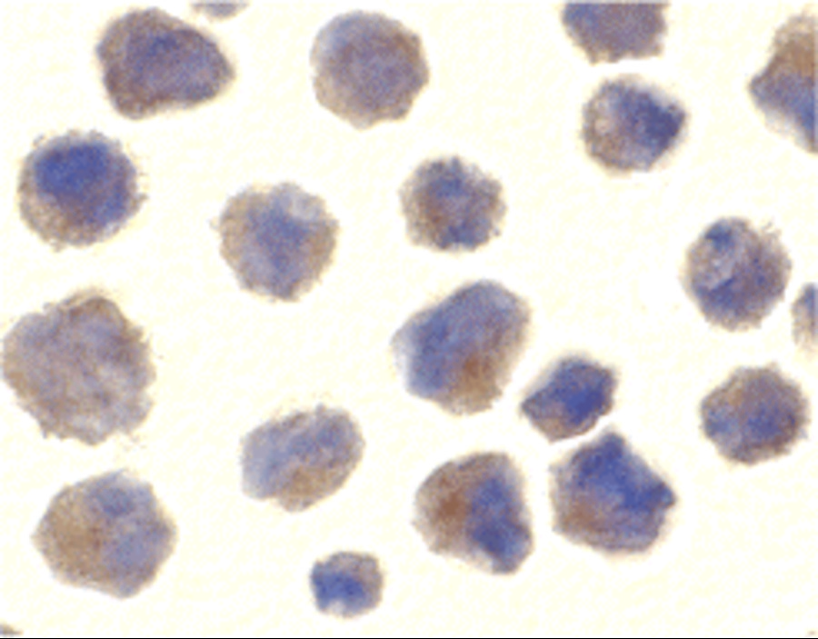
Immunocytochemical analysis of 3T3/NIH cells using anti-BACE antibody (2253) at 10 ug/ml. Cells was fixed with formaldehyde and blocked with 10% serum for 1 h at RT; antigen retrieval was by heat mediation with a citrate buffer (pH6). Samples were incubated with primary antibody overnight at 4C. A goat anti-rabbit IgG H&L (HRP) at 1/250 was used as secondary. Counter stained with Hematoxylin.
Immunocytochemical analysis of 3T3/NIH cells using anti-BACE antibody (2253) at 10 ug/ml. Cells was fixed with formaldehyde and blocked with 10% serum for 1 h at RT; antigen retrieval was by heat mediation with a citrate buffer (pH6). Samples were incubated with primary antibody overnight at 4C. A goat anti-rabbit IgG H&L (HRP) at 1/250 was used as secondary. Counter stained with Hematoxylin.

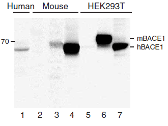
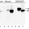
Description KO and Overexpression Validation of BACE in Human and Mouse Brain and 293 Cells. (Singer et al., 2005)
Western blot analysis of the BACE1 (2253) antibody’s ability to recognize human and murine BACE1. The BACE1 antibody recognized both the mouse and human forms of BACE1. Lanes 1–4 are frontal cortex homogenates from human and mouse brains. Lane 1 is from a neurologically unimpaired aged human control case, lane 2 from a BACE1-deficient mouse, lane 3 from a nontransgenic mouse and lane 4 from hBACE1 transgenic mouse. Lanes 5–7 are lysates from HEK293T cells transfected with a plasmid vector expressing eGFP, mBACE1 and hBACE1, respectively.
Western blot analysis of the BACE1 (2253) antibody’s ability to recognize human and murine BACE1. The BACE1 antibody recognized both the mouse and human forms of BACE1. Lanes 1–4 are frontal cortex homogenates from human and mouse brains. Lane 1 is from a neurologically unimpaired aged human control case, lane 2 from a BACE1-deficient mouse, lane 3 from a nontransgenic mouse and lane 4 from hBACE1 transgenic mouse. Lanes 5–7 are lysates from HEK293T cells transfected with a plasmid vector expressing eGFP, mBACE1 and hBACE1, respectively.

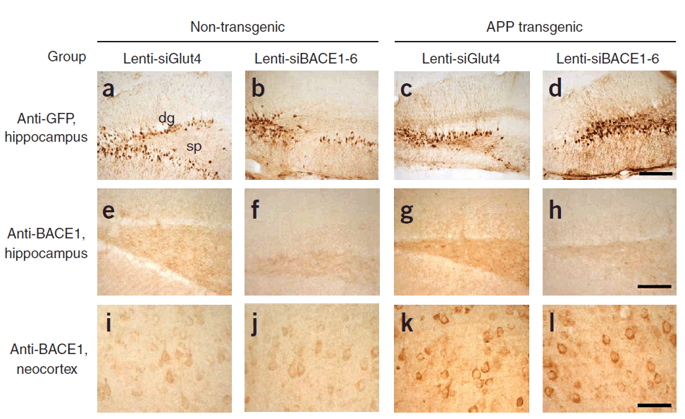
Description KD Validation of BACE in Mouse Brain (Singer et al., 2005)
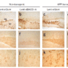
Characterization of the effects of lenti-siBACE1-6 expression in the brains of APP transgenic mice. (a–d) Anti-eGFP immunoreactivity in the hippocampus (the injection site) shows comparable and consistent expression of lenti-siRNA constructs in the dentate gyrus (dg) and stratus polymorphus (sp). (e) Anti-BACE1 immunoreactivity in the hippocampus of nontransgenic mice treated with lenti-siGlut4. (f) Reduced BACE1 immunostaining in the hippocampus of nontransgenic mice treated with lenti-siBACE1-6 vector. (g) Intense BACE1 immunoreactivity in the hippocampus of APP transgenic mice treated with lenti-siGlut4. (h) Reduced BACE1 expression in APP transgenic mice treated with lenti-siBACE1-6 vector. (i,j) Anti-BACE1 reacted with pyramidal cell bodies in the neocortex, which was not injected,
Characterization of the effects of lenti-siBACE1-6 expression in the brains of APP transgenic mice. (a–d) Anti-eGFP immunoreactivity in the hippocampus (the injection site) shows comparable and consistent expression of lenti-siRNA constructs in the dentate gyrus (dg) and stratus polymorphus (sp). (e) Anti-BACE1 immunoreactivity in the hippocampus of nontransgenic mice treated with lenti-siGlut4. (f) Reduced BACE1 immunostaining in the hippocampus of nontransgenic mice treated with lenti-siBACE1-6 vector. (g) Intense BACE1 immunoreactivity in the hippocampus of APP transgenic mice treated with lenti-siGlut4. (h) Reduced BACE1 expression in APP transgenic mice treated with lenti-siBACE1-6 vector. (i,j) Anti-BACE1 reacted with pyramidal cell bodies in the neocortex, which was not injected,

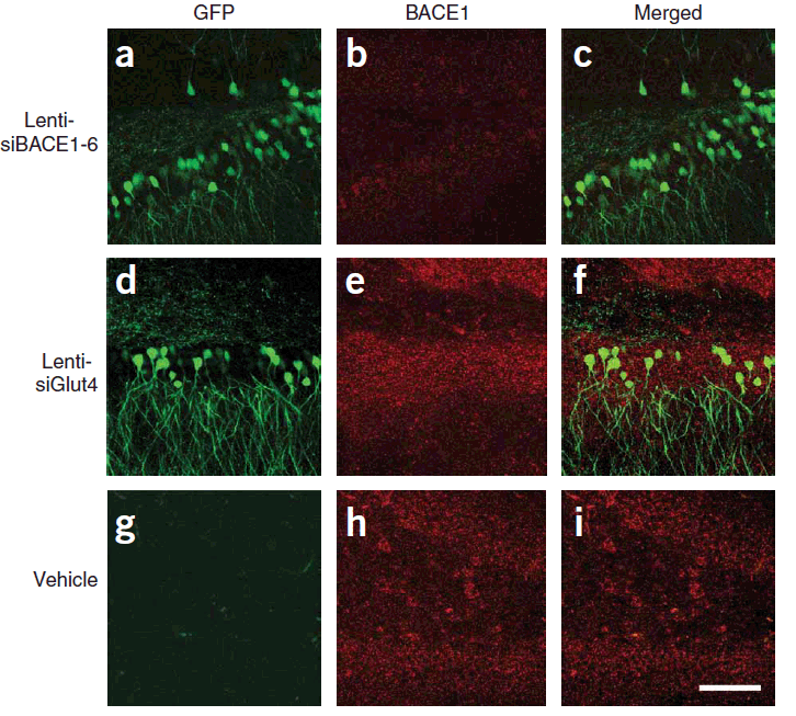
Description KD Validation of BACE in Mouse Brain (Singer et al., 2005)
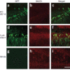
Immunolabeling patterns of BACE1 expression and the lenti-siRNA distribution. Sections from APP transgenic mice treated with the eGFPtagged lenti siRNA vectors (green) were co-immunolabeled with an antibody against BACE1 (red) and imaged with the LSCM. All sections are from the hippocampus of treated mice. (a–c) Lenti-siBACE1-6–treated mice. Areas within the hippocampus expressing the eGFP tagged vector have reduced BACE1 immunolabeling. (d–f) Mice treated with the eGFP-tagged control lenti-siGlut4 show unchanged expression of BACE1 in the hippocampus. (g–i) Mice treated with a saline vehicle show unchanged expression of BACE1 in the hippocampus.
Immunolabeling patterns of BACE1 expression and the lenti-siRNA distribution. Sections from APP transgenic mice treated with the eGFPtagged lenti siRNA vectors (green) were co-immunolabeled with an antibody against BACE1 (red) and imaged with the LSCM. All sections are from the hippocampus of treated mice. (a–c) Lenti-siBACE1-6–treated mice. Areas within the hippocampus expressing the eGFP tagged vector have reduced BACE1 immunolabeling. (d–f) Mice treated with the eGFP-tagged control lenti-siGlut4 show unchanged expression of BACE1 in the hippocampus. (g–i) Mice treated with a saline vehicle show unchanged expression of BACE1 in the hippocampus.

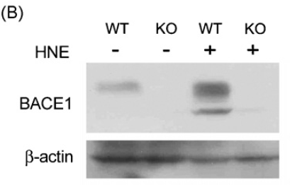
Description KO Validation of BACE in MEF Cells (Jo et al., 2010)
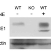
Wildtype and BACE −/− MEFs were exposed to HNE (15_M) for 2 h. BACE1 levels were examined by Western blot with anti-BACE antibodies (2253).
Wildtype and BACE −/− MEFs were exposed to HNE (15_M) for 2 h. BACE1 levels were examined by Western blot with anti-BACE antibodies (2253).

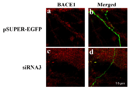
Description KD Validation of BACE in DRG (Hyun, 2416)
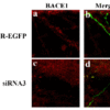
Decreased BACE1 expression in DRG following siRNA3 transfection. DRG neurons were transfected with 1 μg siRNA3 plasmid and incubated for 48 hours in 37°C. DRG neurons were stained for BACE1 us¬ing the Anti-BACE antibody . (a,b) Neurons transfected with the control plas¬mid pSUPER-EGFP (green) did not display any changes in BACE1 expression (red). (c,d) DRG neurons transfected with siR¬NA3 displayed reduced BACE1 expression in the axon.
Decreased BACE1 expression in DRG following siRNA3 transfection. DRG neurons were transfected with 1 μg siRNA3 plasmid and incubated for 48 hours in 37°C. DRG neurons were stained for BACE1 us¬ing the Anti-BACE antibody . (a,b) Neurons transfected with the control plas¬mid pSUPER-EGFP (green) did not display any changes in BACE1 expression (red). (c,d) DRG neurons transfected with siR¬NA3 displayed reduced BACE1 expression in the axon.
Handling
Storage This antibody is stable for at least one (1) year at -20°C. Avoid multiple freeze-thaw cycles.
Dilution Instructions Dilute in PBS or medium which is identical to that used in the assay system.
Application Instructions Immunoblotting: use at 1:500-1:1,000 dilution.
Positive control: Human brain tissue lysate.
Positive control: Human brain tissue lysate.
References & Data Sheet
Data Sheet  Download PDF Data Sheet
 Download PDF Data Sheet

