Anti-Survivin (CT) Antibody (2235)
$445.00
SKU: 2235
Categories: Antibody Products, Apoptosis Antibodies, Products
Overview
Product Name Anti-Survivin (CT) Antibody (2235)
Description Anti-Survivin (CT) Rabbit Polyclonal Antibody
Target Survivin (CT)
Species Reactivity Human
Applications ELISA,WB,ICC,IF
Host Rabbit
Clonality Polyclonal
Isotype IgG
Immunogen Peptide corresponding to aa 129- 142 of human Survivin (accession no. NP_001159).
Properties
Form Liquid
Concentration Lot Specific
Formulation PBS, pH 7.4.
Buffer Formulation Phosphate Buffered Saline
Buffer pH pH 7.4
Format Purified
Purification Purified by peptide immuno-affinity chromatography
Specificity Information
Specificity This antibody recognizes human Survivin (17kDa).
Target Name Baculoviral IAP repeat-containing protein 5
Target ID Survivin (CT)
Uniprot ID O15392
Alternative Names Apoptosis inhibitor 4, Apoptosis inhibitor survivin
Gene Name BIRC5
Gene ID 332
Accession Number NP_001159
Sequence Location Cytoplasm, Nucleus, Chromosome, Chromosome, centromere, Cytoplasm, cytoskeleton, spindle, Chromosome, centromere, kinetochore, Midbody
Biological Function Multitasking protein that has dual roles in promoting cell proliferation and preventing apoptosis (PubMed:9859993, PubMed:21364656, PubMed:20627126, PubMed:25778398, PubMed:28218735). Component of a chromosome passage protein complex (CPC) which is essential for chromosome alignment and segregation during mitosis and cytokinesis (PubMed:16322459). Acts as an important regulator of the localization of this complex; directs CPC movement to different locations from the inner centromere during prometaphase to midbody during cytokinesis and participates in the organization of the center spindle by associating with polymerized microtubules (PubMed:20826784). Involved in the recruitment of CPC to centromeres during early mitosis via association with histone H3 phosphorylated at 'Thr-3' (H3pT3) during mitosis (PubMed:20929775). The complex with RAN plays a role in mitotic spindle formation by serving as a physical scaffold to help deliver the RAN effector molecule TPX2 to microtubules (PubMed:18591255). May counteract a default induction of apoptosis in G2/M phase (PubMed:9859993). The acetylated form represses STAT3 transactivation of target gene promoters (PubMed:20826784). May play a role in neoplasia (PubMed:10626797). Inhibitor of CASP3 and CASP7 (PubMed:21536684). Essential for the maintenance of mitochondrial integrity and function (PubMed:25778398). Isoform 2 and isoform 3 do not appear to play vital roles in mitosis (PubMed:12773388, PubMed:16291752). Isoform 3 shows a marked reduction in its anti-apoptotic effects when compared with the displayed wild-type isoform (PubMed:10626797). {PubMed:10626797, PubMed:12773388, PubMed:16291752, PubMed:16322459, PubMed:18591255, PubMed:20627126, PubMed:20826784, PubMed:20929775, PubMed:21364656, PubMed:21536684, PubMed:25778398, PubMed:28218735, PubMed:9859993}.
Research Areas Apoptosis
Background Apoptosis can be prevented by the inhibitor of apoptosis proteins (IAPs). IAPs constitute a conserved gene family that binds to and inhibits cell death proteases. A novel IAP has been identified and designated Survivin. Survivin interacts with the processed form of caspase-3 and inhibits its proteolytic activity. Survivin is expressed predominantly in tissues of embryos, transformed cell lines, and many human cancers.
Application Images


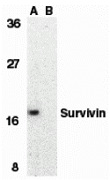
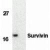
Description Western Blot Validation in Human MOLT4 Cell Lysate with the absence (A) or presence (B) of blocking peptide
Loading: 15 ug of lysates per lane. Antibodies: Survivin 2235 (1 ug/mL), 1h incubation at RT in 5% NFDM/TBST.Secondary: Goat anti-rabbit IgG HRP conjugate at 1:10000 dilution.
Loading: 15 ug of lysates per lane. Antibodies: Survivin 2235 (1 ug/mL), 1h incubation at RT in 5% NFDM/TBST.Secondary: Goat anti-rabbit IgG HRP conjugate at 1:10000 dilution.

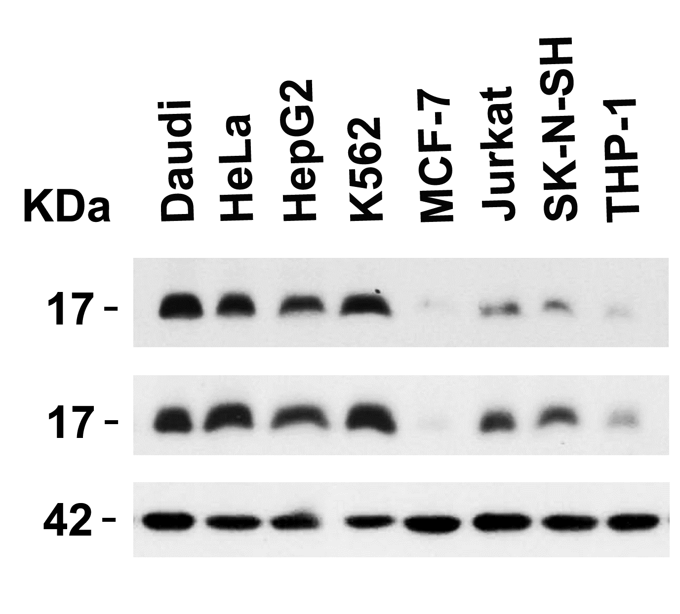
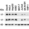
Description Independent Antibody Validation (IAV) via Protein Expression Profile in Human Cell Lines
Loading: 15 ug of lysates per lane. Antibodies: Survivin 2233 (5 ug/mL), Survivin 2235 (4 ug/mL) and beta-actin 3779 (1 ug/mL), 1h incubation at RT in 5% NFDM/TBST.Secondary: Goat anti-rabbit IgG HRP conjugate at 1:10000 dilution.
Loading: 15 ug of lysates per lane. Antibodies: Survivin 2233 (5 ug/mL), Survivin 2235 (4 ug/mL) and beta-actin 3779 (1 ug/mL), 1h incubation at RT in 5% NFDM/TBST.Secondary: Goat anti-rabbit IgG HRP conjugate at 1:10000 dilution.

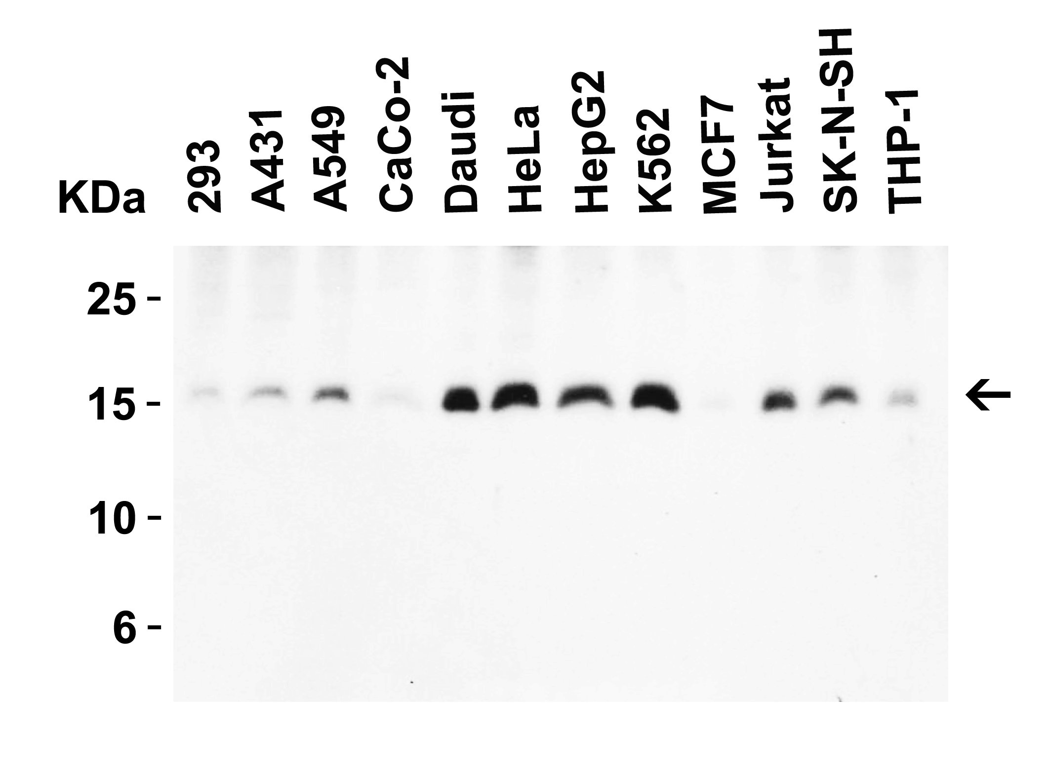
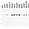
Description Western Blot Validation in Human Cell Lines
Loading: 15 ug of lysates per lane. Antibodies: Survivin 2235 (4 ug/mL), 1h incubation at RT in 5% NFDM/TBST.Secondary: Goat anti-rabbit IgG HRP conjugate at 1:10000 dilution.
Loading: 15 ug of lysates per lane. Antibodies: Survivin 2235 (4 ug/mL), 1h incubation at RT in 5% NFDM/TBST.Secondary: Goat anti-rabbit IgG HRP conjugate at 1:10000 dilution.

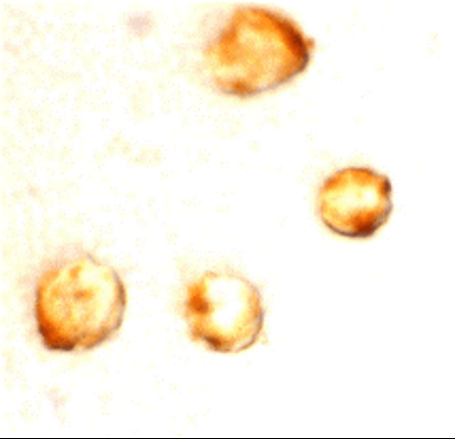
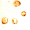
Description Immunocytochemistry Validation of Survivin in MOLT4 Cells
Immunocytochemical analysis of MOLT4 cells using anti-Survivin antibody (2235) at 10 ug/ml. Cells was fixed with formaldehyde and blocked with 10% serum for 1 h at RT; antigen retrieval was by heat mediation with a citrate buffer (pH6). Samples were incubated with primary antibody overnight at 4C. A goat anti-rabbit IgG H&L (HRP) at 1/250 was used as secondary. Counter stained with Hematoxylin.
Immunocytochemical analysis of MOLT4 cells using anti-Survivin antibody (2235) at 10 ug/ml. Cells was fixed with formaldehyde and blocked with 10% serum for 1 h at RT; antigen retrieval was by heat mediation with a citrate buffer (pH6). Samples were incubated with primary antibody overnight at 4C. A goat anti-rabbit IgG H&L (HRP) at 1/250 was used as secondary. Counter stained with Hematoxylin.

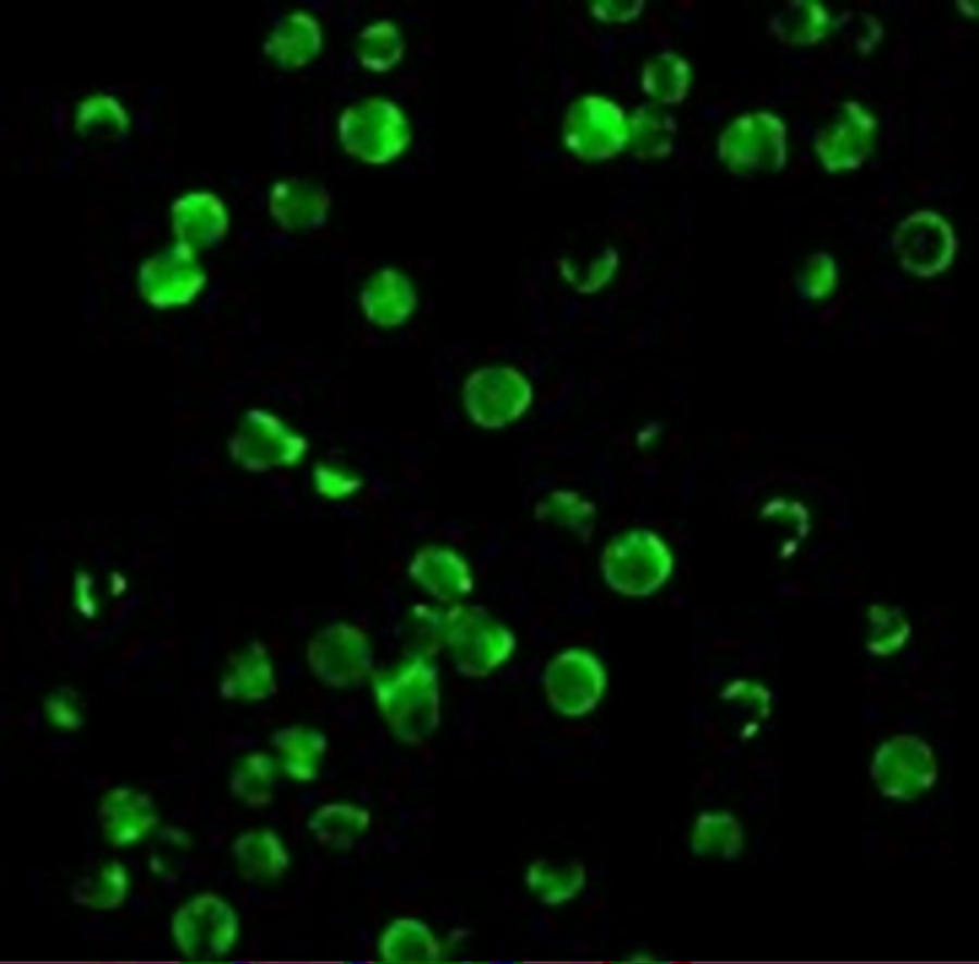
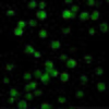
Description Immunofluorescence Validation of Survivin in MOLT4 Cells
Immunofluorescent analysis of 4% paraformaldehyde-fixed MOLT4 cells labeling Survivin with 2235 at 10 ug/mL, followed by goat anti-rabbit IgG secondary antibody at 1/500 dilution (green).
Immunofluorescent analysis of 4% paraformaldehyde-fixed MOLT4 cells labeling Survivin with 2235 at 10 ug/mL, followed by goat anti-rabbit IgG secondary antibody at 1/500 dilution (green).

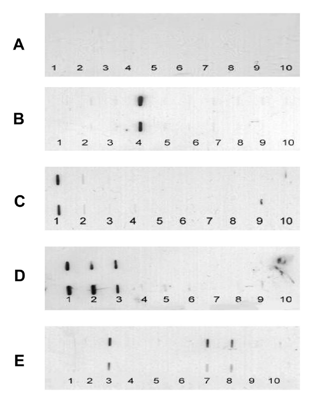
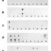
Description Slot Blot Validation of Survivin in Hepatocellular Carcinoma (HCC) Patients (Zhang et al., 2416)
Slot blot analysis of Survivin recombinant protein expression with anti-Survivin antibodies (2235) in the four representative HCC sera. Survivin (Lane 7) was detected in the fourth HCC serum (E), but not in the normal human serum (A) and the other three HCC sera (B-D). Phosphate bufferedsaline (PBS) (Lanes 9 and 10) was used as a negative control.
Slot blot analysis of Survivin recombinant protein expression with anti-Survivin antibodies (2235) in the four representative HCC sera. Survivin (Lane 7) was detected in the fourth HCC serum (E), but not in the normal human serum (A) and the other three HCC sera (B-D). Phosphate bufferedsaline (PBS) (Lanes 9 and 10) was used as a negative control.

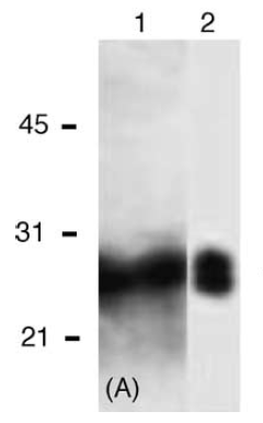
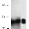
Description Western Blot Validation of Survivin with Recombinant Protein (Megliorino et al., 2005)
Lane 1: A 28 kDa peptide that corresponded to the size of ORF of survivin was detected in SDS-PAGE with Coomassie blue staining. Lane 2: WB analysis of Survivin recombinant protein expression with anti-Survivin antibodies (2235).
Lane 1: A 28 kDa peptide that corresponded to the size of ORF of survivin was detected in SDS-PAGE with Coomassie blue staining. Lane 2: WB analysis of Survivin recombinant protein expression with anti-Survivin antibodies (2235).

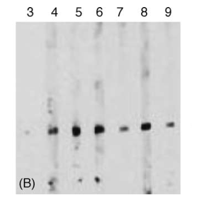
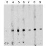
Description Western Blot Validation of Survivin with Recombinant Protein in Sera from Cancer Patients (Megliorino et al., 2005)
WB analysis of Survivin recombinant protein expression with anti-Survivin antibodies (2235) in six cancer sera. 6 x His-tagged Survivin recombinant protein (Lanes 4-9) was strongly detected at 28kD, but not in the normal human serum (Lane 3).
WB analysis of Survivin recombinant protein expression with anti-Survivin antibodies (2235) in six cancer sera. 6 x His-tagged Survivin recombinant protein (Lanes 4-9) was strongly detected at 28kD, but not in the normal human serum (Lane 3).
Handling
Storage This antibody is stable for at least one (1) year at -20°C. Avoid multiple freeze-thaw cycles.
Dilution Instructions Dilute in PBS or medium which is identical to that used in the assay system.
Application Instructions Immunoblotting: use at 1ug/mL.
Positive control: MOLT4 cell lysate.
Immunocytochemistry: use at 10ug/mL.
These are recommended concentrations.
Enduser should determine optimal concentrations for their applications.
Positive control: MOLT4 cell lysate.
Immunocytochemistry: use at 10ug/mL.
These are recommended concentrations.
Enduser should determine optimal concentrations for their applications.
References & Data Sheet
Data Sheet  Download PDF Data Sheet
 Download PDF Data Sheet

