Anti-Apaf1 (CT) Antibody (2015)
$445.00
| Host | Quantity | Applications | Species Reactivity | Data Sheet | |
|---|---|---|---|---|---|
| Rabbit | 100ug | ELISA,WB,IHC-P | Human, Mouse, Rat |  |
SKU: 2015
Categories: Antibody Products, Apoptosis Antibodies, Products
Overview
Product Name Anti-Apaf1 (CT) Antibody (2015)
Description Anti-Apaf-1 (CT) Rabbit Polyclonal Antibody
Target Apaf1 (CT)
Species Reactivity Human, Mouse, Rat
Applications ELISA,WB,IHC-P
Host Rabbit
Clonality Polyclonal
Isotype IgG
Immunogen Peptide corresponding to aa 1158- 1177 of human Apaf1. This sequence differs from that of mouse Apaf1 by one amino acid.
Properties
Form Liquid
Concentration Lot Specific
Formulation PBS, pH 7.4.
Buffer Formulation Phosphate Buffered Saline
Buffer pH pH 7.4
Format Purified
Purification Purified by peptide immuno-affinity chromatography
Specificity Information
Specificity This antibody recognizes human, mouse, and rat Apaf1 (130kDa).
Target Name Apoptotic protease-activating factor 1
Target ID Apaf1 (CT)
Uniprot ID O14727
Alternative Names APAF-1
Gene Name APAF1
Gene ID 317
Accession Number NP_001151
Sequence Location Cytoplasm
Biological Function Oligomeric Apaf-1 mediates the cytochrome c-dependent autocatalytic activation of pro-caspase-9 (Apaf-3), leading to the activation of caspase-3 and apoptosis. This activation requires ATP. Isoform 6 is less effective in inducing apoptosis. {PubMed:10393175, PubMed:12804598}.
Research Areas Apoptosis
Background The mammalian homologues of the key cell death gene CED-4 in C. elegans has been identified recently from human and mouse and designated Apaf1 (for apoptosis protease- activating factor 1). Apaf1 binds to cytochrome c (Apaf2) and caspase-9 (Apaf3), which leads to caspase-9 activation. Activated caspase-9 in turn cleaves and activates caspase-3, one of the key proteases responsible for the proteolytic cleavage of many key proteins in apoptosis. Apaf1 can also associate with caspase-4 and caspase-8. Apaf1 is ubiquitously expressed in human tissues.
Application Images


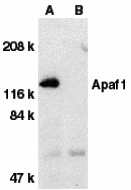
Description Western blot analysis of Apaf1 in human heart tissue lysate with Apaf1 antibody at 1 ug/mL dilution in the absence (A) or presence (B) of blocking peptide.

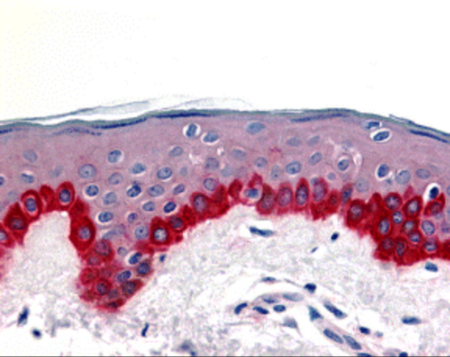
Description Immunohistochemistry of Apaf1 in human skin tissue with Apaf1 antibody at 20 ug/mL.
Handling
Storage This antibody is stable for at least one (1) year at -20°C. Avoid multiple freeze-thaw cycles.
Dilution Instructions Dilute in PBS or medium which is identical to that used in the assay system.
Application Instructions Immunoblotting: use at 1-10ug/mL
Immunohistochemistry: use at 10-20ug/mL
These are recommended concentrations.
Endusers should determine optimal concentrations for their applications.
Positive control: Human heart tissue lysate.
Immunohistochemistry: use at 10-20ug/mL
These are recommended concentrations.
Endusers should determine optimal concentrations for their applications.
Positive control: Human heart tissue lysate.
References & Data Sheet
Data Sheet  Download PDF Data Sheet
 Download PDF Data Sheet


