Anti-Heme Oxygenase-1 Antibody (13061)
$346.00
SKU: 13061
Categories: Antibody Products, Enzymes and Enzyme Inhibitor Antibodies, Products
Overview
Product Name Anti-Heme Oxygenase-1 Antibody (13061)
Description Anti-Heme Oxygenase-1 Mouse Monoclonal Antibody
Target Heme Oxygenase-1
Species Reactivity Human, Mouse, Bovine
Applications WB,ELISA
Host Mouse
Clonality Monoclonal
Clone ID 1F12.A6
Isotype IgG1
Immunogen Synthetic peptide corresponding to aa 1-30 of human HO-1.
Properties
Form Liquid
Concentration 1.0 mg/mL
Formulation PBS, pH 7.4.
Buffer Formulation Phosphate Buffered Saline
Buffer pH pH 7.4
Format Purified
Purification Purified by Protein G affinity chromatography
Specificity Information
Specificity This antibody recognizes human, mouse, and bovine HO-1. It does not cross-react with HO-2.
Target Name Heme oxygenase 1
Target ID Heme Oxygenase-1
Uniprot ID P09601
Alternative Names HO-1, EC 1.14.14.18
Gene Name HMOX1
Accession Number NP_002124.1
Sequence Location Microsome, Endoplasmic reticulum membrane, Peripheral membrane protein, Cytoplasmic side
Biological Function Heme oxygenase cleaves the heme ring at the alpha methene bridge to form biliverdin. Biliverdin is subsequently converted to bilirubin by biliverdin reductase. Under physiological conditions, the activity of heme oxygenase is highest in the spleen, where senescent erythrocytes are sequestrated and destroyed. Exhibits cytoprotective effects since excess of free heme sensitizes cells to undergo apoptosis.
Research Areas Enzymes
Background Heme-oxygenase is an enzyme that catalyzes heme catabolism to yield biliverdin, iron, and carbon monoxide (CO). There are three isoforms of heme-oxygenase: HO-1, HO-2, and HO-3. HO-1 and HO-2 have been identified as the two major isoforms in mammals. HO-1, also known as heat shock protein 32 (Hsp32) is induced by most oxidative stress inducers, cytokines, inflammatory agents, and heat shock. HO-1 deficiency appears to cause reduced stress defense, a pro-inflammatory tendency, susceptibility to atherosclerotic lesion formation, endothelial cell injury, and growth retardation. Therefore, up-regulation of HO-1 is one of the major defense mechanisms against oxidative stress.
Application Images


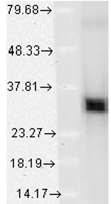
Description Immunoblotting: use at 0.5-1ug/ml. A band of ~32kDa is detected.

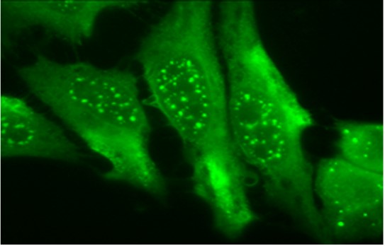
Description Immunofluorescence: use at 5-10ug/ml. Detection of HO-1 in formaldehyde-fixed HeLa cells.
Handling
Storage This antibody is stable for at least one (1) year at -20°C.
Dilution Instructions Dilute in PBS or medium that is identical to that used in the assay system.
Application Instructions ELISA: use at 1ug/ml with HO-1 on the solid phase.
Immunoblotting: use at 0.5-1ug/ml. A band of ~32kDa is detected.
Immunofluorescence: use at 5-10ug/ml
Positive control: Human cell line lysates such as A431, A549, HeLa, HepG2, Jurkat, or MCF7.
These are recommended concentrations.
Enduser should determine optimal concentrations for their applications.
References & Data Sheet
Data Sheet  Download PDF Data Sheet
 Download PDF Data Sheet


