Anti-TACE / ADAM17 Antibody (1131)
$469.00
| Host | Quantity | Applications | Species Reactivity | Data Sheet | |
|---|---|---|---|---|---|
| Rabbit | 100ug | ELISA,WB,ICC,IF | Human, Mouse, Rat |  |
SKU: 1131
Categories: Antibody Products, Apoptosis Antibodies, Products
Overview
Product Name Anti-TACE / ADAM17 Antibody (1131)
Description Anti-TACE (CT) Rabbit Polyclonal Antibody
Target TACE / ADAM17
Species Reactivity Human, Mouse, Rat
Applications ELISA,WB,ICC,IF
Host Rabbit
Clonality Polyclonal
Isotype IgG
Immunogen Peptide corresponding to aa 807-823 of human TACE (accession no. NP_003174). This sequence differs from mouse and rat TACE by one amino acid.
Properties
Form Liquid
Concentration Lot Specific
Formulation PBS, pH 7.4.
Buffer Formulation Phosphate Buffered Saline
Buffer pH pH 7.4
Format Purified
Purification Purified by peptide immuno-affinity chromatography
Specificity Information
Specificity This antibody recognizes human, mouse, and rat TACE, and 80-130kDa bands are detected in immunoblots. These bands may represent mature protein, precursor, and glycosylated TACE.
Target Name Disintegrin and metalloproteinase domain-containing protein 17
Target ID TACE / ADAM17
Uniprot ID P78536
Alternative Names ADAM 17, EC 3.4.24.86, Snake venom-like protease, TNF-α convertase, TNF-α-converting enzyme, CD antigen CD156b
Gene Name ADAM17
Gene ID 6868
Accession Number NP_003174
Sequence Location Membrane; Single-pass type I membrane protein.
Biological Function Cleaves the membrane-bound precursor of TNF-alpha to its mature soluble form (PubMed:9034191). Responsible for the proteolytical release of soluble JAM3 from endothelial cells surface (PubMed:20592283). Responsible for the proteolytic release of several other cell-surface proteins, including p75 TNF-receptor, interleukin 1 receptor type II, p55 TNF-receptor, transforming growth factor-alpha, L-selectin, growth hormone receptor, MUC1 and the amyloid precursor protein (PubMed:12441351). Acts as an activator of Notch pathway by mediating cleavage of Notch, generating the membrane-associated intermediate fragment called Notch extracellular truncation (NEXT) (PubMed:24226769). Plays a role in the proteolytic processing of ACE2 (PubMed:24227843). Plays a role in hemostasis through shedding of GP1BA, the platelet glycoprotein Ib alpha chain (By similarity). Mediates the proteolytic cleavage of LAG3, leading to release the secreted form of LAG3 (By similarity). Mediates the proteolytic cleavage of IL6R, leading to the release of secreted form of IL6R (PubMed:26876177, PubMed:28060820). Mediates the proteolytic cleavage and shedding of FCGR3A upon NK cell stimulation, a mechanism that allows for increased NK cell motility and detachment from opsonized target cells. {UniProtKB:Q9Z0F8, PubMed:12441351, PubMed:20592283, PubMed:24226769, PubMed:24227843, PubMed:24337742, PubMed:26876177, PubMed:28060820, PubMed:9034191}.
Research Areas Apoptosis
Background TNFalpha is synthesized as a 26kDa type II membrane-bound precursor that is cleaved by a convertase to generate secreted 17kDa mature TNFalpha. TNFalpha converting enzyme (TACE) has been identified, and human and mouse TACE cDNAs have been cloned. TACE is a membrane-bound metalloprotease-disintegrin in the family of mammalian ADAM (for a disintegrin and metalloprotease). TACE also processes other cell surface proteins, including TNF receptor, TGFalpha, L-selectin, and alpha-cleavage of amyloid protein precursor (APP). TACE mRNA is expressed in a variety of human and mouse tissues.
Application Images


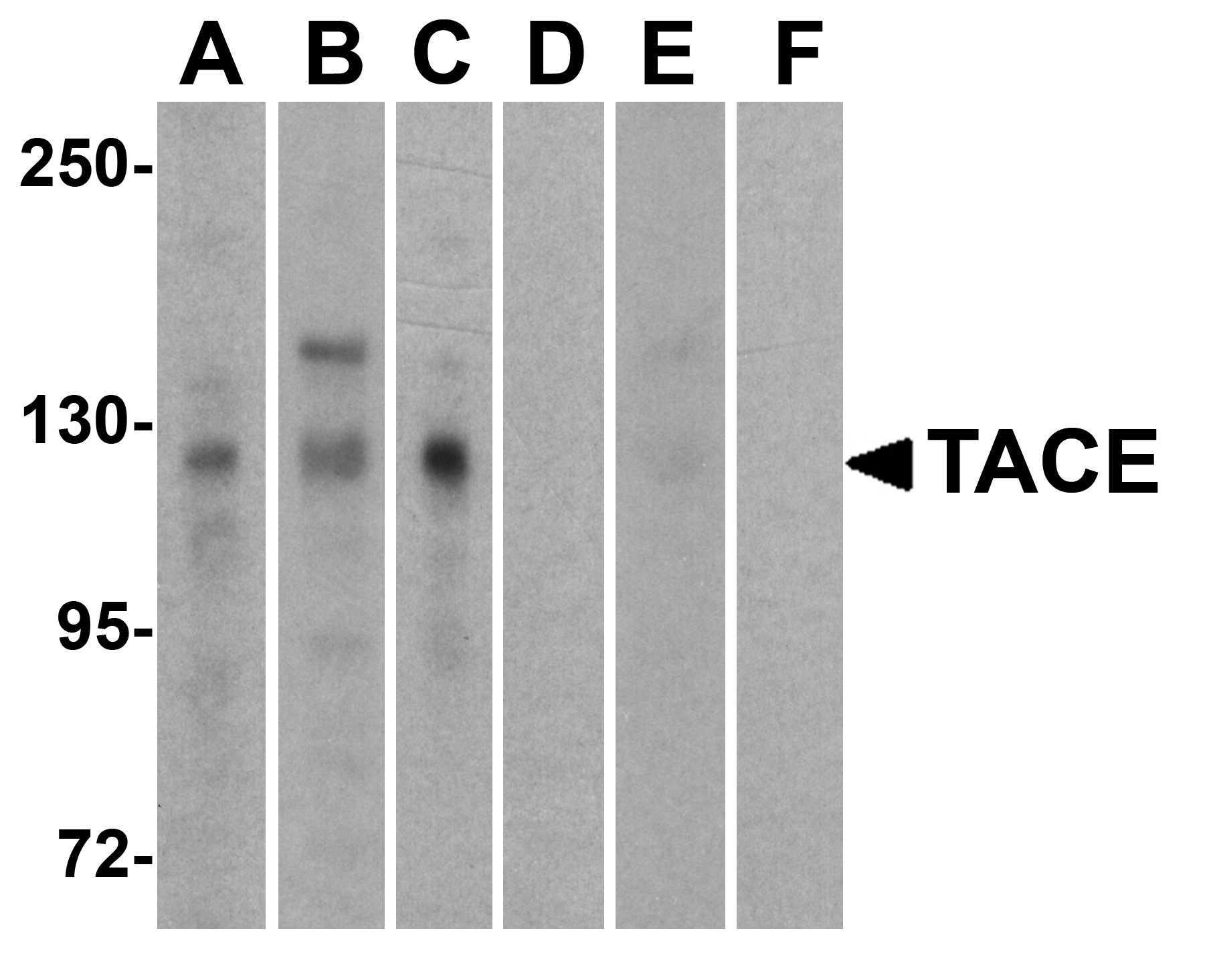
Description Western Blot Validation of TACE in Human Cell Lines
Loading: 15 ug of lysates per lane. Antibodies: TACE (1 µg/mL), 1h incubation at RT in 5% NFDM/TBST. Secondary: Goat anti-rabbit IgG HRP conjugate at 1:10000 dilution. Lanes: HeLa (A,D), Jurkat (B, E), Raji (C,F) in the absence (A-C) or presence (E-F) of blocking peptide.
Loading: 15 ug of lysates per lane. Antibodies: TACE (1 µg/mL), 1h incubation at RT in 5% NFDM/TBST. Secondary: Goat anti-rabbit IgG HRP conjugate at 1:10000 dilution. Lanes: HeLa (A,D), Jurkat (B, E), Raji (C,F) in the absence (A-C) or presence (E-F) of blocking peptide.

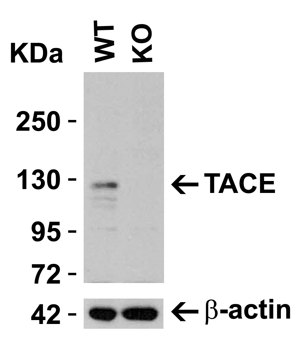
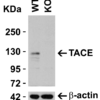
Description KO Validation in HeLa Cells
Loading: 10 ug of HeLa WT cell lysates or TACE KO cell lysates. Antibodies: TACE 1131 (0.25 ug/mL) and beta-actin 3779 (1 ug/mL), 1 h incubation at RT in 5% NFDM/TBST. Secondary: Goat Anti-Rabbit IgG HRP conjugate at 1:10000 dilution.
Loading: 10 ug of HeLa WT cell lysates or TACE KO cell lysates. Antibodies: TACE 1131 (0.25 ug/mL) and beta-actin 3779 (1 ug/mL), 1 h incubation at RT in 5% NFDM/TBST. Secondary: Goat Anti-Rabbit IgG HRP conjugate at 1:10000 dilution.

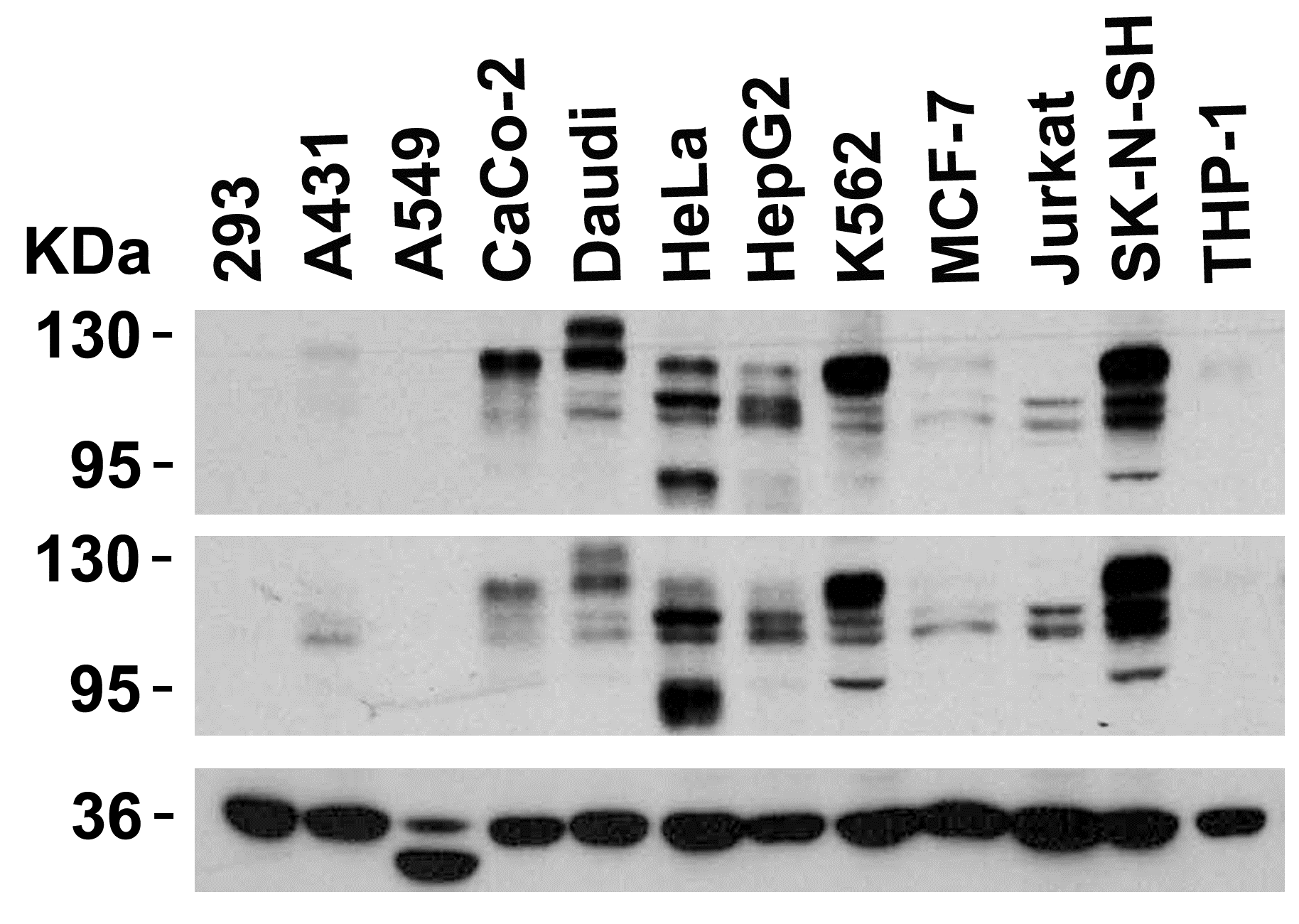
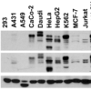
Description Independent Antibody Validation (IAV) via Protein Expression Profile in Cell Lines
Loading: 15 ug of lysates per lane. Antibodies: TACE 1131 (0.5 ug/mL), TACE 22-001 (2 ug/mL), and GAPDH (0.02 ug/mL), 1h incubation at RT in 5% NFDM/TBST. Secondary: Goat anti-rabbit IgG HRP conjugate at 1:10000 dilution.
Loading: 15 ug of lysates per lane. Antibodies: TACE 1131 (0.5 ug/mL), TACE 22-001 (2 ug/mL), and GAPDH (0.02 ug/mL), 1h incubation at RT in 5% NFDM/TBST. Secondary: Goat anti-rabbit IgG HRP conjugate at 1:10000 dilution.

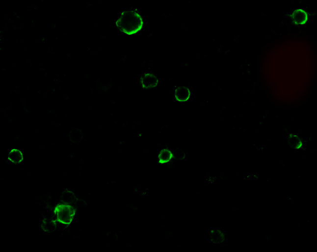
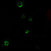
Description Immunofluorescence Validation of TACE in HeLa Cells
Immunofluorescent analysis of 4% paraformaldehyde-fixed HeLa cells labeling TACE with 1131 at 10 ug/mL, followed by goat anti-rabbit IgG secondary antibody at 1/500 dilution (green).
Immunofluorescent analysis of 4% paraformaldehyde-fixed HeLa cells labeling TACE with 1131 at 10 ug/mL, followed by goat anti-rabbit IgG secondary antibody at 1/500 dilution (green).

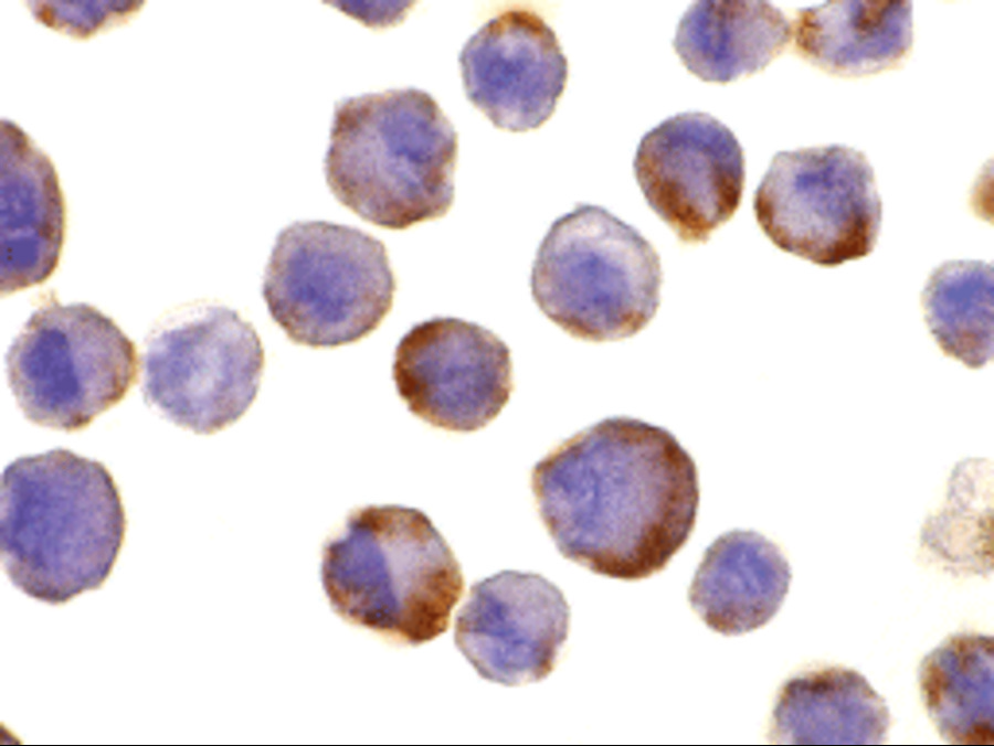
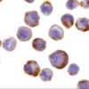
Description Immunocytochemistry Validation of TACE in HeLa Cells
Immunohistochemical analysis of HeLa cells using anti-TACE antibody (1131) at 10 ug/ml. Cells was fixed with formaldehyde and blocked with 10% serum for 1 h at RT; antigen retrieval was by heat mediation with a citrate buffer (pH6). Samples were incubated with primary antibody overnight at 4C. A goat anti-rabbit IgG H&L (HRP) at 1/250 was used as secondary. Counter stained with Hematoxylin.
Immunohistochemical analysis of HeLa cells using anti-TACE antibody (1131) at 10 ug/ml. Cells was fixed with formaldehyde and blocked with 10% serum for 1 h at RT; antigen retrieval was by heat mediation with a citrate buffer (pH6). Samples were incubated with primary antibody overnight at 4C. A goat anti-rabbit IgG H&L (HRP) at 1/250 was used as secondary. Counter stained with Hematoxylin.

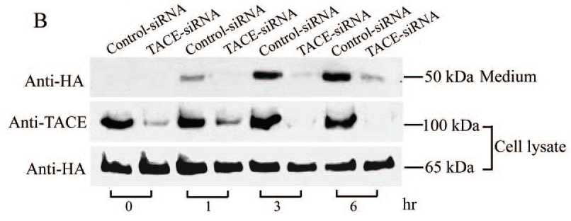
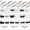
Description KD Validation of TACE in Monkey COS Cells. (Wang et al., 2006)
COS cells stably expressing Pref-1A were transfected with control siRNA or TACE siRNA. TACE was detected in lysates by using the anti-TACE antibody (1131). TACE expression levels were markedly reduced in TACE knockdown cell lysate.
COS cells stably expressing Pref-1A were transfected with control siRNA or TACE siRNA. TACE was detected in lysates by using the anti-TACE antibody (1131). TACE expression levels were markedly reduced in TACE knockdown cell lysate.

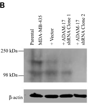
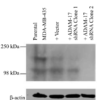
Description KD Validation of TACE in MDA-MB-435 Cells. (McGowan et al., 2416)
ADAM-17 protein expression, following transfection with ADAM-17 shRNA (two clones) or neomycin-resistant negative control vector, was examined by immunoblot analysis with anti-ADAM-17 antibodies (1131).
ADAM-17 protein expression, following transfection with ADAM-17 shRNA (two clones) or neomycin-resistant negative control vector, was examined by immunoblot analysis with anti-ADAM-17 antibodies (1131).

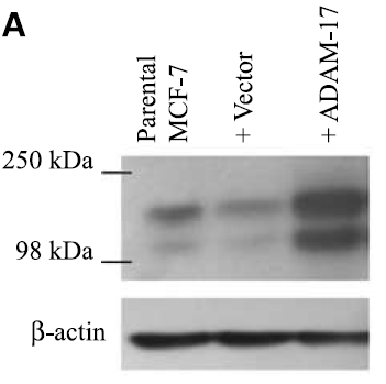
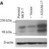
Description Overexpression Validation of TACE in MCF-7 Cells. (McGowan et al., 2416)
ADAM-17 (TACE) protein expression, following transfection of vector and ADAM-17 cDNA, was examined by immunoblot analysis with anti-ADAM-17 (1131) antibodies in MCF-7 cells. Increased ADAM-17 was detected in ADAM-17 transfected cells.
ADAM-17 (TACE) protein expression, following transfection of vector and ADAM-17 cDNA, was examined by immunoblot analysis with anti-ADAM-17 (1131) antibodies in MCF-7 cells. Increased ADAM-17 was detected in ADAM-17 transfected cells.

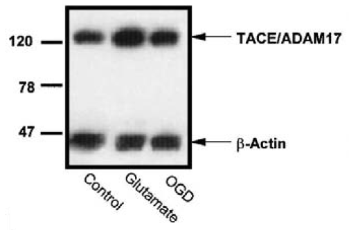
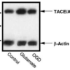
Description Induced Expression Validation of TACE in Rat Cortical Neurons (Hurtado et al., 2002)
Effect of oxygen–glucose deprivation(OGD) or glutamate on the levels of TACE/ADAM17 in rat cortical cultures. Western blot analysis of TACE in homogenates from control, glutamate, and OGD-exposed cultures from a representative experiment.
Effect of oxygen–glucose deprivation(OGD) or glutamate on the levels of TACE/ADAM17 in rat cortical cultures. Western blot analysis of TACE in homogenates from control, glutamate, and OGD-exposed cultures from a representative experiment.

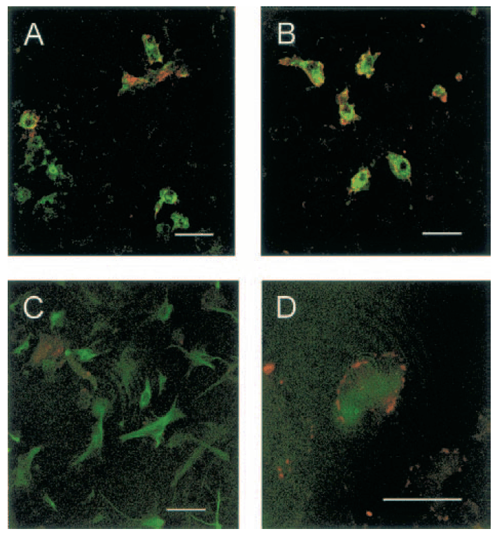
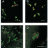
Description Immunofluorescence Validation of TACE in Rat Cortical Neurons (Hurtado et al., 2002)
Double immunostaining of control and glutamate-exposed rat cortical cultures. (A) Control cultures show TACE immunoreactivity at the cellular membrane of some microglial cells (B) Glutamate-exposed cultures show that most microglial cells express TACE immunoreactivity.(C) Control cultures show that TACE immunostaining does not colocalize with astrocytes [glial fibrillary acidic protein (GFAP)-positive cells]. (D) Astrocyte (GFAP-positive cell) showing TACE immunoreactivity in its surface after treatment with glutamate.
Double immunostaining of control and glutamate-exposed rat cortical cultures. (A) Control cultures show TACE immunoreactivity at the cellular membrane of some microglial cells (B) Glutamate-exposed cultures show that most microglial cells express TACE immunoreactivity.(C) Control cultures show that TACE immunostaining does not colocalize with astrocytes [glial fibrillary acidic protein (GFAP)-positive cells]. (D) Astrocyte (GFAP-positive cell) showing TACE immunoreactivity in its surface after treatment with glutamate.

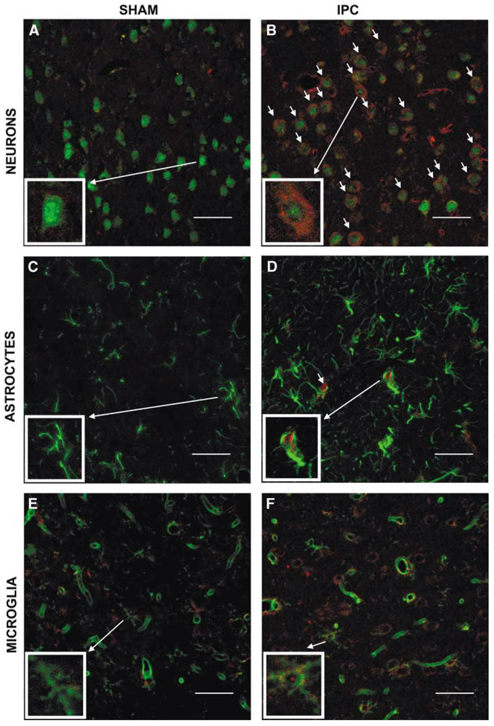
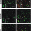
Description Immunofluorescence Validation of TACE in Rat Brain (Pradillo et al, 2005)
Cellular localization of TACE. Double immunofluorescence staining of brain sections from sham-operated (SHAM; A, C, E) and IPC-exposed animals (IPC; B, D, F) of TACE (red) and the cellular markers (green) NeuN (neurons; A, B), GFAP (astrocytes; C, D) and L. esculentum lectin (microglia and endothelium; E, F). White arrows indicate TACE-positive cells.
Cellular localization of TACE. Double immunofluorescence staining of brain sections from sham-operated (SHAM; A, C, E) and IPC-exposed animals (IPC; B, D, F) of TACE (red) and the cellular markers (green) NeuN (neurons; A, B), GFAP (astrocytes; C, D) and L. esculentum lectin (microglia and endothelium; E, F). White arrows indicate TACE-positive cells.
Handling
Storage This antibody is stable for at least one (1) year at -20°C. Avoid multiple freeze-thaw cycles.
Dilution Instructions Dilute in PBS or medium which is identical to that used in the assay system.
Application Instructions Immunoblotting: use at 1ug/mL.
Positive control: HeLa or Jurkat cell lysate.
Immunocytochemistry: use at 10ug/mL.
These are recommended concentrations.
Enduser should determine optimal concentrations for their applications.
Positive control: HeLa or Jurkat cell lysate.
Immunocytochemistry: use at 10ug/mL.
These are recommended concentrations.
Enduser should determine optimal concentrations for their applications.
References & Data Sheet
Data Sheet  Download PDF Data Sheet
 Download PDF Data Sheet

