Anti-p62dok Antibody (1107)
$445.00
SKU: 1107
Categories: Antibody Products, Neuroscience and Signal Transduction Antibodies, Products
Overview
Product Name Anti-p62dok Antibody (1107)
Description Anti-DOK (CT) Rabbit Polyclonal Antibody
Target p62dok
Species Reactivity Human
Applications ELISA,WB,ICC,IF
Host Rabbit
Clonality Polyclonal
Isotype IgG
Immunogen Peptide corresponding to aa 425- 439 of human p62dok.
Properties
Form Liquid
Concentration Lot Specific
Formulation PBS, pH 7.4.
Buffer Formulation Phosphate Buffered Saline
Buffer pH pH 7.4
Format Purified
Purification Purified by peptide immuno-affinity chromatography
Specificity Information
Specificity This antibody recognizes human p62dok (62kD).
Target Name Docking protein 1
Target ID p62dok
Uniprot ID Q99704
Alternative Names Downstream of tyrosine kinase 1, p62(dok, pp62
Gene Name DOK1
Gene ID 1796
Accession Number NP_001184189
Sequence Location [Isoform 1]: Cytoplasm. Nucleus.; [Isoform 3]: Cytoplasm, perinuclear region.
Biological Function DOK proteins are enzymatically inert adaptor or scaffolding proteins. They provide a docking platform for the assembly of multimolecular signaling complexes. DOK1 appears to be a negative regulator of the insulin signaling pathway. Modulates integrin activation by competing with talin for the same binding site on ITGB3. {PubMed:18156175}.
Research Areas Neuroscience
Background Signals from most growth factors and cytokines are transduced by receptor tyrosine kinases or non-receptor tyrosine kinases. Activated tyrosine kinases phosphorylate their substrates which mediates the cellular response to extracellular stimuli. A long-sought major substrate termed p62dok (downstream of tyrosine kinase) for many tyrosine kinases including c-kit, v-abl, v-Fps, v-Src, v-Fms, and activated EGF, PDGF, IGF, VEGF, and insulin receptors, has been identified in human and mouse. Upon phosphorylation, p62dok forms a complex with the ras GTPase-activating protein (RasGAP). p62dok, along with p56dok, represents a new family of RasGAP-binding proteins.
Application Images
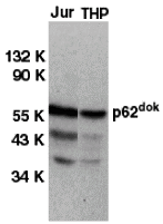


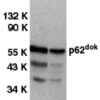
Description Western blot analysis of DOK1 in Jurkat (Jur) and THP-1 (THP) cell lysates with DOK1 antibody at 1 ug/mL.

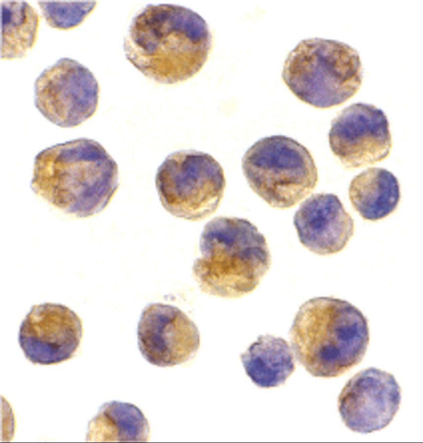
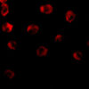
Description Immunocytochemistry of DOK1 in K562 cells with DOK1 antibody at 2 ug/mL.

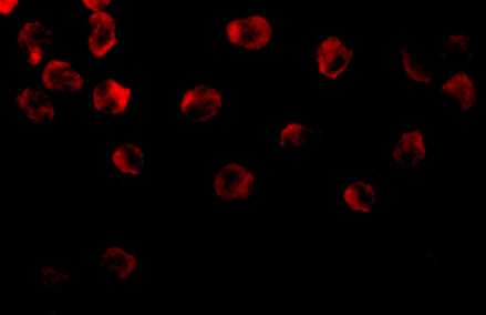
Description Immunofluorescence of DOK1 in K562 cells with DOK1 antibody at 10 ug/ml.
Handling
Storage This antibody is stable for at least one (1) year at -20°C. Avoid multiple freeze-thaw cycles.
Dilution Instructions Dilute in PBS or medium which is identical to that used in the assay system.
Application Instructions Immunoblotting: use at 1:1,000-1:2,000 dilution.
Positive control: Whole cell lysate of Jurkat cells.
Positive control: Whole cell lysate of Jurkat cells.
References & Data Sheet
Data Sheet  Download PDF Data Sheet
 Download PDF Data Sheet


