Anti-TrpC5 Ca+2 Channel Antibody (11541)
$466.00
SKU: 11541
Categories: Antibody Products, Ion Channel Antibodies, Products
Overview
Product Name Anti-TrpC5 Ca+2 Channel Antibody (11541)
Description Anti-TrpC5 Ca+2 Channel Mouse Monoclonal Antibody
Target TrpC5 Ca+2 Channel
Species Reactivity Human, Mouse, Rat
Applications WB,IHC,IP,AM
Host Mouse
Clonality Monoclonal
Clone ID S67-15
Isotype IgG2b
Immunogen Synthetic peptide corresponding to aa 827-845 of human TrpC5 (accession no. Q9UL62).
Properties
Form Liquid
Concentration Lot Specific
Formulation PBS, pH 7.4; 50% glycerol, 0.09% sodium azide.
Buffer Formulation Phosphate Buffered Saline
Buffer pH pH 7.4
Buffer Anti-Microbial 0.09% Sodium Azide
Buffer Cryopreservative 50% Glycerol
Format Purified
Purification Purified by Protein G affinity chromatography
Specificity Information
Specificity This antibody recognizes human, mouse, and rat TrpC5.
Target Name Short transient receptor potential channel 5
Target ID TrpC5 Ca+2 Channel
Uniprot ID Q9UL62
Alternative Names TrpC5, Transient receptor protein 5, TRP-5, hTRP-5, hTRP5
Gene Name TRPC5
Accession Number NP_036603.1
Sequence Location Cell membrane, Multi-pass membrane protein
Biological Function Thought to form a receptor-activated non-selective calcium permeant cation channel. Probably is operated by a phosphatidylinositol sUniProtKB:Q9QX29, PubMed:16284075}.
Research Areas Ion Channels
Background Ion channels are integral membrane proteins that help establish and control the small voltage gradient across the plasma membrane of living cells by allowing the flow of ions down their electrochemical gradient. TrpC5 (transient receptor potential cation channel, subfamily C, member 5) is a subtype of the TrpC family of mammalian transient receptor potential ion channels. Homomultimeric TrpC5 and heteromultimeric TrpC5-TrpC1 channels are activated by extracellular reduced thioredoxin. This activation has been associated with rheumatoid arthritis as well as with action on anesthetics such as chloroform, halothane, and propofol.
Application Images


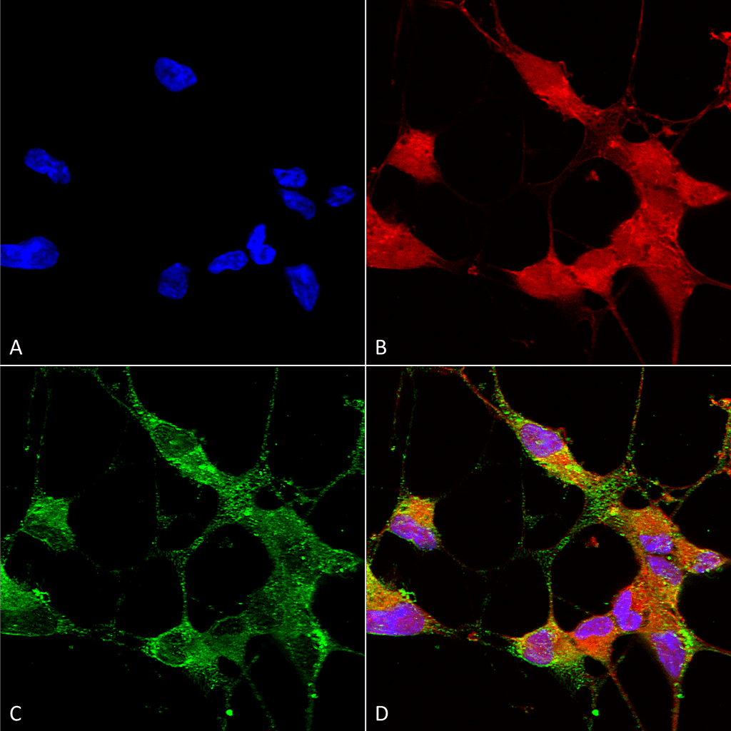
Description Immunocytochemistry/Immunofluorescence analysis using Mouse Anti-TrpC5 Monoclonal Antibody, Clone N67/15 (11541). Tissue: Neuroblastoma cells (SH-SY5Y). Species: Human. Fixation: 4% PFA for 15 min. Primary Antibody: Mouse Anti-TrpC5 Monoclonal Antibody (11541) at 1:50 for overnight at 4°C with slow rocking. Secondary Antibody: AlexaFluor 488 at 1:1000 for 1 hour at RT. Counterstain: Phalloidin-iFluor 647 (red) F-Actin stain; Hoechst (blue) nuclear stain at 1:800, 1.6mM for 20 min at RT. (A) Hoechst (blue) nuclear stain. (B) Phalloidin-iFluor 647 (red) F-Actin stain. (C) TrpC5 Antibody (D) Composite.

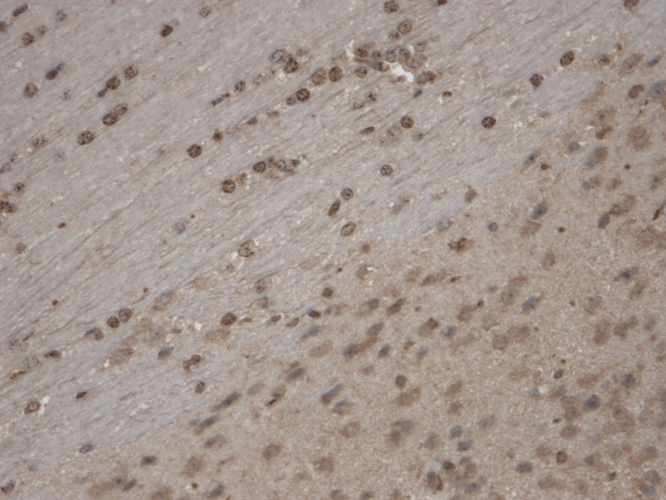
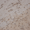
Description Immunohistochemistry analysis using Mouse Anti-TrpC5 Monoclonal Antibody, Clone N67/15 (11541). Tissue: Brain Slice. Species: Mouse. Fixation: 10% Formalin Solution for 12-24 hours at RT. Primary Antibody: Mouse Anti-TrpC5 Monoclonal Antibody (11541) at 1:1000 for 1 hour at RT. Secondary Antibody: HRP/DAB Detection System: Biotinylated Goat Anti-Mouse, Streptavidin Peroxidase, DAB Chromogen (brown) for 30 minutes at RT. Counterstain: Mayer Hematoxylin (purple/blue) nuclear stain at 250-500 µl for 5 minutes at RT. Localization: Nuclear staining. Magnification: 10X.

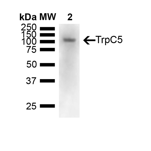
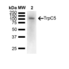
Description Western Blot analysis of Mouse brain showing detection of 110 kDa TrpC5 protein using Mouse Anti-TrpC5 Monoclonal Antibody, Clone N67/15 (11541). Lane 1: Molecular Weight Ladder (MW). Lane 2: Mouse Brain. Load: 15 ug. Block: 5% Skim Milk powder in TBST. Primary Antibody: Mouse Anti-TrpC5 Monoclonal Antibody (11541) at 1:1000 for Overnight at 4C. Secondary Antibody: Goat anti-mouse IgG:HRP at 1:7000 for 1 hour at RT with shaking. Color Development: Chemiluminescent for HRP (Moss) for 5 min in RT. Predicted/Observed Size: 110 kDa.
Handling
Storage This antibody is stable for at least one (1) year at -20°C.
Dilution Instructions Dilute in PBS or medium which is identical to that used in the assay system.
Application Instructions Immunoblotting: use at 1-10ug/mL. A band of ~110kDa is detected.Immunohistochemistry and
Immunocytochemistry: use at 0.1-1ug/mL
Immunofluorescence: use at 1-10ug/mL
These are recommended concentrations. User should determine optimal concentrations for their application.
Positive control: Rat brain lysate.
Immunocytochemistry: use at 0.1-1ug/mL
Immunofluorescence: use at 1-10ug/mL
These are recommended concentrations. User should determine optimal concentrations for their application.
Positive control: Rat brain lysate.
References & Data Sheet
Data Sheet  Download PDF Data Sheet
 Download PDF Data Sheet

