Anti-Smac (CT) Antibody (1409)
$445.00
| Host | Quantity | Applications | Species Reactivity | Data Sheet | |
|---|---|---|---|---|---|
| Rabbit | 100ug | ELISA,WB,IHC-P,IP,IF | Human, Mouse, Rat |  |
SKU: 1409
Categories: Antibody Products, Apoptosis Antibodies, Products
Overview
Product Name Anti-Smac (CT) Antibody (1409)
Description Anti-Smac (CT) Rabbit Polyclonal Antibody
Target Smac (CT)
Species Reactivity Human, Mouse, Rat
Applications ELISA,WB,IHC-P,IP,IF
Host Rabbit
Clonality Polyclonal
Isotype IgG
Immunogen Synthetic peptide corresponding to aa 225-239 of human Smac (accession no. AAF87716).
Properties
Form Liquid
Concentration Lot Specific
Formulation PBS, pH 7.4.
Buffer Formulation Phosphate Buffered Saline
Buffer pH pH 7.4
Format Purified
Purification Purified by peptide immuno-affinity chromatography
Specificity Information
Specificity This antibody recognizes human, mouse, and rat Smac (25kDa).
Target Name Diablo IAP-binding mitochondrial protein
Target ID Smac (CT)
Uniprot ID Q9NR28
Alternative Names Diablo homolog, mitochondrial, Direct IAP-binding protein with low pI, Second mitochondria-derived activator of caspase, Smac
Gene Name DIABLO
Gene ID 56616
Accession Number NP_063940
Sequence Location Mitochondrion
Biological Function Promotes apoptosis by activating caspases in the cytochrome c/Apaf-1/caspase-9 pathway. Acts by opposing the inhibitory activity of inhibitor of apoptosis proteins (IAP). Inhibits the activity of BIRC6/bruce by inhibiting its binding to caspases. Isoform 3 attenuates the stability and apoptosis-inhibiting activity of XIAP/BIRC4 by promoting XIAP/BIRC4 ubiquitination and degradation through the ubiquitin-proteasome pathway. Isoform 3 also disrupts XIAP/BIRC4 interacting with processed caspase-9 and promotes caspase-3 activation. Isoform 1 is defective in the capacity to down-regulate the XIAP/BIRC4 abundance. {PubMed:10929711, PubMed:14523016, PubMed:15200957}.
Research Areas Apoptosis
Background Inhibitors of apoptosis proteins (IAPs) regulate programmed cell death by inhibiting members of the caspase family of enzymes. A novel mammalian protein that binds to IAPs and neutralizes the inhibitory effect of IAPs on caspases has been identified and designated Smac/DIABLO. Smac is a mitochondrial protein that is released along with cytochrome c during apoptosis and activates the cytochrome c/Apaf- 1/caspase-9 pathway. The N-terminal amino acids of Smac are required for binding to IAPs and for activation of caspases. Smac is expressed in a variety of human and mouse tissues.
Application Images




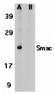
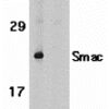
Description Western Blot Validation in Human Heart Tissue Lysate
Loading: 15 ug of lysates per lane. Antibodies: Smac 1409 (1 ug/mL), 1h incubation at RT in 5% NFDM/TBST.Secondary: Goat anti-rabbit IgG HRP conjugate at 1:10000 dilution.(A) Without blocking peptide(B) With blocking peptide
Loading: 15 ug of lysates per lane. Antibodies: Smac 1409 (1 ug/mL), 1h incubation at RT in 5% NFDM/TBST.Secondary: Goat anti-rabbit IgG HRP conjugate at 1:10000 dilution.(A) Without blocking peptide(B) With blocking peptide

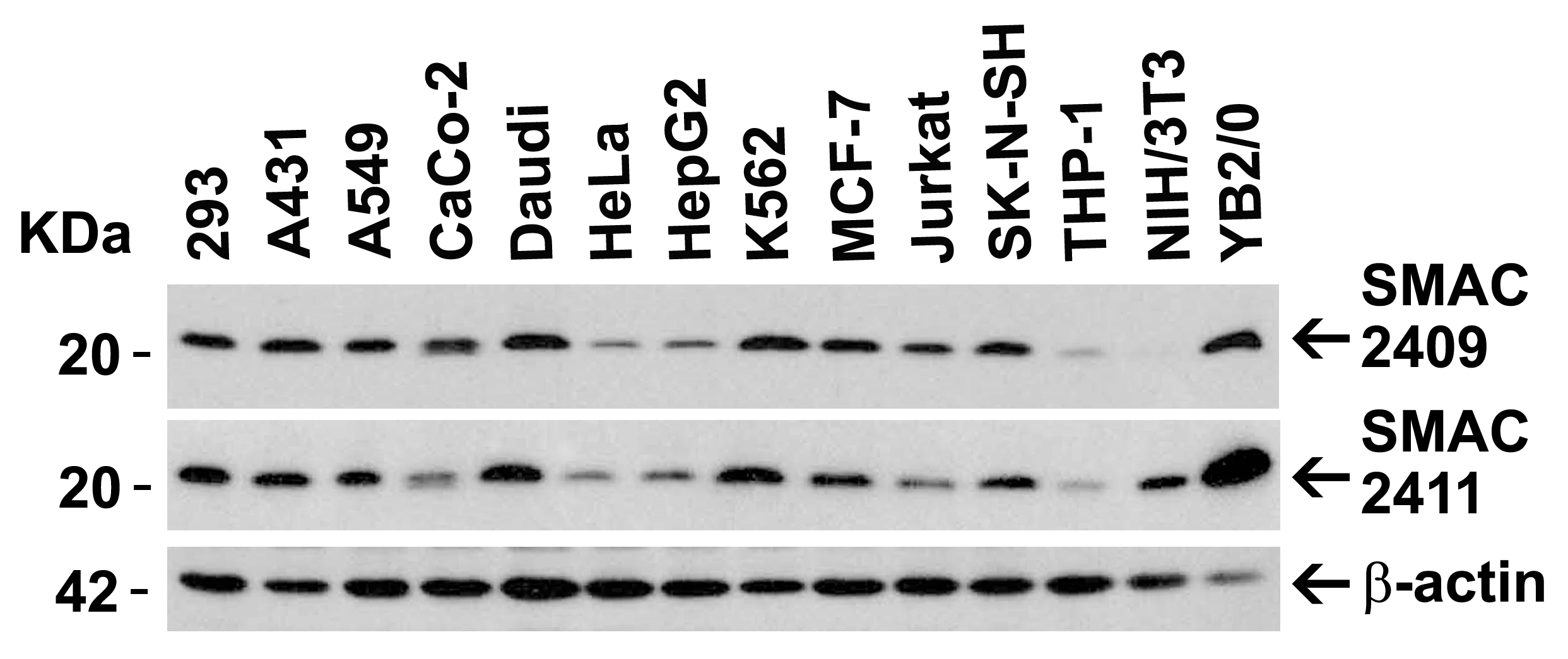
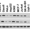
Description Independent Antibody Validation (IAV) via Protein Expression Profile in Cell Lines
Loading: 15 ug of lysates per lane. Antibodies: Smac 1409 (1 ug/mL), Smac 1411 (1 ug/mL), and beta-actin (1 ug/mL), 1h incubation at RT in 5% NFDM/TBST.Secondary: Goat anti-rabbit IgG HRP conjugate at 1:10000 dilution.
Loading: 15 ug of lysates per lane. Antibodies: Smac 1409 (1 ug/mL), Smac 1411 (1 ug/mL), and beta-actin (1 ug/mL), 1h incubation at RT in 5% NFDM/TBST.Secondary: Goat anti-rabbit IgG HRP conjugate at 1:10000 dilution.

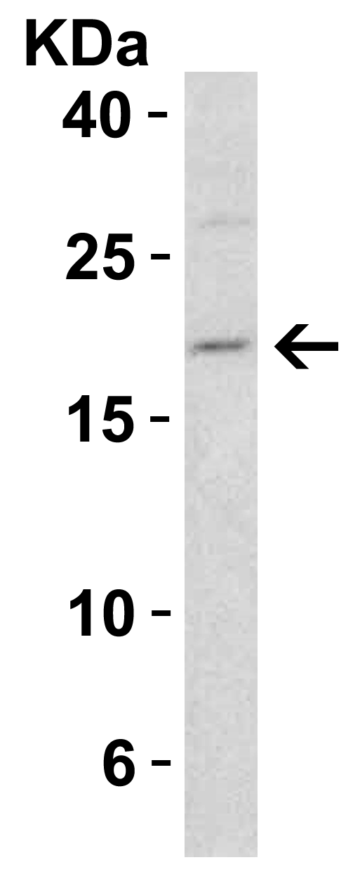
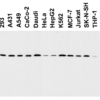
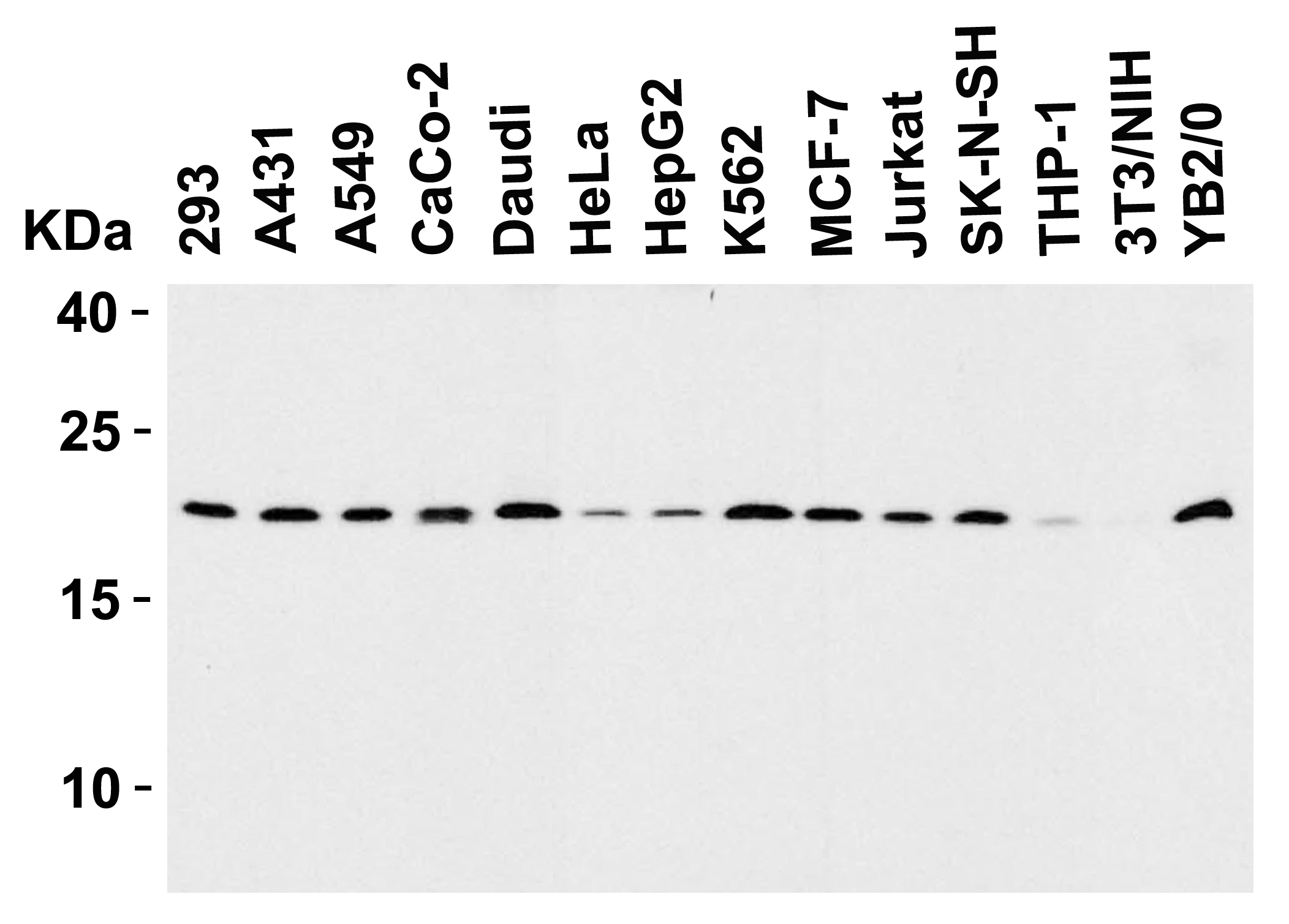
Description Western Blot Validation in Human, Mouse and Rat Cell Lines
Loading: 15 ug of lysates per lane. Antibodies: Smac 1409 (1 ug/mL), 1h incubation at RT in 5% NFM/TBST.Secondary: Goat anti-rabbit IgG HRP conjugate at 1:10000 dilution.
Loading: 15 ug of lysates per lane. Antibodies: Smac 1409 (1 ug/mL), 1h incubation at RT in 5% NFM/TBST.Secondary: Goat anti-rabbit IgG HRP conjugate at 1:10000 dilution.

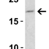
Description Western Blot Validation in Mouse 3T3/NIH Cells
Loading: 15 ug of lysate. Antibodies: Smac 1409 (1 ug/mL), 1h incubation at RT in 5% NFDM/TBST.Secondary: Goat anti-rabbit IgG HRP conjugate at 1:10000 dilution.
Loading: 15 ug of lysate. Antibodies: Smac 1409 (1 ug/mL), 1h incubation at RT in 5% NFDM/TBST.Secondary: Goat anti-rabbit IgG HRP conjugate at 1:10000 dilution.

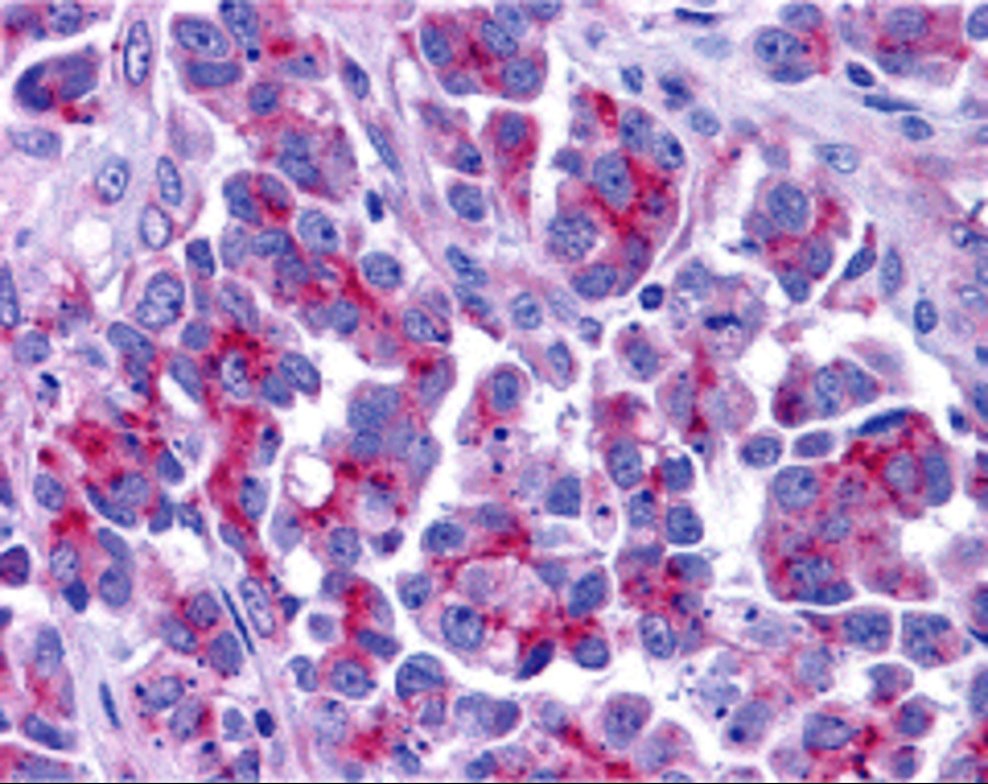
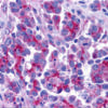
Description Immunohistochemistry Validation of Smac in Human Ovary Tissue
Immunohistochemical analysis of paraffin-embedded Human Ovary tissue using anti-Smac antibody (1409) at 5 ug/ml. Tissue was fixed with formaldehyde and blocked with 10% serum for 1 h at RT; antigen retrieval was by heat mediation with a citrate buffer (pH6). Samples were incubated with primary antibody overnight at 4C. A goat anti-rabbit IgG H&L (HRP) at 1/250 was used as secondary. Counter stained with Hematoxylin.
Immunohistochemical analysis of paraffin-embedded Human Ovary tissue using anti-Smac antibody (1409) at 5 ug/ml. Tissue was fixed with formaldehyde and blocked with 10% serum for 1 h at RT; antigen retrieval was by heat mediation with a citrate buffer (pH6). Samples were incubated with primary antibody overnight at 4C. A goat anti-rabbit IgG H&L (HRP) at 1/250 was used as secondary. Counter stained with Hematoxylin.

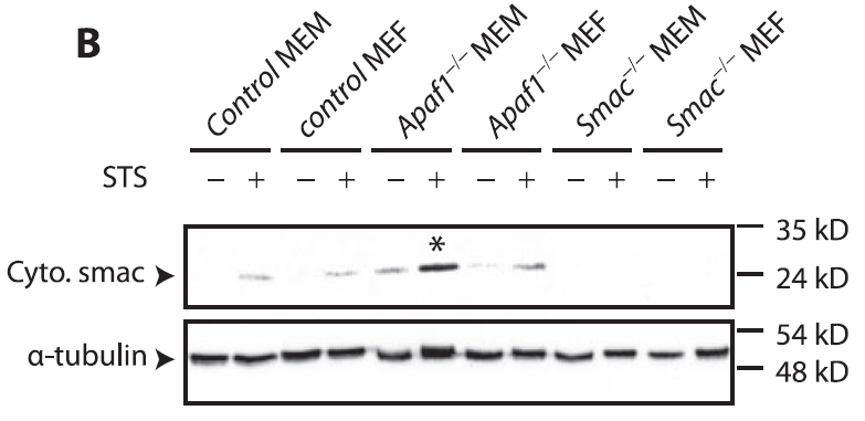
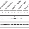
Description KO Validation in Mouse Fibroblasts and Myoblasts (Ho et al., 2416)
The indicated MEFs or MEMs were exposed to 2 uM STS for 4 h and analyzed by Western blot. Accumulation of Smac/Diablo in mitochondrion-depleted cytosol fractions fromSTS-treated Apaf-1 KO cells were detected by anti-smac antibodies. Smac expression was not detected in smac KO mice.
The indicated MEFs or MEMs were exposed to 2 uM STS for 4 h and analyzed by Western blot. Accumulation of Smac/Diablo in mitochondrion-depleted cytosol fractions fromSTS-treated Apaf-1 KO cells were detected by anti-smac antibodies. Smac expression was not detected in smac KO mice.

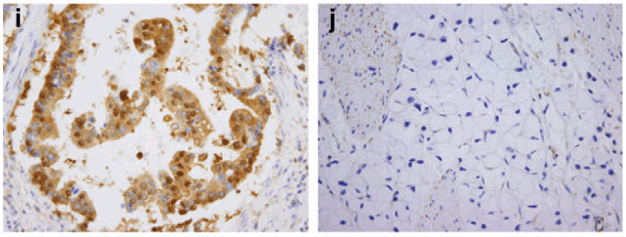
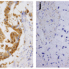
Description Immunohistochemistry Validation of Smac in Human gastric carcinoma (Kim et al., 2011)
Smac was highly expressed in gastric mucosa of patients with gastric carcinoma.
Smac was highly expressed in gastric mucosa of patients with gastric carcinoma.

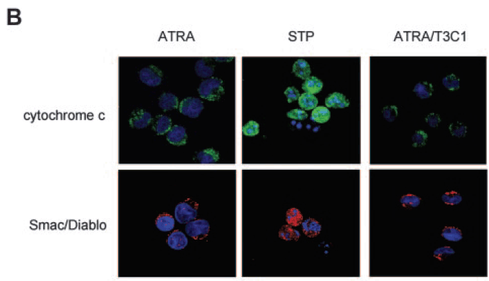
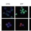
Description Immunofluorescence Analysis of Smac in NB4-LR1 Cells (Saumet et al., 2005)
NB4-LR1 cells were either treated with ATRA(1 μM) for 3 days without or with the T3C1 recombinant fragment (3 uM) or treated with staurosporine (STP; 5 uM ) for 3.5 hours. STP, but not ATRA or AYRA/T3C1 induced the release of smac.
NB4-LR1 cells were either treated with ATRA(1 μM) for 3 days without or with the T3C1 recombinant fragment (3 uM) or treated with staurosporine (STP; 5 uM ) for 3.5 hours. STP, but not ATRA or AYRA/T3C1 induced the release of smac.

Description Induced Expression Validation in Rat Liver (Genestier et al., 2005)
Mitochondria from rat liver were treated with increasing concentrations of rPVL (A), rLukS (B), or Bax alpha (C) for 1 hour at 30C. rPVL induces the release of the apoptogenic proteins cytochrome c and Smac/DIABLO from isolated mitochondria.
Mitochondria from rat liver were treated with increasing concentrations of rPVL (A), rLukS (B), or Bax alpha (C) for 1 hour at 30C. rPVL induces the release of the apoptogenic proteins cytochrome c and Smac/DIABLO from isolated mitochondria.

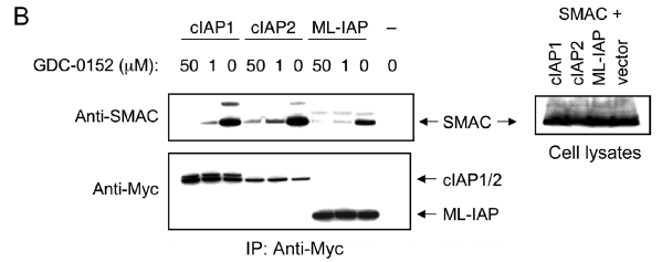
Description Overxpression Validation in HEK293T Cells (Flygare et al., 2012)
HEK293T cells were transiently transfected with Smac and Myc-tagged cIAP1, cIAP2, ML-IAP, or empty vector. Cells were lysed, and lysates were incubated with the indicated concentrations of 1 and immunoprecipitated with anti-Myc antibody (left panels). Samples were then immunoblotted with anti-Smac and anti-Myc antibodies. Whole-cell lysates are shown in the right panel.
HEK293T cells were transiently transfected with Smac and Myc-tagged cIAP1, cIAP2, ML-IAP, or empty vector. Cells were lysed, and lysates were incubated with the indicated concentrations of 1 and immunoprecipitated with anti-Myc antibody (left panels). Samples were then immunoblotted with anti-Smac and anti-Myc antibodies. Whole-cell lysates are shown in the right panel.
Handling
Storage This antibody is stable for at least one (1) year at -20°C. Avoid multiple freeze-thaw cycles.
Dilution Instructions Dilute in PBS or medium which is identical to that used in the assay system.
Application Instructions Immunoblotting: use at 1ug/mL.
Positive control: Human heart tissue lysate.
Immunohistochemistry: use at 5ug/mL
These are recommended concentrations.
Enduser should determine optimal concentrations for their applications.
Positive control: Human heart tissue lysate.
Immunohistochemistry: use at 5ug/mL
These are recommended concentrations.
Enduser should determine optimal concentrations for their applications.
References & Data Sheet
Data Sheet  Download PDF Data Sheet
 Download PDF Data Sheet

