Anti-RPA32 Antibody (56216)
$503.00
SKU: 56216
Categories: Antibody Products, Cancer Research Antibodies, Products
Overview
Product Name Anti-RPA32 Antibody (56216)
Description Anti-RPA32 Mouse Monoclonal Antibody
Target RPA32
Species Reactivity Human, Mouse
Applications WB,ICC/IF,IP
Host Mouse
Clonality Monoclonal
Clone ID 12F3.3
Isotype IgG2a
Immunogen N-terminally 6x-His tagged fusion protein corresponding to full-length human RPA32 expressed in E. coli.
Properties
Form Liquid
Concentration Lot Specific
Formulation PBS, pH 7.4
Buffer Formulation Phosphate Buffered Saline
Buffer pH pH 7.4
Format Purified
Purification Purified by Protein G affinity chromatography
Specificity Information
Specificity This antibody recognizes human and mouse RPA32. RPA (Replication Protein A) is a single-stranded DNA binding protein. Human RPA is a heterotrimeric protein containing subunits of 70, 32, and 14 kD. This protein complex is highly conserved in eukaryotes and is essential in DNA replication, homologous recombination, and nucleotide excision repair. RPA32 is phosphorylated during the S-phase of the cell cycle, in response to DNA damage, and during apoptosis of Jurkat T-lymphocytes.
Target Name Replication protein A 32 kDa subunit
Target ID RPA32
Uniprot ID P15927
Alternative Names RP-A p32, Replication factor A protein 2, RF-A protein 2, Replication protein A 34 kDa subunit, RP-A p34
Gene Name RPA2
Sequence Location Nucleus, Nucleus, PML body
Biological Function As part of the heterotrimeric replication protein A complex (RPA/RP-A), binds and stabilizes single-stranded DNA intermediates, that form during DNA replication or upon DNA stress. It prevents their reannealing and in parallel, recruits and activates different proteins and complexes involved in DNA metabolism. Thereby, it plays an essential role both in DNA replication and the cellular response to DNA damage. In the cellular response to DNA damage, the RPA complex controls DNA repair and DNA damage checkpoint activation. Through recruitment of ATRIP activates the ATR kinase a master regulator of the DNA damage response. It is required for the recruitment of the DNA double-strand break repair factors RAD51 and RAD52 to chromatin in response to DNA damage. Also recruits to sites of DNA damage proteins like XPA and XPG that are involved in nucleotide excision repair and is required for this mechanism of DNA repair. Plays also a role in base excision repair (BER) probably through interaction with UNG. Also recruits SMARCAL1/HARP, which is involved in replication fork restart, to sites of DNA damage. May also play a role in telomere maintenance. {PubMed:15205463, PubMed:17765923, PubMed:17959650, PubMed:19116208, PubMed:20154705, PubMed:21504906, PubMed:2406247, PubMed:24332808, PubMed:7697716, PubMed:7700386, PubMed:8702565, PubMed:9430682, PubMed:9765279}.
Research Areas Cancer research
Application Images


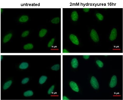
Description RPA32 antibody [12F3.3] detects RPA32 protein at DNA damage foci by immunofluorescent analysis.
Sample: 2mM hydroxyurea treated (right) or untreated (left) HeLa cells were fixed in ice-cold MeOH for 5 min.
Green: RPA32 protein stained by RPA32 antibody [12F3.3] (56216) diluted at 1:1000.
Blue: Hoechst 33342 staining.
Scale bar = 10 um.
Sample: 2mM hydroxyurea treated (right) or untreated (left) HeLa cells were fixed in ice-cold MeOH for 5 min.
Green: RPA32 protein stained by RPA32 antibody [12F3.3] (56216) diluted at 1:1000.
Blue: Hoechst 33342 staining.
Scale bar = 10 um.

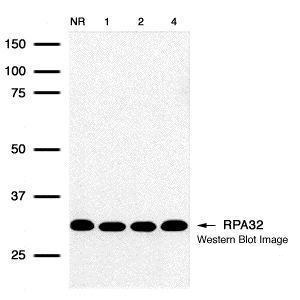
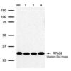
Description Detection of human RPA32 protein using RPA32 12F3.3 monoclonal antibody (56216) in HeLa (IR treated) whole cell extract. No radiation, 1hr, 2hr, and 4hr exposure corresponding to lanes 1 through 4 respectively.

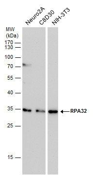
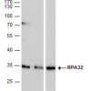
Description RPA32 antibody detects RPA32 protein by western blot analysis. Various whole cell extracts (30 ug) were separated by 12% SDS-PAGE, and blotted with RPA32 antibody (56216) diluted by 1:500. The HRP-conjugated anti-mouse IgG antibody was used to detect the primary antibody.
Handling
Storage This antibody is stable for at least one (1) year at -70°C. Avoid multiple freeze-thaw cycles.
Dilution Instructions Dilute in PBS or medium which is identical to that used in the assay system.
Application Instructions Immunoblotting,
Immunoprecipitation: use at 0.2-1 ug/mL.
Positive control: HeLa, Raji, and 3T3 cells and recombinant protein.
Immunoprecipitation: use at 0.2-1 ug/mL.
Positive control: HeLa, Raji, and 3T3 cells and recombinant protein.
References & Data Sheet
References Iftode, C et al. 1999. Crit Rev Biochem Mol Biol 34: 141-180
Bochkarev, A et al. 1999. EMBO 18: 4498-4504
Dong, J et al. 1999. Biochem J 337: 311-317
Sibenaller, ZA et al. 1998. Biochemistry 37: 12496-12506
Stigger, E et al. 1998. J Biol Chem 273: 9337-9343
Bochkareva, E et al. 1998. J Biol Chem 273: 3932-3936.
Bochkarev, A et al. 1999. EMBO 18: 4498-4504
Dong, J et al. 1999. Biochem J 337: 311-317
Sibenaller, ZA et al. 1998. Biochemistry 37: 12496-12506
Stigger, E et al. 1998. J Biol Chem 273: 9337-9343
Bochkareva, E et al. 1998. J Biol Chem 273: 3932-3936.
Data Sheet  Download PDF Data Sheet
 Download PDF Data Sheet

