Anti-RIP3 Antibody (2283)
$445.00
| Host | Quantity | Applications | Species Reactivity | Data Sheet | |
|---|---|---|---|---|---|
| Rabbit | 100ug | ELISA,IF,IHC-P,WB,IP | Mouse, Rat |  |
SKU: 2283
Categories: Antibody Products, Apoptosis Antibodies, Products
Overview
Product Name Anti-RIP3 Antibody (2283)
Description Anti-RIP3 Rabbit Polyclonal Antibody
Target RIP3
Species Reactivity Mouse, Rat
Applications ELISA,IF,IHC-P,WB,IP
Host Rabbit
Clonality Polyclonal
Isotype IgG
Immunogen Synthetic peptide corresponding to aa 473-486 of mouse RIP3 (accession no. AAF03133).
Properties
Form Liquid
Concentration Lot Specific
Formulation PBS, pH 7.4.
Buffer Formulation Phosphate Buffered Saline
Buffer pH pH 7.4
Format Purified
Purification Purified by peptide immuno-affinity chromatography
Specificity Information
Specificity This antibody recognizes mouse and rat RIP3 (57kDa).
Target Name Receptor-interacting serine/threonine-protein kinase 3
Target ID RIP3
Uniprot ID Q9QZL0
Alternative Names EC 2.7.11.1, RIP-like protein kinase 3, Receptor-interacting protein 3, RIP-3, mRIP3
Gene Name Ripk3
Gene ID 56532
Accession Number NP_064339
Sequence Location Cytoplasm, cytosol, Nucleus
Biological Function Serine/threonine-protein kinase that activates necroptosis and apoptosis, two parallel forms of cell death (PubMed:27321907, PubMed:27746097, PubMed:27917412, PubMed:28607035, PubMed:32200799, PubMed:32296175). Necroptosis, a programmed cell death process in response to death-inducing TNF-alpha family members, is triggered by RIPK3 following activation by ZBP1 (PubMed:19590578, PubMed:22423968, PubMed:24012422, PubMed:24019532, PubMed:24557836, PubMed:27746097, PubMed:27819681, PubMed:27819682, PubMed:24095729, PubMed:32200799, PubMed:27321907, PubMed:32296175). Activated RIPK3 forms a necrosis-inducing complex and mediates phosphorylation of MLKL, promoting MLKL localization to the plasma membrane and execution of programmed necrosis characterized by calcium influx and plasma membrane damage (PubMed:24813849, PubMed:24813850, PubMed:27321907). In addition to TNF-induced necroptosis, necroptosis can also take place in the nucleus in response to orthomyxoviruses infection: following ZBP1 activation, which senses double-stranded Z-RNA structures, nuclear RIPK3 catalyzes phosphorylation and activation of MLKL, promoting disruption of the nuclear envelope and leakage of cellular DNA into the cytosol (PubMed:32200799, PubMed:32296175). Also regulates apoptosis: apoptosis depends on RIPK1, FADD and CASP8, and is independent of MLKL and RIPK3 kinase activity (PubMed:27321907). Phosphorylates RIPK1: RIPK1 and RIPK3 undergo reciprocal auto- and trans-phosphorylation (By similarity). In some cell types, also able to restrict viral replication by promoting cell death-independent responses (PubMed:30635240). In response to flavivirus infection in neurons, promotes a cell death-independent pathway that restricts viral replication: together with ZBP1, promotes a death-independent transcriptional program that modifies the cellular metabolism via up-regulation expression of the enzyme ACOD1/IRG1 and production of the metabolite itaconate (PubMed:30635240). Itaconate inhibits the activity of succinate dehydrogenase, generating a metabolic state in neurons that suppresses replication of viral genomes (PubMed:30635240). RIPK3 binds to and enhances the activity of three metabolic enzymes: GLUL, GLUD1, and PYGL (By similarity). These metabolic enzymes may eventually stimulate the tricarboxylic acid cycle and oxidative phosphorylation, which could result in enhanced ROS production (By similarity). {UniProtKB:Q9Y572, PubMed:19590578, PubMed:22423968, PubMed:24012422, PubMed:24019532, PubMed:24095729, PubMed:24557836, PubMed:24813849, PubMed:24813850, PubMed:27321907, PubMed:27746097, PubMed:27819681, PubMed:27819682, PubMed:27917412, PubMed:28607035, PubMed:30635240, PubMed:32200799, PubMed:32296175}.
Research Areas Apoptosis
Background Serine/threonine protein kinases, such as ASK1, RIP, DAP and ZIP kinases, are mediators of apoptosis. Receptor interacting proteins, including RIP and RIP2/RICK mediate apoptosis induced by TNFR1 and Fas, two members of the family of death receptors. A novel member in the RIP kinase family has been identified and designated RIP3. RIP3 contains an N-terminal kinase domain, but, unlike RIP or RIP2, lacks the C-terminal death or CARD domain. RIP3 binds to RIP and TNFR1, mediates TNFR1- induced apoptosis, and attenuates RIP and TNFR1-induced NF- B activation. The messenger RNA of RIP3 is found in some adult tissues.
Application Images


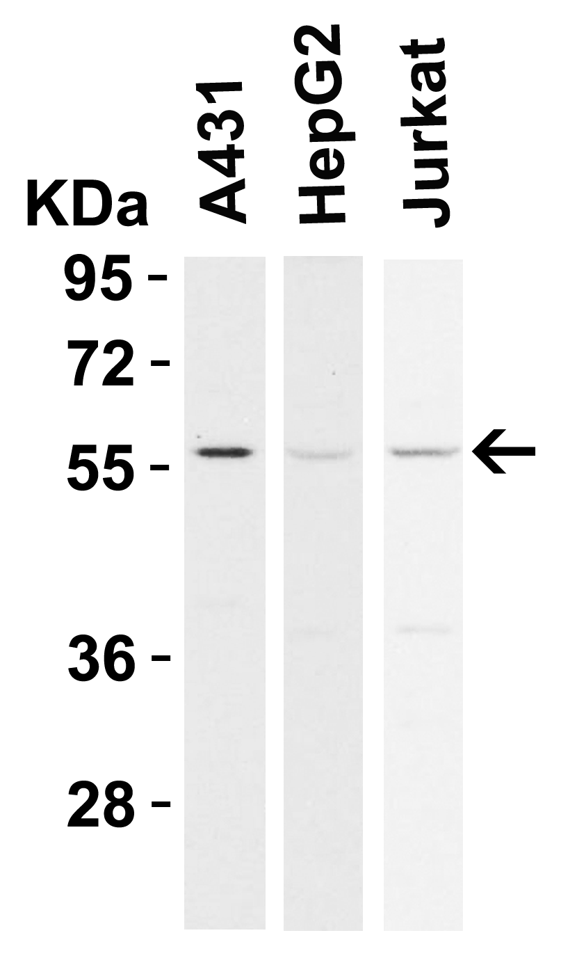
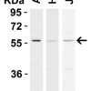
Description Western Blot Validation in Human Cell Lines
Loading: 15 ug of lysates per lane. Antibodies: RIP3 2283, (0.5 ug/mL), 1h incubation at RT in 5% NFDM/TBST.Secondary: Goat anti-rabbit IgG HRP conjugate at 1:10000 dilution.
Loading: 15 ug of lysates per lane. Antibodies: RIP3 2283, (0.5 ug/mL), 1h incubation at RT in 5% NFDM/TBST.Secondary: Goat anti-rabbit IgG HRP conjugate at 1:10000 dilution.

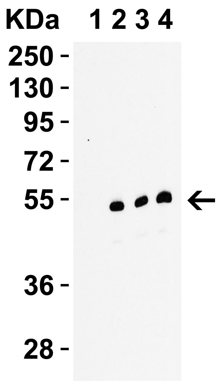
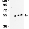
Description Western Blot Validation in C2C12 Cells Mouse
Loading: 15 ug of lysates per lane. Antibodies: RIP3 2283, 1h incubation at RT in 5% NFDM/TBST.Secondary: Goat anti-rabbit IgG HRP conjugate at 1:10000 dilution.Lane 1: 2283, 0.1 ug/mL in the presence of peptide blockingLane 2: 2283, 0.1 ug/mLLane 3: 2283, 0.2 ug/mLLane 4: 2283, 0.5 ug/mL
Loading: 15 ug of lysates per lane. Antibodies: RIP3 2283, 1h incubation at RT in 5% NFDM/TBST.Secondary: Goat anti-rabbit IgG HRP conjugate at 1:10000 dilution.Lane 1: 2283, 0.1 ug/mL in the presence of peptide blockingLane 2: 2283, 0.1 ug/mLLane 3: 2283, 0.2 ug/mLLane 4: 2283, 0.5 ug/mL

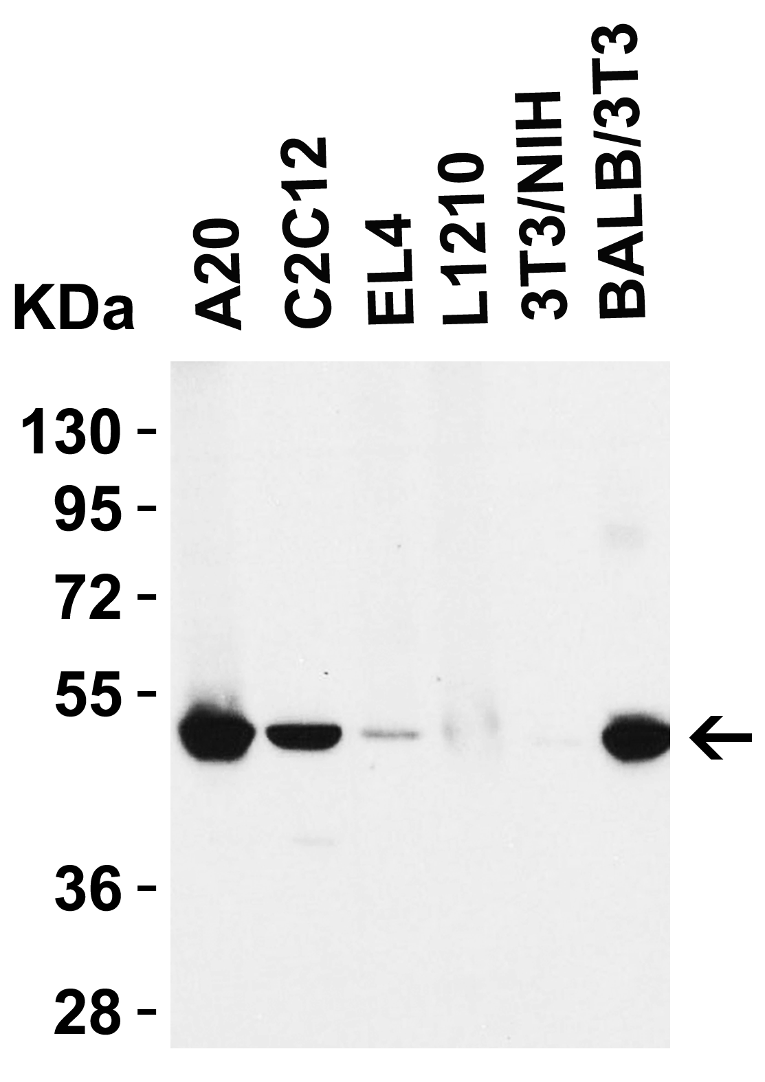
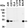
Description Western Blot Validation in Mouse Cell lines
Loading: 15 ug of lysates per lane. Antibodies: RIP3 2283, (0.5 ug/mL), 1h incubation at RT in 5% NFDM/TBST.Secondary: Goat anti-rabbit IgG HRP conjugate at 1:10000 dilution.
Loading: 15 ug of lysates per lane. Antibodies: RIP3 2283, (0.5 ug/mL), 1h incubation at RT in 5% NFDM/TBST.Secondary: Goat anti-rabbit IgG HRP conjugate at 1:10000 dilution.

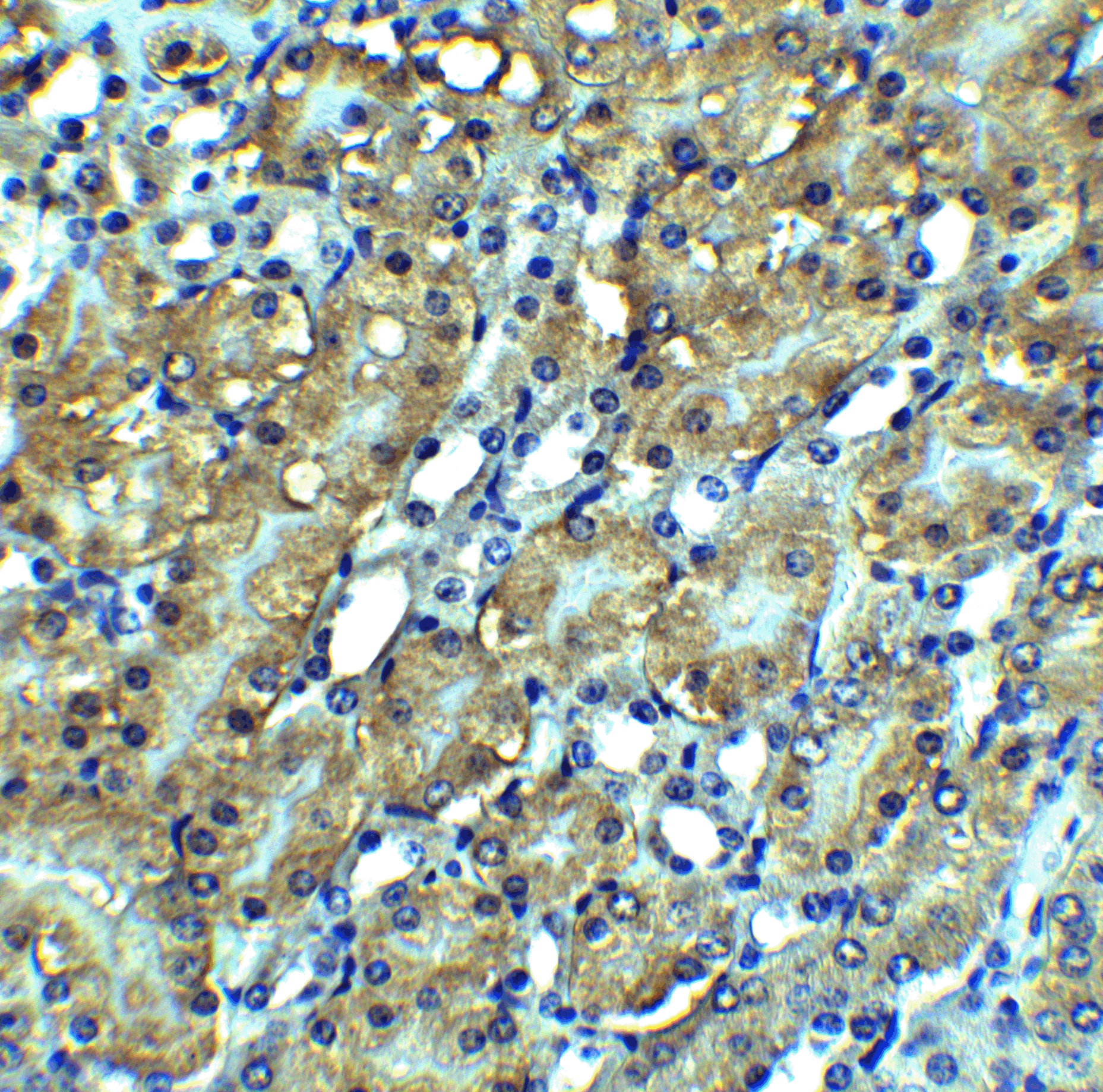
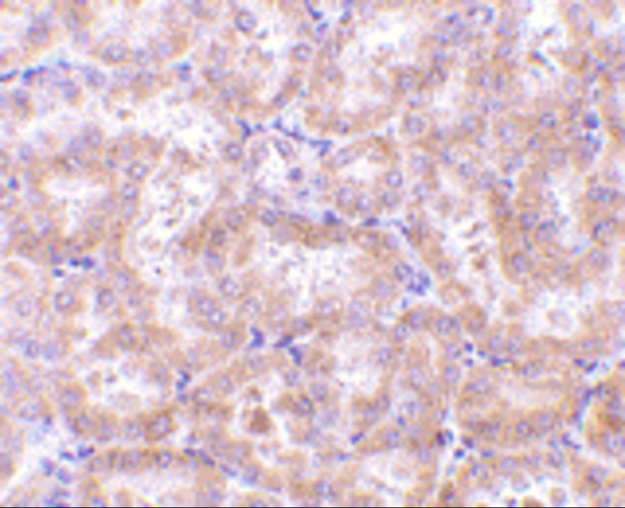
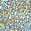
Description Immunohistochemistry Validation of RIP3 in Mouse Kidney Tissue
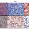
Immunohistochemical analysis of paraffin-embedded mouse kidney tissue using anti-RIP3 antibody (2283) at 2.5 ug/ml. Tissue was fixed with formaldehyde and blocked with 10% serum for 1 h at RT; antigen retrieval was by heat mediation with a citrate buffer (pH6). Samples were incubated with primary antibody overnight at 4C. A goat anti-rabbit IgG H&L (HRP) at 1/250 was used as secondary. Counter stained with Hematoxylin.
Immunohistochemical analysis of paraffin-embedded mouse kidney tissue using anti-RIP3 antibody (2283) at 2.5 ug/ml. Tissue was fixed with formaldehyde and blocked with 10% serum for 1 h at RT; antigen retrieval was by heat mediation with a citrate buffer (pH6). Samples were incubated with primary antibody overnight at 4C. A goat anti-rabbit IgG H&L (HRP) at 1/250 was used as secondary. Counter stained with Hematoxylin.

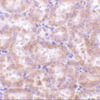
Description Immunohistochemistry Validation of RIP3 in Rat Kidney Tissue
Immunohistochemical analysis of paraffin-embedded rat kidney tissue using anti-RIP3 antibody (2283) at 5 ug/ml. Tissue was fixed with formaldehyde and blocked with 10% serum for 1 h at RT; antigen retrieval was by heat mediation with a citrate buffer (pH6). Samples were incubated with primary antibody overnight at 4C. A goat anti-rabbit IgG H&L (HRP) at 1/250 was used as secondary. Counter stained with Hematoxylin.
Immunohistochemical analysis of paraffin-embedded rat kidney tissue using anti-RIP3 antibody (2283) at 5 ug/ml. Tissue was fixed with formaldehyde and blocked with 10% serum for 1 h at RT; antigen retrieval was by heat mediation with a citrate buffer (pH6). Samples were incubated with primary antibody overnight at 4C. A goat anti-rabbit IgG H&L (HRP) at 1/250 was used as secondary. Counter stained with Hematoxylin.

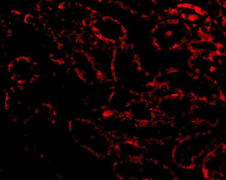
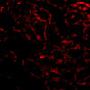
Description Immunofluorescence Validation of RIP3 in Rat Kidney Tissue
Immunofluorescent analysis of 4% paraformaldehyde-fixed Rat Kidney tissue labeling RIP3 with 2283 at 20 ug/mL, followed by goat anti-rabbit IgG secondary antibody at 1/500 dilution (red).
Immunofluorescent analysis of 4% paraformaldehyde-fixed Rat Kidney tissue labeling RIP3 with 2283 at 20 ug/mL, followed by goat anti-rabbit IgG secondary antibody at 1/500 dilution (red).

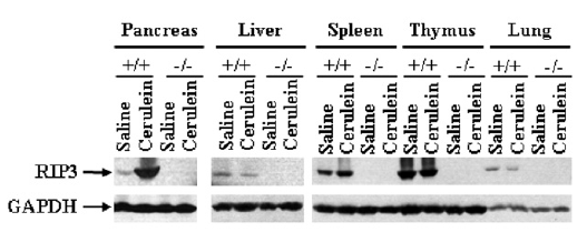
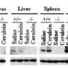
Description KO Validation of RIP3 in RIP3 KO Mice (He et al., 2009)
Western blot analysis of RIP3 with anti-RIP3 antibodies shows disrupted RIP3 expression in pancreas, liver spleen thymus and lung of RIP3 KO mice. Cerulein treatment upregulated RIP3 expression in the pancreas of WT mice.
Western blot analysis of RIP3 with anti-RIP3 antibodies shows disrupted RIP3 expression in pancreas, liver spleen thymus and lung of RIP3 KO mice. Cerulein treatment upregulated RIP3 expression in the pancreas of WT mice.

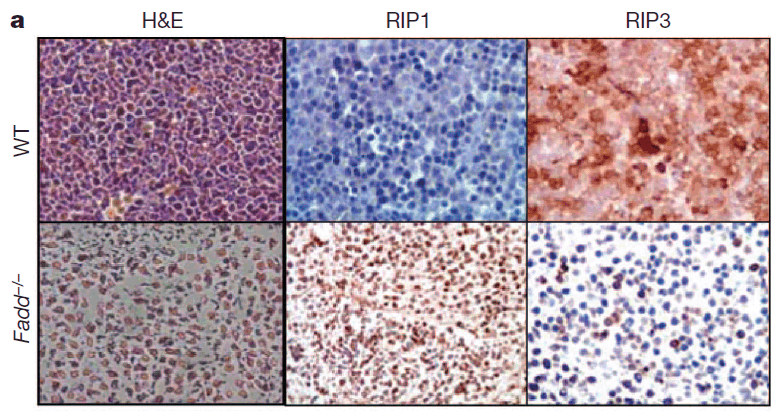
Description Immunohistochemistry Validation of RIP3 in Fadd KO mice (Zhang et al., 2011)
RIP3 expression detected by anti-RIP3 antibodies (2283) was decreased in Fadd KO mice as compared to WT mice.
RIP3 expression detected by anti-RIP3 antibodies (2283) was decreased in Fadd KO mice as compared to WT mice.

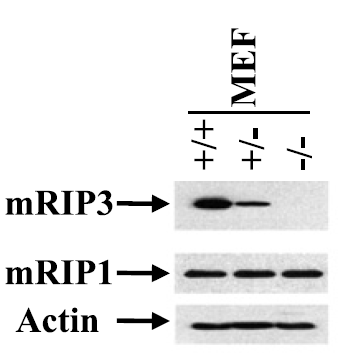
Description KO Validation of RIP3 in MEF Cells (He et al., 2012)
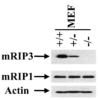
Western blot analysis of RIP3 with anti-RIP3 antibodies shows disrupted RIP3 expression in RIP3 KO MEFs, but not in WT MEF cells.
Western blot analysis of RIP3 with anti-RIP3 antibodies shows disrupted RIP3 expression in RIP3 KO MEFs, but not in WT MEF cells.

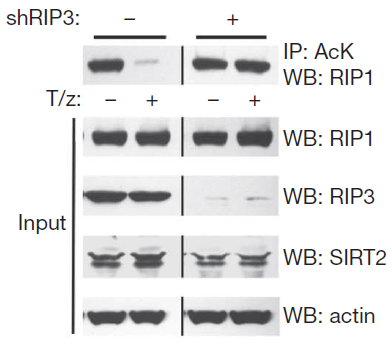
Description KD Validation of RIP3 in L929 Cells (Narayan et al., 2012)
Immunoblot analysis of RIP3 with anti-RIP3 antibodies (2283) shows RIP3 expression was disrupted in RIP3 knockdown L929 cells.
Immunoblot analysis of RIP3 with anti-RIP3 antibodies (2283) shows RIP3 expression was disrupted in RIP3 knockdown L929 cells.

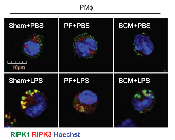
Description Regulated Expression Validation of RIP3 in Macrophages (Li et al., 2016)
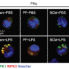
Confocal microscopy shows colocalization of RIPK1 (green) and RIPK3 (red) in PMϕ and RIPK3 was detected by anti-RIPK3 antibodies (2283). PF or BCM suppressed LPS-induced upregulation of RIP3.
Confocal microscopy shows colocalization of RIPK1 (green) and RIPK3 (red) in PMϕ and RIPK3 was detected by anti-RIPK3 antibodies (2283). PF or BCM suppressed LPS-induced upregulation of RIP3.

Description Overexpression Validation of RIP3 in Cancer Cell Lines (Yang et al., 2017)
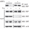
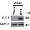
The cancer cell lines were stably expressing flag-tagged RIP3 and RIP expression was detected by anti-RIP3 antibodies (2283) in RIP3-overexpressed cells.
The cancer cell lines were stably expressing flag-tagged RIP3 and RIP expression was detected by anti-RIP3 antibodies (2283) in RIP3-overexpressed cells.
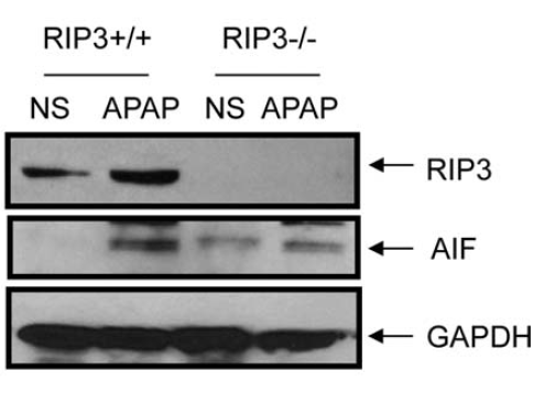
Description Induced Expression of RIP3 by Acetaminophen in WT and RIP3 KO Mice (Ramachandran et al., 2412)
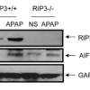
Wild-type and RIP3 KO mice were treated with300 mg/kg acetaminophen or saline. RIP3 expression detected by anti-RIP3 antibodies (2283) were up-regulated after acetaminophen treatment in the liver, while this effect was not observed in RIP3 KO mice.
Wild-type and RIP3 KO mice were treated with300 mg/kg acetaminophen or saline. RIP3 expression detected by anti-RIP3 antibodies (2283) were up-regulated after acetaminophen treatment in the liver, while this effect was not observed in RIP3 KO mice.

Description Immunogold EM Validation in MEF Cells (Li et al., 2012)
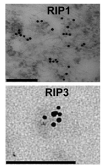
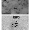
Clustering of RIP3 in necrotic MEFs shown by immunogold EM with anti-RIP3 antibodies (2283). Scale bars, 100 nm (RIP3).
Clustering of RIP3 in necrotic MEFs shown by immunogold EM with anti-RIP3 antibodies (2283). Scale bars, 100 nm (RIP3).
Handling
Storage This antibody is stable for at least one (1) year at -20°C. Avoid multiple freeze-thaw cycles.
Dilution Instructions Dilute in PBS or medium which is identical to that used in the assay system.
Application Instructions Immunoblotting: use at 1ug/mL.
Positive control: NIH/3T3 cell lysate.
Immunohistochemistry: use at 5ug/mL.
These are recommended concentrations.
Enduser should determine optimal concentrations for their applications.
Positive control: NIH/3T3 cell lysate.
Immunohistochemistry: use at 5ug/mL.
These are recommended concentrations.
Enduser should determine optimal concentrations for their applications.
References & Data Sheet
Data Sheet  Download PDF Data Sheet
 Download PDF Data Sheet

