Anti-RbAp48 Antibody (3111)
$503.00
SKU: 3111
Categories: Antibody Products, Cancer Research Antibodies, Products
Overview
Product Name Anti-RbAp48 Antibody (3111)
Description Anti-RbAp48 Mouse Monoclonal Antibody
Target RbAp48
Species Reactivity Human
Applications WB,ICC/IF,IHC-P,IP,EMSA
Host Mouse
Clonality Monoclonal
Clone ID 15G12
Isotype IgG2a
Immunogen GST fusion protein corresponding to aa 1-425 of human RbAp48.
Properties
Form Liquid
Concentration Lot Specific
Formulation PBS, pH 7.4.
Buffer Formulation Phosphate Buffered Saline
Buffer pH pH 7.4
Format Purified
Purification Purified by Protein G affinity chromatography
Specificity Information
Specificity This antibody recognizes human and mouse RbAp48 and RbAp46.
Target Name Retinoblastoma-associated protein
Target ID RbAp48
Uniprot ID P06400
Alternative Names p105-Rb, p110-RB1, pRb, Rb, pp110
Gene Name RB1
Gene ID 5925
Accession Number NP_000312
Sequence Location Nucleus
Biological Function Tumor suppressor that is a key regulator of the G1/S transition of the cell cycle (PubMed:10499802). The hypophosphorylated form binds transcription regulators of the E2F family, preventing transcription of E2F-responsive genes (PubMed:10499802). Both physically blocks E2Fs transactivating domain and recruits chromatin-modifying enzymes that actively repress transcription (PubMed:10499802). Cyclin and CDK-dependent phosphorylation of RB1 induces its dissociation from E2Fs, thereby activating transcription of E2F responsive genes and triggering entry into S phase (PubMed:10499802). RB1 also promotes the G0-G1 transition upon phosphorylation and activation by CDK3/cyclin-C (PubMed:15084261). Directly involved in heterochromatin formation by maintaining overall chromatin structure and, in particular, that of constitutive heterochromatin by stabilizing histone methylation. Recruits and targets histone methyltransferases SUV39H1, KMT5B and KMT5C, leading to epigenetic transcriptional repression. Controls histone H4 'Lys-20' trimethylation. Inhibits the intrinsic kinase activity of TAF1. Mediates transcriptional repression by SMARCA4/BRG1 by recruiting a histone deacetylase (HDAC) complex to the c-FOS promoter. In resting neurons, transcription of the c-FOS promoter is inhibited by BRG1-dependent recruitment of a phospho-RB1-HDAC1 repressor complex. Upon calcium influx, RB1 is dephosphorylated by calcineurin, which leads to release of the repressor complex (By similarity). {UniProtKB:P13405, UniProtKB:P33568, PubMed:10499802, PubMed:15084261}.; (Microbial infection) In case of viral infections, interactions with SV40 large T antigen, HPV E7 protein or adenovirus E1A protein induce the disassembly of RB1-E2F1 complex thereby disrupting RB1's activity. {PubMed:1316611, PubMed:17974914, PubMed:18701596, PubMed:2839300, PubMed:8892909}.
Research Areas Cancer Research
Application Images


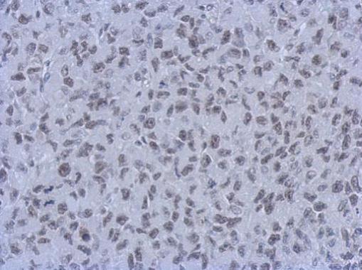
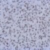
Description Immunohistochemical analysis of paraffin-embedded H1299 xenograft, using RbAp48 + RbAp46(3111) antibody at 1:200 dilution.

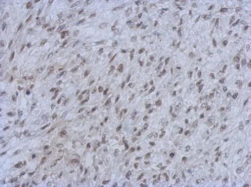
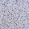
Description Immunohistochemical analysis of paraffin-embedded C2C12 xenograft, using RbAp48 + RbAp46(3111) antibody at 1:200 dilution.

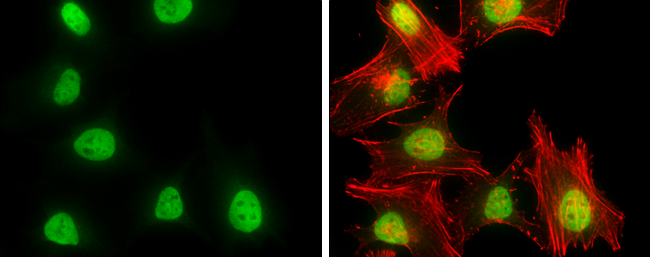
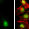
Description RbAp48 + RbAp46 antibody [15G12] detects RbAp48 + RbAp46 protein at nucleus by immunofluorescent analysis.Sample: HeLa cells were fixed in 4% paraformaldehyde at RT for 15 min.Green: RbAp48 + RbAp46 stained by RbAp48 + RbAp46 antibody [15G12] (3111) diluted at 1:500.Red: phalloidin, a cytoskeleton marker, diluted at 1:100.

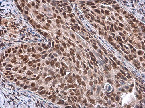
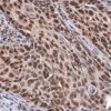
Description RbAp48 + RbAp46 antibody [15G12] detects RbAp48 + RbAp46 protein at nucleus by immunohistochemical analysis.Sample: Paraffin-embedded human cervical carcinoma.RbAp48 + RbAp46 stained by RbAp48 + RbAp46 antibody [15G12] (3111) diluted at 1:100.Antigen Retrieval: Citrate buffer, pH 6.0, 15 min

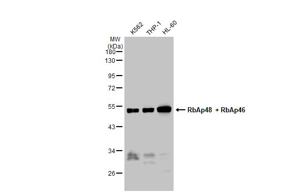
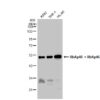
Description Various whole cell extracts (30 ug) were separated by 10% SDS-PAGE, and the membrane was blotted with RbAp48 + RbAp46 antibody [15G12] (3111) diluted at 1:500. The HRP-conjugated anti-mouse IgG antibody was used to detect the primary antibody, and the signal was developed with Trident ECL plus-Enhanced.
Handling
Storage This antibody is stable at -20°C for at least one (1) year; store in appropriate aliquots to avoid multiple freeze-thaw cycles.
Dilution Instructions Dilute in PBS or medium which is identical to that used in the assay system.
Application Instructions Immunoblotting: use at 1-5ug/mL. A band of ~48kDa is detected. Detection of RbAp48 in K562, THP-1, and HL-60 whole cell extracts (30ug), separated by 10% SDS-PAGE, with #3111 at 2ug/mL.
These are recommended concentrations.
Endusers should determine optimal concentrations for their applications.
Immunohistochemistry: use at 5-10ug/mL. Detection of RbAp48 in paraffin-embedded human cervical carcinoma with #3111 at 10ug/mL.
These are recommended concentrations.
Endusers should determine optimal concentrations for their applications.
Immunohistochemistry: use at 5-10ug/mL. Detection of RbAp48 in paraffin-embedded human cervical carcinoma with #3111 at 10ug/mL.
References & Data Sheet
Data Sheet  Download PDF Data Sheet
 Download PDF Data Sheet

