Anti-PUMA (NT) Antibody (11008)
$445.00
| Host | Quantity | Applications | Species Reactivity | Data Sheet | |
|---|---|---|---|---|---|
| Rabbit | 100ug | ELISA,WB,IHC-P,IF | Human, Mouse |  |
SKU: 11008
Categories: Antibody Products, Apoptosis Antibodies, Products
Overview
Product Name Anti-PUMA (NT) Antibody (11008)
Description Anti-PUMA (NT) Rabbit Polyclonal Antibody
Target PUMA (NT)
Species Reactivity Human, Mouse
Applications ELISA,WB,IHC-P,IF
Host Rabbit
Clonality Polyclonal
Isotype IgG
Immunogen Synthetic peptide corresponding to aa 2-16 of human PUMA-a (accession no. NP_055232).. This sequence is identical in mouse PUMA-a.
Properties
Form Liquid
Concentration Lot Specific
Formulation PBS, pH 7.4.
Buffer Formulation Phosphate Buffered Saline
Buffer pH pH 7.4
Format Purified
Purification Purified by peptide immuno-affinity chromatography
Specificity Information
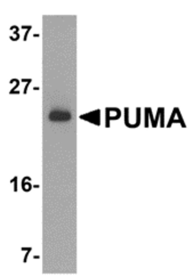
Specificity This antibody recognizes human and mouse PUMA (23kDa).
Target Name Bcl-2-binding component 3, isoforms 1/2
Target ID PUMA (NT)
Uniprot ID Q9BXH1
Alternative Names JFY-1, p53 up-regulated modulator of apoptosis
Gene Name BBC3
Gene ID 27113
Accession Number NP_055232
Sequence Location Mitochondrion
Biological Function Essential mediator of p53/TP53-dependent and p53/TP53-independent apoptosis (PubMed:11463391, PubMed:23340338). Promotes partial unfolding of BCL2L1 and dissociation of BCL2L1 from p53/TP53, releasing the bound p53/TP53 to induce apoptosis (PubMed:23340338). Regulates ER stress-induced neuronal apoptosis (By similarity). {UniProtKB:Q99ML1, PubMed:11463391, PubMed:23340338}.
Research Areas Apoptosis
Background PUMA (p53 Upregulated Modulator of Apoptosis) is a pro-apoptotic Bcl-2 family member that encodes two BH3 domain- containing proteins, PUMA-alpha and PUMA-beta. PUMA proteins bind Bcl-2, localize to mitochondria, and induce cytochrome c release and apoptosis in response to p53.
Application Images


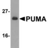
Description Western Blot Validation of PUMA in K562 Cells
Loading: 15 ug of lysates per lane. Antibodies: 11008 (2 ug/mL), 1 h incubation at RT in 5% NFDM/TBST.Secondary: Goat anti-rabbit IgG HRP conjugate at 1:10000 dilution
Loading: 15 ug of lysates per lane. Antibodies: 11008 (2 ug/mL), 1 h incubation at RT in 5% NFDM/TBST.Secondary: Goat anti-rabbit IgG HRP conjugate at 1:10000 dilution

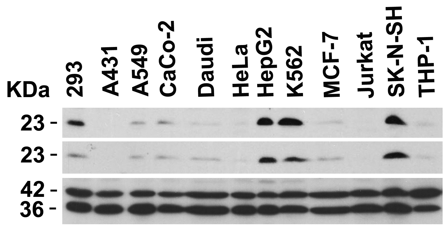
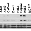
Description Independent Antibody Validation (IAV) via Protein Expression Profile in Human Cells
Loading: 20 ug of lysates per lane. Antibodies: 11007 (3 ug/mL), 11008 (2 ug/mL), beta-actin (1 ug/mL) and GAPDH (0.02 ug/mL), 1 h incubation at RT in 5% NFDM/TBST.Secondary: Goat anti-rabbit IgG HRP conjugate at 1:10000 dilution.
Loading: 20 ug of lysates per lane. Antibodies: 11007 (3 ug/mL), 11008 (2 ug/mL), beta-actin (1 ug/mL) and GAPDH (0.02 ug/mL), 1 h incubation at RT in 5% NFDM/TBST.Secondary: Goat anti-rabbit IgG HRP conjugate at 1:10000 dilution.

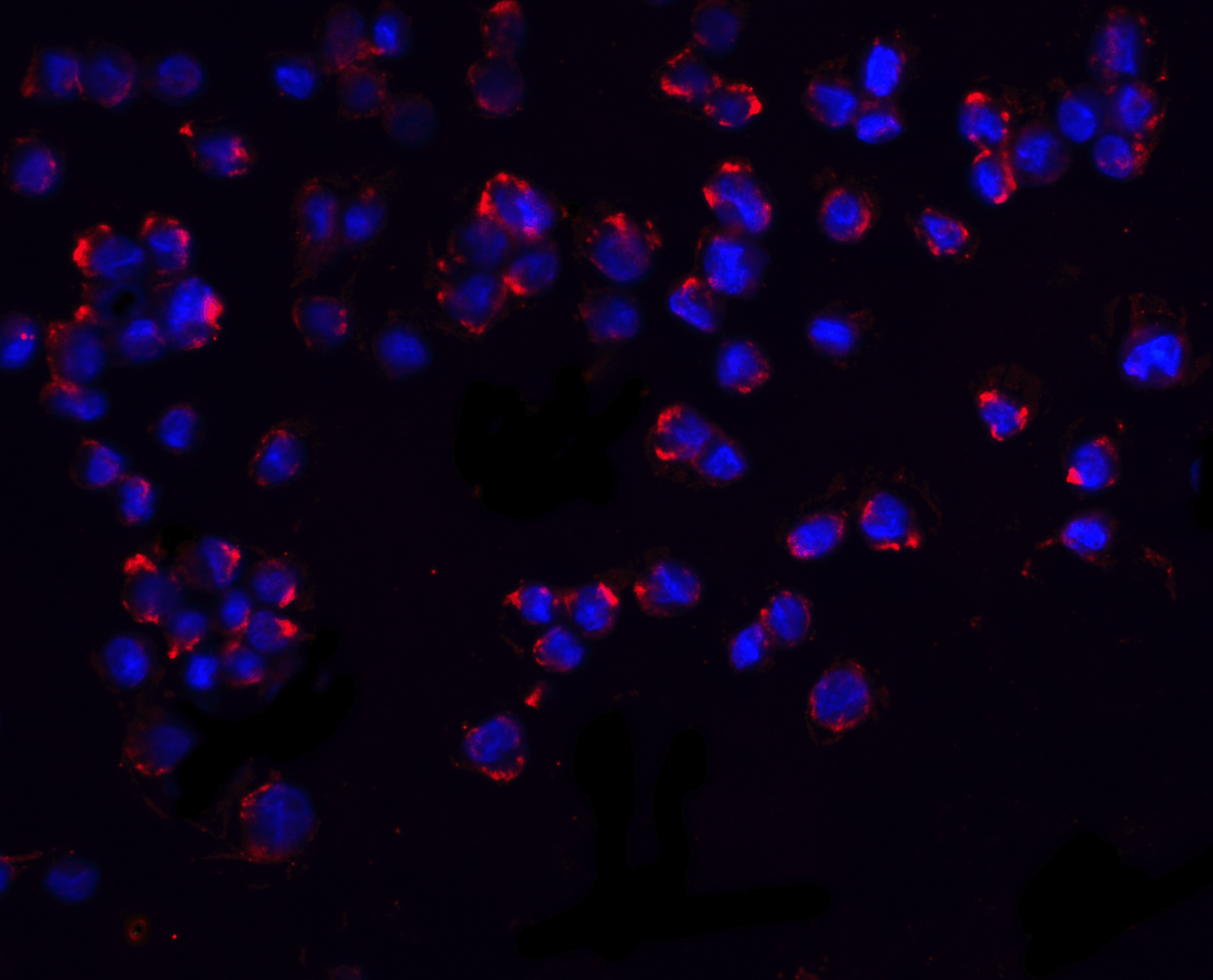
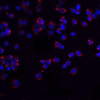
Description Immunofluorescence Validation of PUMA in K562 Cells
Immunofluorescent analysis of 4% paraformaldehyde-fixed K562 cells labeling PUMA with 11008 at 20 ug/mL, followed by goat anti-rabbit IgG secondary antibody at 1/500 dilution (red) and DAPI staining (blue).
Immunofluorescent analysis of 4% paraformaldehyde-fixed K562 cells labeling PUMA with 11008 at 20 ug/mL, followed by goat anti-rabbit IgG secondary antibody at 1/500 dilution (red) and DAPI staining (blue).

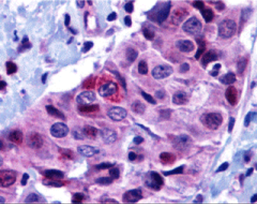
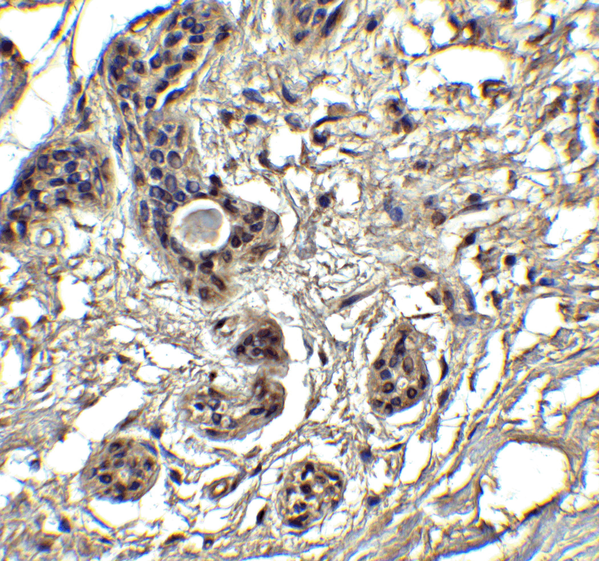
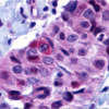
Description Immunohistochemistry Validation of PUMA in Human Breast Carcinoma
Immunohistochemical analysis of paraffin-embedded human breast carcinoma tissue using anti-PUMA antibody (11008) at 10 ug/ml. Tissue was fixed with formaldehyde and blocked with 10% serum for 1 h at RT; antigen retrieval was by heat mediation with a citrate buffer (pH6). Samples were incubated with primary antibody overnight at 4C. A goat anti-rabbit IgG H&L (HRP) at 1/250 was used as secondary. Counter stained with Hematoxylin.
Immunohistochemical analysis of paraffin-embedded human breast carcinoma tissue using anti-PUMA antibody (11008) at 10 ug/ml. Tissue was fixed with formaldehyde and blocked with 10% serum for 1 h at RT; antigen retrieval was by heat mediation with a citrate buffer (pH6). Samples were incubated with primary antibody overnight at 4C. A goat anti-rabbit IgG H&L (HRP) at 1/250 was used as secondary. Counter stained with Hematoxylin.

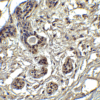
Description Immunohistochemistry Validation of PUMA in Human Breast Tissue
Immunohistochemical analysis of paraffin-embedded human breast tissue using anti-PUMA antibody (11008) at 2.5 ug/ml. Tissue was fixed with formaldehyde and blocked with 10% serum for 1 h at RT; antigen retrieval was by heat mediation with a citrate buffer (pH6). Samples were incubated with primary antibody overnight at 4C. A goat anti-rabbit IgG H&L (HRP) at 1/250 was used as secondary. Counter stained with Hematoxylin.
Immunohistochemical analysis of paraffin-embedded human breast tissue using anti-PUMA antibody (11008) at 2.5 ug/ml. Tissue was fixed with formaldehyde and blocked with 10% serum for 1 h at RT; antigen retrieval was by heat mediation with a citrate buffer (pH6). Samples were incubated with primary antibody overnight at 4C. A goat anti-rabbit IgG H&L (HRP) at 1/250 was used as secondary. Counter stained with Hematoxylin.

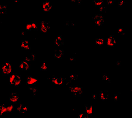
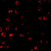
Description Immunofluorescence Validation of PUMA in K562
Immunofluorescent analysis of 4% paraformaldehyde-fixed K562 cells labeling PUMA with 11008 at 10 ug/mL, followed by goat anti-rabbit IgG secondary antibody at 1/500 dilution (red). Image showing cytosol staining on K562 cells
Immunofluorescent analysis of 4% paraformaldehyde-fixed K562 cells labeling PUMA with 11008 at 10 ug/mL, followed by goat anti-rabbit IgG secondary antibody at 1/500 dilution (red). Image showing cytosol staining on K562 cells

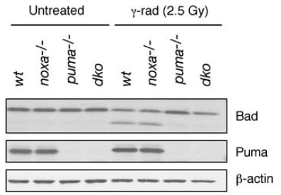
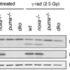
Description KO Validation of PUMA in Mouse Thymocytes (Michalak et al., 2008)
Western blot analysis ofthymocytes from wt, noxa knockout, puma knockout and noxa/puma double knockout mice cultured for 7 h in the presence or absence of 2.5 Gy g-irradiation.PUMA expression was not detected in puma KO and double KO mice with anti-PUMA antibodies (11008).
Western blot analysis ofthymocytes from wt, noxa knockout, puma knockout and noxa/puma double knockout mice cultured for 7 h in the presence or absence of 2.5 Gy g-irradiation.PUMA expression was not detected in puma KO and double KO mice with anti-PUMA antibodies (11008).

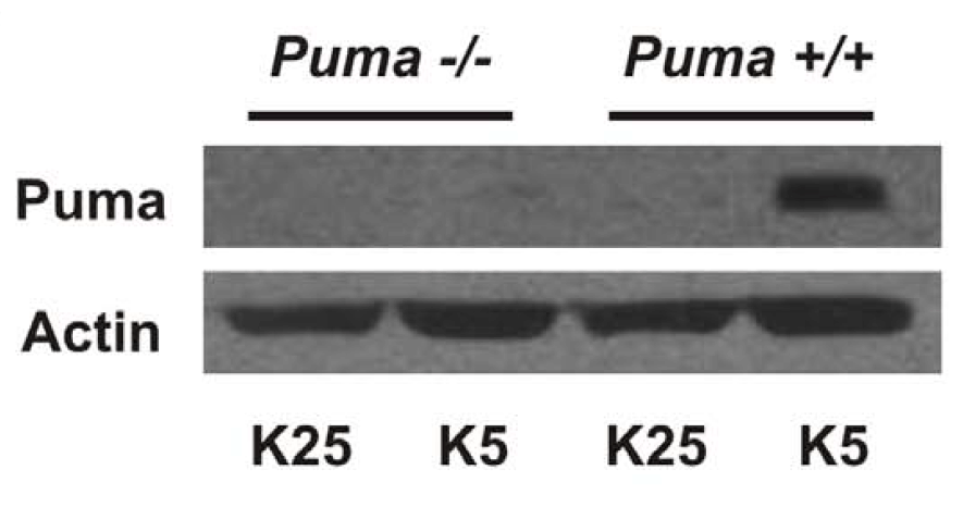
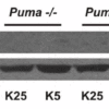
Description KO Validation of PUMA in Mouse Cerebellar Neurons (Ambacher et al., 2012)
Puma expression is induced by potassium withdrawal in cerebellar granule neurons. After 7 days in culture CGNs were either maintained in media containing 25 mM potassium (K25) or switched to low potassium medium containing 5 mM potassium (K5). PUMA protein levels were analyzed by western blot with anti-PUMA antibodies (11008). PUMA expression was not detected in PUMA KO mice and was increased after treatment in WT.
Puma expression is induced by potassium withdrawal in cerebellar granule neurons. After 7 days in culture CGNs were either maintained in media containing 25 mM potassium (K25) or switched to low potassium medium containing 5 mM potassium (K5). PUMA protein levels were analyzed by western blot with anti-PUMA antibodies (11008). PUMA expression was not detected in PUMA KO mice and was increased after treatment in WT.

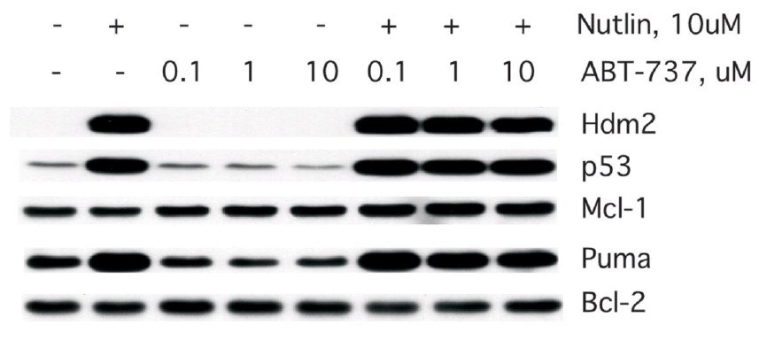
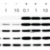
Description Induced Expression of PUMA in MCF7 cells (Wade et al., 2008)
Western analysis of MCF7 treated with the indicated dose of Nutlin-3a orABT-737 for 24h. Note that Puma is induced following Nutlin-3a treatment in these cells and PUMA expression was detected by anti-PUMA antibodies (11008)
Western analysis of MCF7 treated with the indicated dose of Nutlin-3a orABT-737 for 24h. Note that Puma is induced following Nutlin-3a treatment in these cells and PUMA expression was detected by anti-PUMA antibodies (11008)

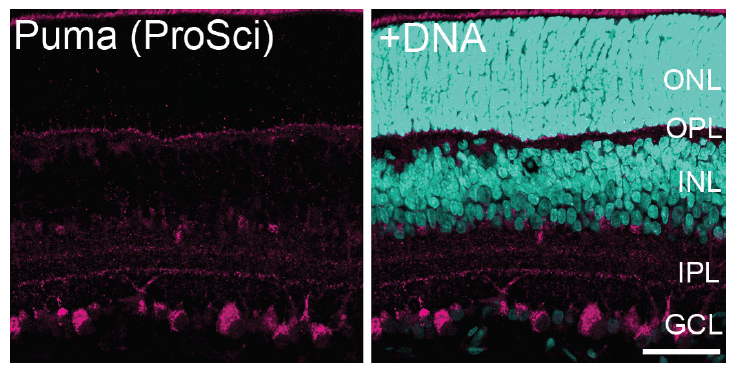
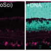
Description Immunofluorescence Validation of PUMA in Rat Retina (Wakabayashi et al., 2012)
PUMA expression in the rat retina detected by anti-PUMA antibodies (11008). The specimens were counterstained with Hoechst 33258 to visualize nuclei (+DNA). GCL, ganglion cell layer; INL, inner nuclear layer; IPL, inner plexiform layer; ONL, outer nuclear layer; OPL, outer plexiform layer; P, postnatal day.
PUMA expression in the rat retina detected by anti-PUMA antibodies (11008). The specimens were counterstained with Hoechst 33258 to visualize nuclei (+DNA). GCL, ganglion cell layer; INL, inner nuclear layer; IPL, inner plexiform layer; ONL, outer nuclear layer; OPL, outer plexiform layer; P, postnatal day.
Handling
Storage This antibody is stable for at least one (1) year at -20°C. Avoid multiple freeze-thaw cycles.
Dilution Instructions Dilute in PBS or medium which is identical to that used in the assay system.
Application Instructions Immunoblotting: use at 2ug/mL.
Positive control: K562 cell lysate.
Immunohistochemistry: use at 10ug/mL.
These are recommended concentrations.
Enduser should determine optimal concentrations for their applications.
Positive control: K562 cell lysate.
Immunohistochemistry: use at 10ug/mL.
These are recommended concentrations.
Enduser should determine optimal concentrations for their applications.
References & Data Sheet
Data Sheet  Download PDF Data Sheet
 Download PDF Data Sheet

