Anti-PHAP III Antibody (11084)
$445.00
| Host | Quantity | Applications | Species Reactivity | Data Sheet | |
|---|---|---|---|---|---|
| Rabbit | 100ug | ELISA,WB,IHC-P,IF | Human, Mouse, Rat |  |
SKU: 11084
Categories: Antibody Products, Apoptosis Antibodies, Products
Overview
Product Name Anti-PHAP III Antibody (11084)
Description Anti-PHAP III Rabbit Polyclonal Antibody
Target PHAP III
Species Reactivity Human, Mouse, Rat
Applications ELISA,WB,IHC-P,IF
Host Rabbit
Clonality Polyclonal
Isotype IgG
Immunogen Synthetic peptide corresponding to amino acids near the carboxy terminus of human PHAP III (accession no. NP_112182)
Properties
Form Liquid
Concentration Lot Specific
Formulation PBS, pH 7.4.
Buffer Formulation Phosphate Buffered Saline
Buffer pH pH 7.4
Format Purified
Purification Purified by peptide immuno-affinity chromatography
Specificity Information
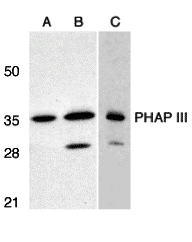
Specificity This antibody recognizes human, mouse and rat PHAP III (35kDa).
Target Name Acidic leucine-rich nuclear phosphoprotein 32 family member E
Target ID PHAP III
Uniprot ID Q9BTT0
Alternative Names LANP-like protein, LANP-L
Gene Name ANP32E
Gene ID 81611
Accession Number NP_112182
Sequence Location Cytoplasm, Nucleus
Biological Function Histone chaperone that specifically mediates the genome-wide removal of histone H2A.Z/H2AZ1 from the nucleosome: removes H2A.Z/H2AZ1 from its normal sites of deposition, especially from enhancer and insulator regions. Not involved in deposition of H2A.Z/H2AZ1 in the nucleosome. May stabilize the evicted H2A.Z/H2AZ1-H2B dimer, thus shifting the equilibrium towards dissociation and the off-chromatin state (PubMed:24463511). Inhibits activity of protein phosphatase 2A (PP2A). Does not inhibit protein phosphatase 1. May play a role in cerebellar development and synaptogenesis. {PubMed:24463511}.
Research Areas Apoptosis
Background The PHAP proteins (tumor suppressor putative HLA-DR associated proteins) are important regulators of mitochondrial apoptosis. PHAP facilitates apoptosome- mediated caspase-9 activation to stimulate the mitochondrial apoptosis pathway. In addition, PHAP opposes both Ras- and myc- mediated cell transformation.
Application Images


Description Western blot analysis of PHAP III expression in human A549 (A) and HepG2 (B) cells, and rat testis (C) with PHAP antibody III at 1 ug/mL.

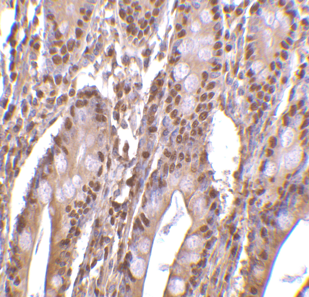
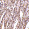
Description Immunohistochemistry of PHAP III in human small intestine tissue with PHAP III antibody at 2 ug/mL.

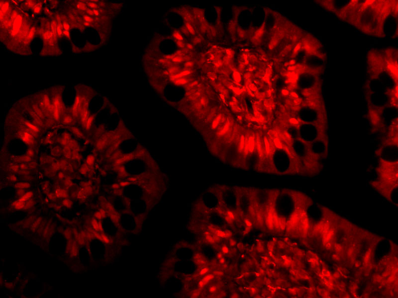
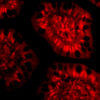
Description Immunofluorescence of PHAP III in Human Small Intestine cells with PHAP III antibody at 10 ug/mL.
Handling
Storage This antibody is stable for at least one (1) year at -20°C. Avoid multiple freeze-thaw cycles.
Dilution Instructions Dilute in PBS or medium which is identical to that used in the assay system.
Application Instructions Western Blot: Use at 1ug/ml.
Positive control: A549 cell lysate.
Immunohistochemistry: use at 2ug/ml.
These are recommended concentrations.
Enduser should determine optimal concentrations for their applications.
Positive control: A549 cell lysate.
Immunohistochemistry: use at 2ug/ml.
These are recommended concentrations.
Enduser should determine optimal concentrations for their applications.
References & Data Sheet
Data Sheet  Download PDF Data Sheet
 Download PDF Data Sheet

