Anti-Protein Disulfide Isomerase (PDI) Antibody (11081)
$473.00
| Host | Quantity | Applications | Species Reactivity | Data Sheet | |
|---|---|---|---|---|---|
| Rabbit | 100ul | WB,IHC,ICC/IF,IP | Human, Monkey, Mouse, Rat, Hamster, Bovine, Ovine, Porcine, Canine, Guinea Pig, Xenopus |  |
SKU: 11081
Categories: Antibody Products, Neuroscience and Signal Transduction Antibodies, Products
Overview
Product Name Anti-Protein Disulfide Isomerase (PDI) Antibody (11081)
Description Anti-PDI Rabbit Polyclonal Antibody
Target Protein Disulfide Isomerase (PDI)
Species Reactivity Human, Monkey, Mouse, Rat, Hamster, Bovine, Ovine, Porcine, Canine, Guinea Pig, Xenopus
Applications WB,IHC,ICC/IF,IP
Host Rabbit
Clonality Polyclonal
Immunogen Synthetic peptide derived from rat PDI conjugated to KLH.
Properties
Form Liquid
Concentration Lot Specific
Formulation Whole antiserum
Buffer Formulation Whole Antiserum
Format Whole Antiserum
Purification Whole antiserum
Specificity Information
Specificity This antibody recognizes human, monkey, mouse, rat, hamster, bovine, ovine, porcine, canine, guinea pig, and Xenopus PDI.
Target Name Protein disulfide-isomerase
Target ID Protein Disulfide Isomerase (PDI)
Uniprot ID P04785
Alternative Names PDI, EC 5.3.4.1, Cellular thyroid hormone-binding protein, Prolyl 4-hydroxylase subunit β
Gene Name P4hb
Gene ID 287164
Accession Number NP_001099245.2
Sequence Location Endoplasmic reticulum, Endoplasmic reticulum lumen, Melanosome, Cell membrane
Biological Function This multifunctional protein catalyzes the formation, breakage and rearrangement of disulfide bonds. At the cell surface, seems to act as a reductase that cleaves disulfide bonds of proteins attached to the cell. May therefore cause structural modifications of exofacial proteins. Inside the cell, seems to form/rearrange disulfide bonds of nascent proteins. At high concentrations, functions as a chaperone that inhibits aggregation of misfolded proteins. At low concentrations, facilitates aggregation (anti-chaperone activity). May be involved with other chaperones in the structural modification of the TG precursor in hormone biogenesis. Also acts a structural subunit of various enzymes such as prolyl 4-hydroxylase and microsomal triacylglycerol transfer protein MTTP. Receptor for LGALS9; the interaction retains P4HB at the cell surface of Th2 T helper cells, increasing disulfide reductase activity at the plasma membrane, altering the plasma membrane redox state and enhancing cell migration. {UniProtKB:P07237}.
Research Areas Neuroscience
Background A microsomal enzyme known as Protein Disulfide Isomerase (PDI) is involved in the formation of disulfide bonds in many extracellular proteins through its oxidase and isomerase activities. It is also involved in the reduction of disulfide bonds in proteins. PDI is located primarily in the endoplasmic reticulum (ER) but may also be found in the cytosol. PDI can reside in the ER due to the highly conserved KDEL sequence at its carboxy terminus which serves as an ER-retention signal.
Application Images


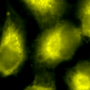
Description Immunocytochemistry/Immunofluorescence analysis using Rabbit Anti-PDI Polyclonal Antibody (11081). Tissue: Cervical cancer cell line (HeLa). Species: Human. Fixation: 2% Formaldehyde for 20 min at RT. Primary Antibody: Rabbit Anti-PDI Polyclonal Antibody (11081) at 1:100 for 12 hours at 4°C. Secondary Antibody: R-PE Goat Anti-Rabbit (yellow) at 1:200 for 2 hours at RT. Counterstain: DAPI (blue) nuclear stain at 1:40000 for 2 hours at RT. Localization: Endoplasmic reticulum lumen. Melanosome. Magnification: 100x. (A) DAPI (blue) nuclear stain. (B) Anti-PDI Antibody. (C) Composite.

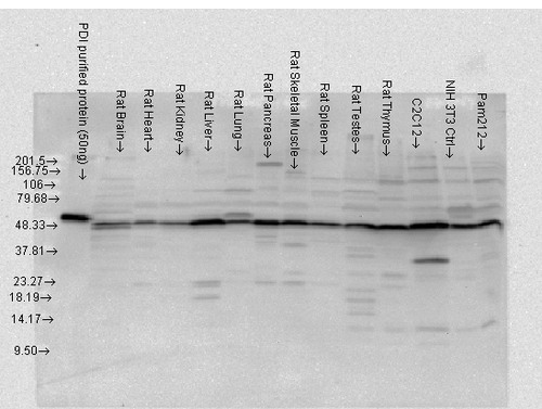
Description Western blot analysis of Rat tissue mix showing detection of PDI protein using Rabbit Anti-PDI Polyclonal Antibody (11081). Load: 15 µgprotein. Block: 1.5% BSA. Primary Antibody: Rabbit Anti-PDI Polyclonal Antibody (11081) at 1:4000 for 2 hours at RT. Secondary Antibody: Donkey Anti-Rabbit IgG: HRP for 1 hour at RT.

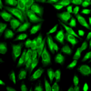
Description Immunocytochemistry/Immunofluorescence analysis using Rabbit Anti-PDI Polyclonal Antibody (11081). Tissue: Cervical cancer cell line (HeLa). Species: Human. Fixation: 2% Formaldehyde for 20 min at RT. Primary Antibody: Rabbit Anti-PDI Polyclonal Antibody (11081) at 1:100 for 12 hours at 4°C. Secondary Antibody: FITC Goat Anti-Rabbit (green) at 1:200 for 2 hours at RT. Counterstain: DAPI (blue) nuclear stain at 1:40000 for 2 hours at RT. Localization: Endoplasmic reticulum lumen. Melanosome. Magnification: 20x. (A) DAPI (blue) nuclear stain. (B) Anti-PDI Antibody. (C) Composite.
Handling
Storage Store frozen product at or below -20°C. For optimal storage, aliquot to smaller portions and store at -20°C to -70°C. Avoid repeated freeze/thaw cycles.
Dilution Instructions Dilute in PBS or medium that is identical to that used in the assay system.
Application Instructions Western blot: Use at 1:4,000 dilution. A band of approx. 58 kD is detected.Immunocytochemistry/Immunohisto- chemistry: Use at 2-5 ug/ml.
Immunoprecipitation: Use at 10 ug/ml.
Immunoprecipitation: Use at 10 ug/ml.
References & Data Sheet
Data Sheet  Download PDF Data Sheet
 Download PDF Data Sheet


