Anti-p84 Antibody (56248)
$503.00
| Host | Quantity | Applications | Species Reactivity | Data Sheet | |
|---|---|---|---|---|---|
| Mouse | 100ug | WB,ICC/IF,IHC-P,IP,ChIP,IHC | Human |  |
SKU: 56248
Categories: Antibody Products, Cancer Research Antibodies, Products
Overview
Product Name Anti-p84 Antibody (56248)
Description Anti-p84 Mouse Monoclonal Antibody
Target p84
Species Reactivity Human
Applications WB,ICC/IF,IHC-P,IP,ChIP,IHC
Host Mouse
Clonality Monoclonal
Clone ID 5.00E+10
Isotype IgG2b
Immunogen Fusion protein containing amino acids 15-374 of human p84 expressed in E. coli.
Properties
Form Liquid
Concentration Lot Specific
Formulation PBS, pH 7.4.
Buffer Formulation Phosphate Buffered Saline
Buffer pH pH 7.4
Format Purified
Purification Purified by Protein G affinity chromatography
Specificity Information
Specificity This antibody recognizes an 84 kD nuclear matrix protein that was first discovered through its association with the N-terminal region of retinoblastoma protein.
Target Name THO complex subunit 1
Target ID p84
Uniprot ID Q96FV9
Alternative Names Tho1, Nuclear matrix protein p84, p84N5, hTREX84
Gene Name THOC1
Sequence Location [Isoform 1]: Nucleus speckle. Nucleus, nucleoplasm. Nucleus matrix. Cytoplasm. Note=Can shuttle between the nucleus and cytoplasm. Nuclear localization is required for induction of apoptotic cell death. Translocates to the cytoplasm during the early phase of apoptosis execution.; [Isoform 2]: Cytoplasm.
Biological Function Required for efficient export of polyadenylated RNA. Acts as component of the THO subcomplex of the TREX complex which is thought to couple mRNA transcription, processing and nuclear export, and which specifically associates with spliced mRNA and not with unspliced pre-mRNA. TREX is recruited to spliced mRNAs by a transcription-independent mechanism, binds to mRNA upstream of the exon-junction complex (EJC) and is recruited in a splicing- and cap-dependent manner to a region near the 5' end of the mRNA where it functions in mRNA export to the cytoplasm via the TAP/NFX1 pathway. The TREX complex is essential for the export of Kaposi's sarcoma-associated herpesvirus (KSHV) intronless mRNAs and infectious virus production. Regulates transcriptional elongation of a subset of genes. Involved in genome stability by preventing co-transcriptional R-loop formation.; Participates in an apoptotic pathway which is characterized by activation of caspase-6, increases in the expression of BAK1 and BCL2L1 and activation of NF-kappa-B. This pathway does not require p53/TP53, nor does the presence of p53/TP53 affect the efficiency of cell killing. Activates a G2/M cell cycle checkpoint prior to the onset of apoptosis. Apoptosis is inhibited by association with RB1.
Research Areas Cancer research
Application Images


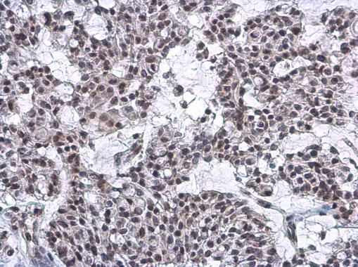
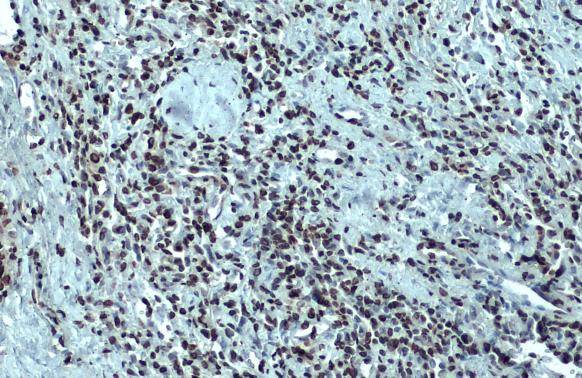
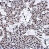
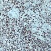
Description Nuclear Matrix Protein p84 antibody [5E10] detects Nuclear Matrix Protein p84 protein at nucleus by immunohistochemical analysis.Sample: Paraffin-embedded human breast carcinoma.Nuclear Matrix Protein p84 stained by Nuclear Matrix Protein p84 antibody [5E10] (56248) diluted at 1:200.Antigen Retrieval: Citrate buffer, pH 6.0, 15 min

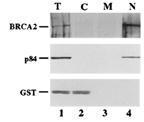
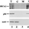
Description Detection of human p84/N5 in nuclear fraction by anti-p84/N5 5E10 monoclonal antibody (56248) in western blot experiment. Lane 1: total lysate, Lane 2: cytoplasmic fraction, Lane 3: membrane fraction, Lane 4: nuclear fraction.

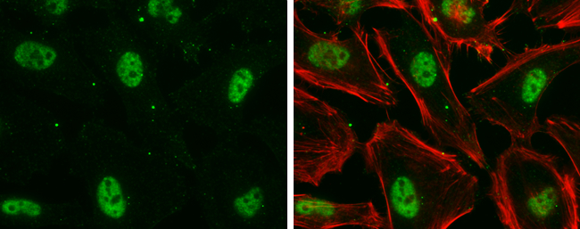
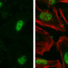
Description Nuclear Matrix Protein p84 antibody [5E10] detects Nuclear Matrix Protein p84 protein at nucleus by immunofluorescent analysis.Sample: HeLa cells were fixed in 4% paraformaldehyde at RT for 15 min.Green: Nuclear Matrix Protein p84 stained by Nuclear Matrix Protein p84 antibody [5E10] (56248) diluted at 1:500.Red: phalloidin, a cytoskeleton marker, diluted at 1:100.

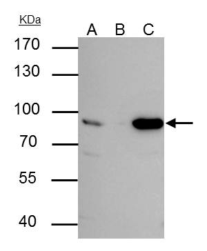
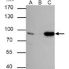
Description p84 antibody [5E10] immunoprecipitates p84 protein in IP experiments. IP Sample: HepG2 whole cell lysate/extract A : 30 ug whole cell lysate/extract of p84 protein expressing HepG2 cells B : Control with 3 ug of pre-immune mouse IgG C : Immunoprecipitation of p84 by 3 ug of p84 antibody [5E10] (56248) 7.5% SDS-PAGE The immunoprecipitated p84 protein was detected by p84 antibody [5E10] (56248) diluted at 1 : 1000. EasyBlot anti-rabbit IgG (HRP) was used as a secondary reagent.

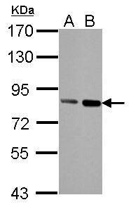
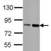
Description Sample (30 ug of whole cell lysate)
A: HeLa
B: HeLa nucleus
7.5% SDS PAGE
56248 diluted at 1:1000
The HRP-conjugated anti-mouse IgG antibody was used to detect the primary antibody.
A: HeLa
B: HeLa nucleus
7.5% SDS PAGE
56248 diluted at 1:1000
The HRP-conjugated anti-mouse IgG antibody was used to detect the primary antibody.

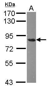
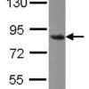
Description Sample (50 ug of whole cell lysate)
A: mouse Cerebellum
7.5% SDS PAGE
56248 diluted at 1:1000
The HRP-conjugated anti-mouse IgG antibody was used to detect the primary antibody.
A: mouse Cerebellum
7.5% SDS PAGE
56248 diluted at 1:1000
The HRP-conjugated anti-mouse IgG antibody was used to detect the primary antibody.

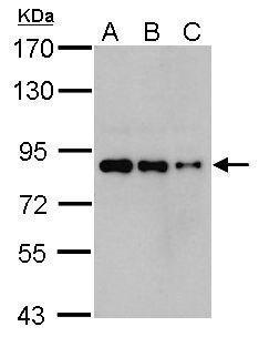
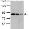
Description Sample (whole cell lysate)
A: 293T 20ug
B: 293T 10ug
C: 293T 5ug
7.5% SDS PAGE
56248 diluted at 1:1000
The HRP-conjugated anti-mouse IgG antibody was used to detect the primary antibody.
A: 293T 20ug
B: 293T 10ug
C: 293T 5ug
7.5% SDS PAGE
56248 diluted at 1:1000
The HRP-conjugated anti-mouse IgG antibody was used to detect the primary antibody.

Description Nuclear Matrix Protein p84 antibody [5E10] detects Nuclear Matrix Protein p84 protein at nucleus by immunohistochemical analysis.Sample: Paraffin-embedded human lung cancer.Nuclear Matrix Protein p84 stained by Nuclear Matrix Protein p84 antibody [5E10] (56248) diluted at 1:100.Antigen Retrieval: Citrate buffer, pH 6.0, 15 min

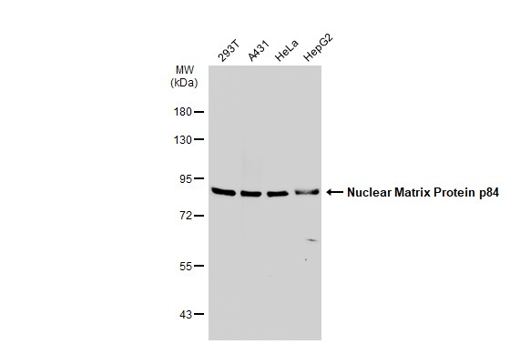
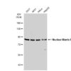
Description Various whole cell extracts (30 ug) were separated by 7.5% SDS-PAGE, and the membrane was blotted with Nuclear Matrix Protein p84 antibody [5E10] (56248) diluted at 1:500. The HRP-conjugated anti-mouse IgG antibody was used to detect the primary antibody.
Handling
Storage This antibody is stable for at least one (1) year at -70°C. Avoid multiple freeze- thaw cycles.
Dilution Instructions Dilute in PBS or medium which is identical to that used in the assay system.
Application Instructions Immunoblotting, immunoprecipitation, and
Immunofluorescence: use at 0.5-2 ug/mL.In immunoblots, a band of 84 kD is detected.
Positive controls: Lysates of Molt 4, HeLa, or Raji cells.
Immunofluorescence: use at 0.5-2 ug/mL.In immunoblots, a band of 84 kD is detected.
Positive controls: Lysates of Molt 4, HeLa, or Raji cells.
References & Data Sheet
Data Sheet  Download PDF Data Sheet
 Download PDF Data Sheet

