Anti-p53 Antibody (3340)
$503.00
| Host | Quantity | Applications | Species Reactivity | Data Sheet | |
|---|---|---|---|---|---|
| Mouse | 100ug | WB,ICC/IF,IHC-P,IHC-Fr,FACS,IP,ELISA | Human |  |
SKU: 3340
Categories: Antibody Products, Cancer Research Antibodies, Products
Overview
Product Name Anti-p53 Antibody (3340)
Description Anti-p53 Mouse Monoclonal Antibody
Target p53
Species Reactivity Human
Applications WB,ICC/IF,IHC-P,IHC-Fr,FACS,IP,ELISA
Host Mouse
Clonality Monoclonal
Clone ID 240
Isotype IgG1
Immunogen Gel-purified p53 containing aa 14-389 of human p53.
Properties
Form Liquid
Concentration Lot Specific
Formulation PBS, pH 7.4
Buffer Formulation Phosphate Buffered Saline
Buffer pH pH 7.4
Format Purified
Purification Purified by Protein G affinity chromatography
Specificity Information
Target Name Cellular tumor antigen p53
Target ID p53
Uniprot ID P04637
Alternative Names Antigen NY-CO-13, Phosphoprotein p53, Tumor suppressor p53
Gene Name TP53
Gene ID 7157
Accession Number NP_000537
Sequence Location Cytoplasm, Nucleus, Nucleus, PML body, Endoplasmic reticulum, Mitochondrion matrix, Cytoplasm, cytoskeleton, microtubule organizing center, centrosome
Biological Function Acts as a tumor suppressor in many tumor types; induces growth arrest or apoptosis depending on the physiological circumstances and cell type. Involved in cell cycle regulation as a trans-activator that acts to negatively regulate cell division by controlling a set of genes required for this process. One of the activated genes is an inhibitor of cyclin-dependent kinases. Apoptosis induction seems to be mediated either by stimulation of BAX and FAS antigen expression, or by repression of Bcl-2 expression. Its pro-apoptotic activity is activated via its interaction with PPP1R13B/ASPP1 or TP53BP2/ASPP2 (PubMed:12524540). However, this activity is inhibited when the interaction with PPP1R13B/ASPP1 or TP53BP2/ASPP2 is displaced by PPP1R13L/iASPP (PubMed:12524540). In cooperation with mitochondrial PPIF is involved in activating oxidative stress-induced necrosis; the function is largely independent of transcription. Induces the transcription of long intergenic non-coding RNA p21 (lincRNA-p21) and lincRNA-Mkln1. LincRNA-p21 participates in TP53-dependent transcriptional repression leading to apoptosis and seems to have an effect on cell-cycle regulation. Implicated in Notch signaling cross-over. Prevents CDK7 kinase activity when associated to CAK complex in response to DNA damage, thus stopping cell cycle progression. Isoform 2 enhances the transactivation activity of isoform 1 from some but not all TP53-inducible promoters. Isoform 4 suppresses transactivation activity and impairs growth suppression mediated by isoform 1. Isoform 7 inhibits isoform 1-mediated apoptosis. Regulates the circadian clock by repressing CLOCK-ARNTL/BMAL1-mediated transcriptional activation of PER2 (PubMed:24051492). {PubMed:11025664, PubMed:12524540, PubMed:12810724, PubMed:15186775, PubMed:15340061, PubMed:17317671, PubMed:17349958, PubMed:19556538, PubMed:20673990, PubMed:20959462, PubMed:22726440, PubMed:24051492, PubMed:9840937}.
Research Areas Cancer Research
Application Images


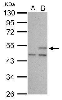
Description Sample (30 ug of whole cell lysate)
A: HCT116 cells with mock treatment for 24 hr
B: HCT116 cells with 30 uM cisplatin treatment for 24 hr
10% SDS PAGE
3340 diluted at 1:1000
The HRP-conjugated anti-mouse IgG antibody was used to detect the primary antibody.
A: HCT116 cells with mock treatment for 24 hr
B: HCT116 cells with 30 uM cisplatin treatment for 24 hr
10% SDS PAGE
3340 diluted at 1:1000
The HRP-conjugated anti-mouse IgG antibody was used to detect the primary antibody.

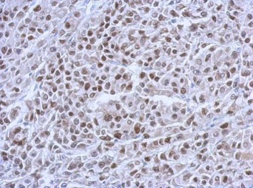
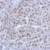
Description Immunohistochemical analysis of paraffin-embedded HBL435 xenograft, using p53(3340) antibody at 1:200 dilution.

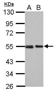
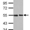
Description Sample (30 ug of whole cell lysate)
A: 293T
B: A431
10% SDS PAGE
3340 diluted at 1:1000
The HRP-conjugated anti-mouse IgG antibody was used to detect the primary antibody.
A: 293T
B: A431
10% SDS PAGE
3340 diluted at 1:1000
The HRP-conjugated anti-mouse IgG antibody was used to detect the primary antibody.
Handling
Storage This antibody is stable for at least one (1) year at -20°C. Avoid multiple freeze-thaw cycles.
Dilution Instructions Dilute in PBS or medium which is identical to that used in the assay system.
Application Instructions ELISA, FACS, IHC (frozen and paraffin), Immunoprecipitation, Immunoblotting.
IHC: use at 2-4ug/ml. Extensive washing may be necessary. Protein digestion and/or microwave antigen retrieval prior to staining is not required. #3340 on paraffin-embedded HBL435 xenograft.
Immunoprecipitation: use at 10ug/mg of lysate. This antibody reacts only with mutant p53 under non-denaturing conditions.
Immunoblotting: use at 5-10ug/ml. A band of 53kDa is detected. #3340 on 30ug of 293T (A) and A431 (B) cell lysates.
These are recommended concentrations.
Enduser should determine optimal concentrations for their applications.
References & Data Sheet
Data Sheet  Download PDF Data Sheet
 Download PDF Data Sheet

