Anti-Otoferlin Antibody (90023)
$520.00
SKU: 90023
Categories: Antibody Products, Neuroscience and Signal Transduction Antibodies, Products
Overview
Product Name Anti-Otoferlin Antibody (90023)
Description Anti-Otoferlin Mouse Monoclonal Antibody
Target Otoferlin
Species Reactivity Human
Applications WB,IF,IHC
Host Mouse
Clonality Monoclonal
Clone ID 13A9
Isotype IgG1
Immunogen GST fusion protein corresponding to aa 1-395 of human otoferlin (accession no. Q9HC10).
Properties
Form Lyophilized
Formulation Lyophilized, 0.1M Tris, 0.1M glycine, 2% sucrose, 1mg/ml.
Buffer Formulation Tris Buffer
Format Purified
Purification Purified by Protein A affinity chromatography
Specificity Information
Specificity This antibody recognizes human otoferlin.
Target Name Otoferlin
Target ID Otoferlin
Uniprot ID Q9HC10
Alternative Names Fer-1-like protein 2
Gene Name OTOF
Sequence Location Cytoplasmic vesicle, secretory vesicle, synaptic vesicle membrane, Basolateral cell membrane, Endoplasmic reticulum membrane, Golgi apparatus membrane, Cell junction, synapse, presynaptic cell membrane, Cell membrane
Biological Function Key calcium ion sensor involved in the Ca(2+)-triggered synaptic vesicle-plasma membrane fusion and in the control of neurotransmitter release at these output synapses. Interacts in a calcium-dependent manner to the presynaptic SNARE proteins at ribbon synapses of cochlear inner hair cells (IHCs) to trigger exocytosis of neurotransmitter. Also essential to synaptic exocytosis in immature outer hair cells (OHCs). May also play a role within the recycling of endosomes (By similarity). {UniProtKB:Q9ESF1}.
Research Areas Neuroscience
Background Otoferlin protein is the product of the OTOF gene. Otoferlin is present in the brain and the cochlea of the inner ear where sound is processed. Hearing relies on Ca2+ influx-driven exocytosis of synaptic vesicles at the ribbon-type active zones of hair cells. The presence of several C2 domains in otoferlin, some of which bind Ca2+, suggest that otoferlin is a Ca2+ sensor for fusion in inner hair cell (IHC) synapses.
Application Images


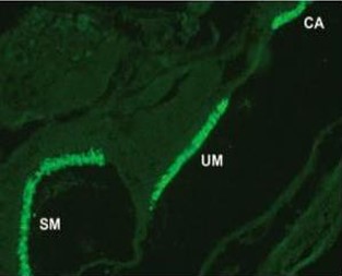
Description Immunocytochemistry: use at 2-10ug/ml. Detection of TG3 in human foreskin with #90022.
Handling
Storage This product is stable for at least one (1) year at -20°C to -70°C. Reconstituted product should be stored in appropriate aliquots to avoid repeated freeze-thaw cycles.
Dilution Instructions Reconstitute lyophilized antibody using ultrapure to a final concentration no less than 100 ug/mL. Reconstitution with PBS or Tris buffer is acceptable if required. Protein carrier, biocide or cryopreservative can be added as needed. Aliquot and store at -20C for longterm storage. Dilute immediately prior to use.
Application Instructions Immunoblotting: use at 1-5ug/mL. A band of ~220kDa is detected.
Immunofluorescence: use at 2-10ug/mL.
These are recommended concentrations.
Enduser should determine optimal concentrations for their applications.
Immunofluorescence: use at 2-10ug/mL.
These are recommended concentrations.
Enduser should determine optimal concentrations for their applications.
References & Data Sheet
Data Sheet  Download PDF Data Sheet
 Download PDF Data Sheet


