Anti-MyD88 (CT) Antibody (2127)
$445.00
SKU: 2127
Categories: Antibody Products, Neuroscience and Signal Transduction Antibodies, Products
Overview
Product Name Anti-MyD88 (CT) Antibody (2127)
Description Anti-MyD88 (CT) Rabbit Polyclonal Antibody
Target MyD88 (CT)
Species Reactivity Human, Mouse
Applications ELISA,WB,IF,IP
Host Rabbit
Clonality Polyclonal
Isotype IgG
Immunogen Peptide corresponding to aa 279-296 of human MyD88. The sequence is identical to that of mouse MyD88.
Properties
Form Liquid
Concentration Lot Specific
Formulation PBS, pH 7.4.
Buffer Formulation Phosphate Buffered Saline
Buffer pH pH 7.4
Format Purified
Purification Purified by peptide immuno-affinity chromatography
Specificity Information
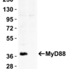
Specificity This antibody recognizes human and mouse MyD88 (35 kD).
Target Name Myeloid differentiation primary response protein MyD88
Target ID MyD88 (CT)
Uniprot ID Q99836
Gene Name MYD88
Gene ID 4615
Accession Number NP_002459
Sequence Location Cytoplasm, Nucleus
Biological Function Adapter protein involved in the Toll-like receptor and IL-1 receptor signaling pathway in the innate immune response (PubMed:15361868, PubMed:18292575, PubMed:33718825). Acts via IRAK1, IRAK2, IRF7 and TRAF6, leading to NF-kappa-B activation, cytokine secretion and the inflammatory response (PubMed:15361868, PubMed:24316379, PubMed:19506249). Increases IL-8 transcription (PubMed:9013863). Involved in IL-18-mediated signaling pathway. Activates IRF1 resulting in its rapid migration into the nucleus to mediate an efficient induction of IFN-beta, NOS2/INOS, and IL12A genes. Upon TLR8 activation by GU-rich single-stranded RNA (GU-rich RNA) derived from viruses such as SARS-CoV-2, SARS-CoV and HIV-1, induces IL1B release through NLRP3 inflammasome activation (PubMed:33718825). MyD88-mediated signaling in intestinal epithelial cells is crucial for maintenance of gut homeostasis and controls the expression of the antimicrobial lectin REG3G in the small intestine (By similarity). {UniProtKB:P22366, PubMed:15361868, PubMed:18292575, PubMed:19506249, PubMed:20855887, PubMed:24316379, PubMed:33718825, PubMed:9013863}.
Research Areas Neuroscience
Background Cellular responses induced by the pro- inflammatory cytokine IL-1 require IL-1 receptor complex (IL-1R1 and IL- 1RacP). Recently, MyD88 was identified as an adapter molecule in the IL-1 signaling pathway. MyD88 associates with and recruits IRAK to the IL-1 receptor. Dominant negative mutants of MyD88 attenuate IL-1R- mediated NF-kappaB activation. MyD88 also functions as a regulator molecule for IL- 18 receptor and human Toll receptor, members of the Toll/IL-1R family of receptors. Targeted disruption of the MyD88 gene results in loss of cellular responses to IL-1 and IL-18, and MyD88-deficient mice lack responses to LPS which require Toll-like receptors 2 and 4 (TLR2 and TLR4) as the signaling receptors. MyD88 is a general adapter protein for the Toll/IL-1R family of receptors and plays an important role in the inflammatory responses induced by cytokines IL-1, IL-18, and LPS. MyD88 is expressed in a variety of tissues.
Application Images


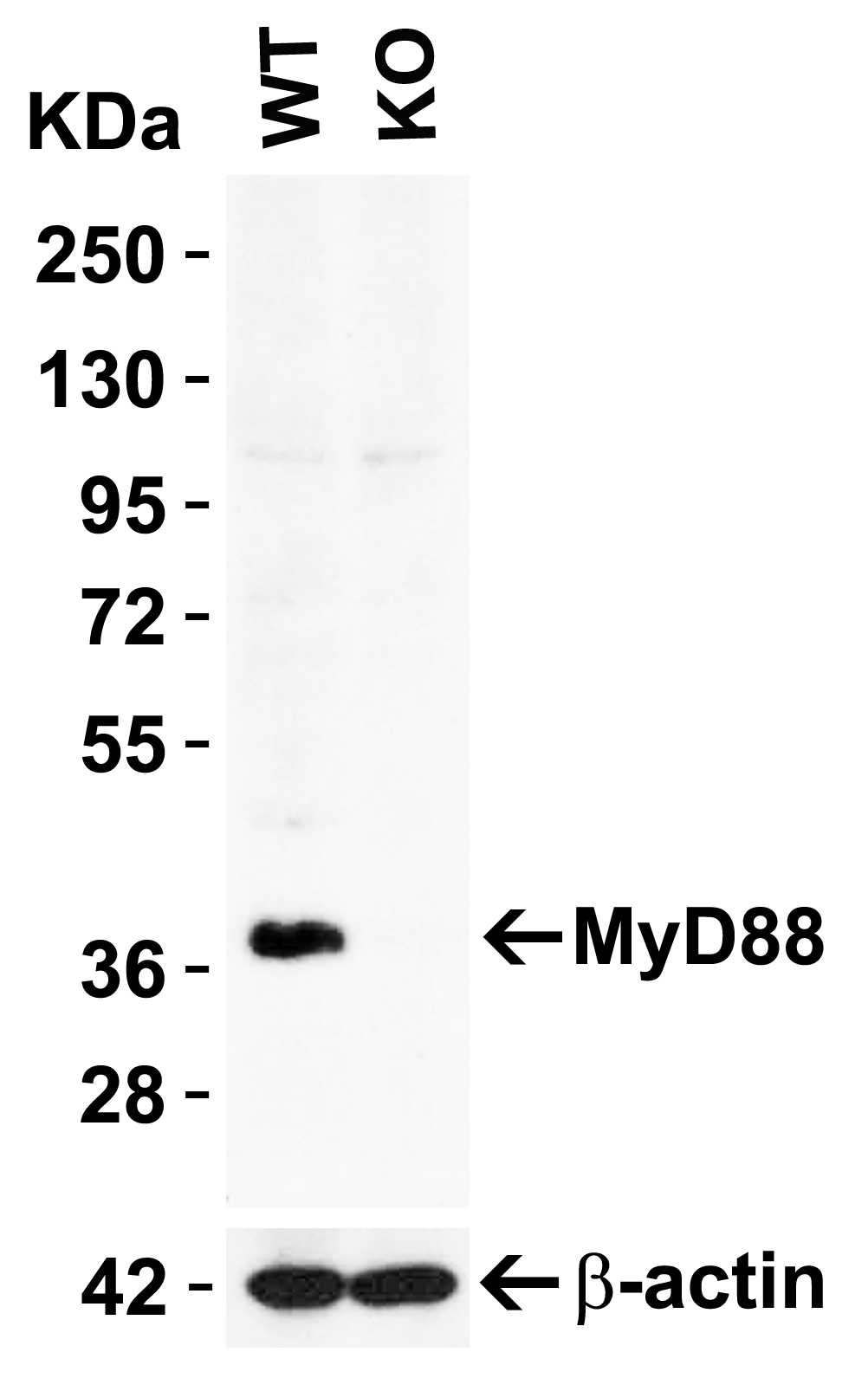
Description KO Validation in HeLa Cells
Loading: 10 ug of HeLa WT cell lysate or MyD88 KO cell lysate. Antibodies: MyD88 2127 (2 ug/mL) and beta-actin 3779 (1 ug/mL), 1 h incubation at RT in 5% NFDM/TBST.Secondary: Goat Anti-Rabbit IgG HRP conjugate at 1:10000 dilution.
Loading: 10 ug of HeLa WT cell lysate or MyD88 KO cell lysate. Antibodies: MyD88 2127 (2 ug/mL) and beta-actin 3779 (1 ug/mL), 1 h incubation at RT in 5% NFDM/TBST.Secondary: Goat Anti-Rabbit IgG HRP conjugate at 1:10000 dilution.

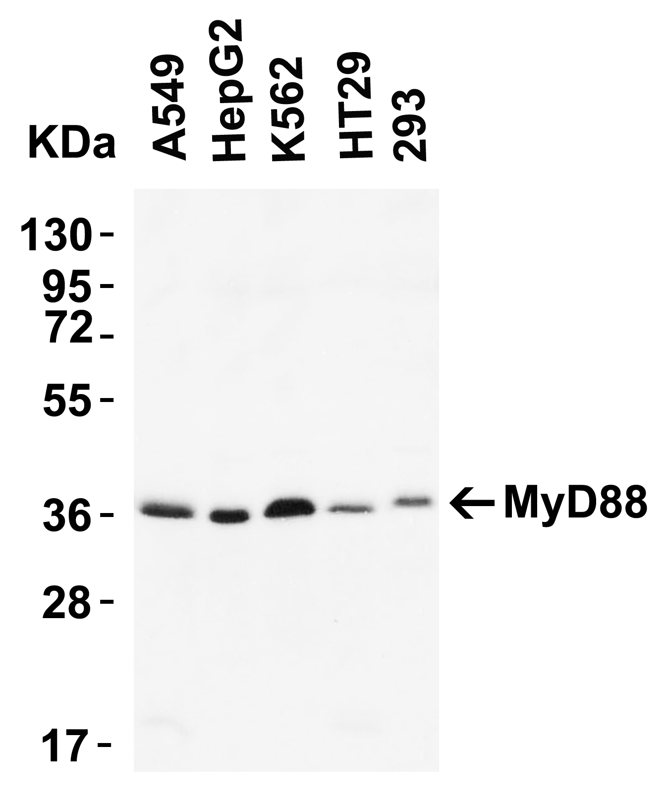
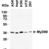
Description Western Blot Validation of MyD88 in human cell lines
Loading: 15 ug of lysates per lane. Antibodies: 2127 (2 ug/mL) 1 h incubation at RT in 5% NFDM/TBST.Secondary: Goat anti-rabbit IgG HRP conjugate at 1:10000 dilution.Predicted band size: 35 kDa
Loading: 15 ug of lysates per lane. Antibodies: 2127 (2 ug/mL) 1 h incubation at RT in 5% NFDM/TBST.Secondary: Goat anti-rabbit IgG HRP conjugate at 1:10000 dilution.Predicted band size: 35 kDa

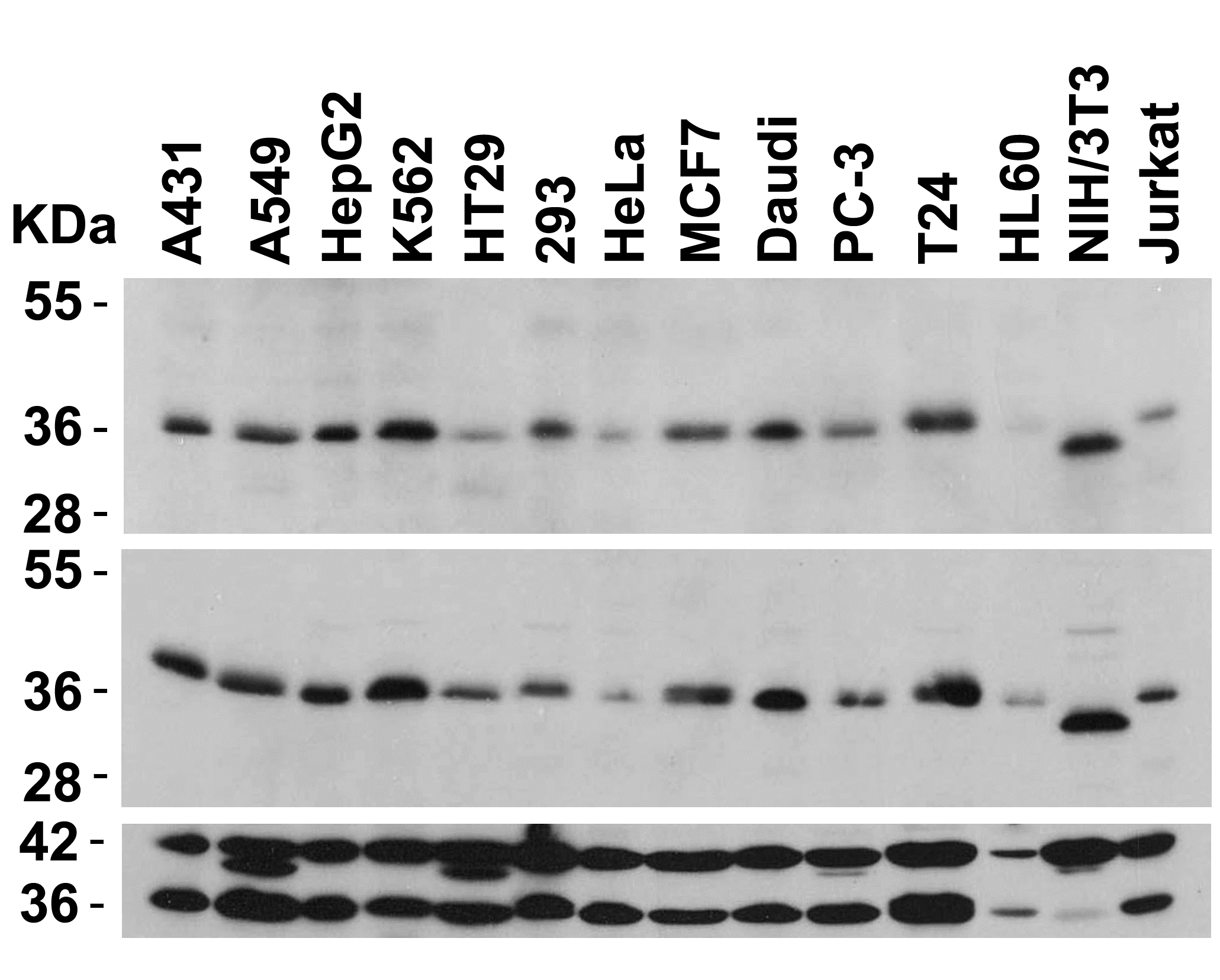
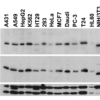
Description Independent Antibody Validation (IAV) via Protein Expression Profile in Cell Lines
Loading: 15 ug of lysates per lane. Antibodies: MyD88 2125 (2 ug/mL), MyD88 2127 (2 ug/mL), beta-actin (1 ug/mL), and GAPDH (0.02 ug/mL), 1 h incubation at RT in 5% NFDM/TBST.Secondary: Goat anti-rabbit IgG HRP conjugate at 1:10000 dilution.
Loading: 15 ug of lysates per lane. Antibodies: MyD88 2125 (2 ug/mL), MyD88 2127 (2 ug/mL), beta-actin (1 ug/mL), and GAPDH (0.02 ug/mL), 1 h incubation at RT in 5% NFDM/TBST.Secondary: Goat anti-rabbit IgG HRP conjugate at 1:10000 dilution.

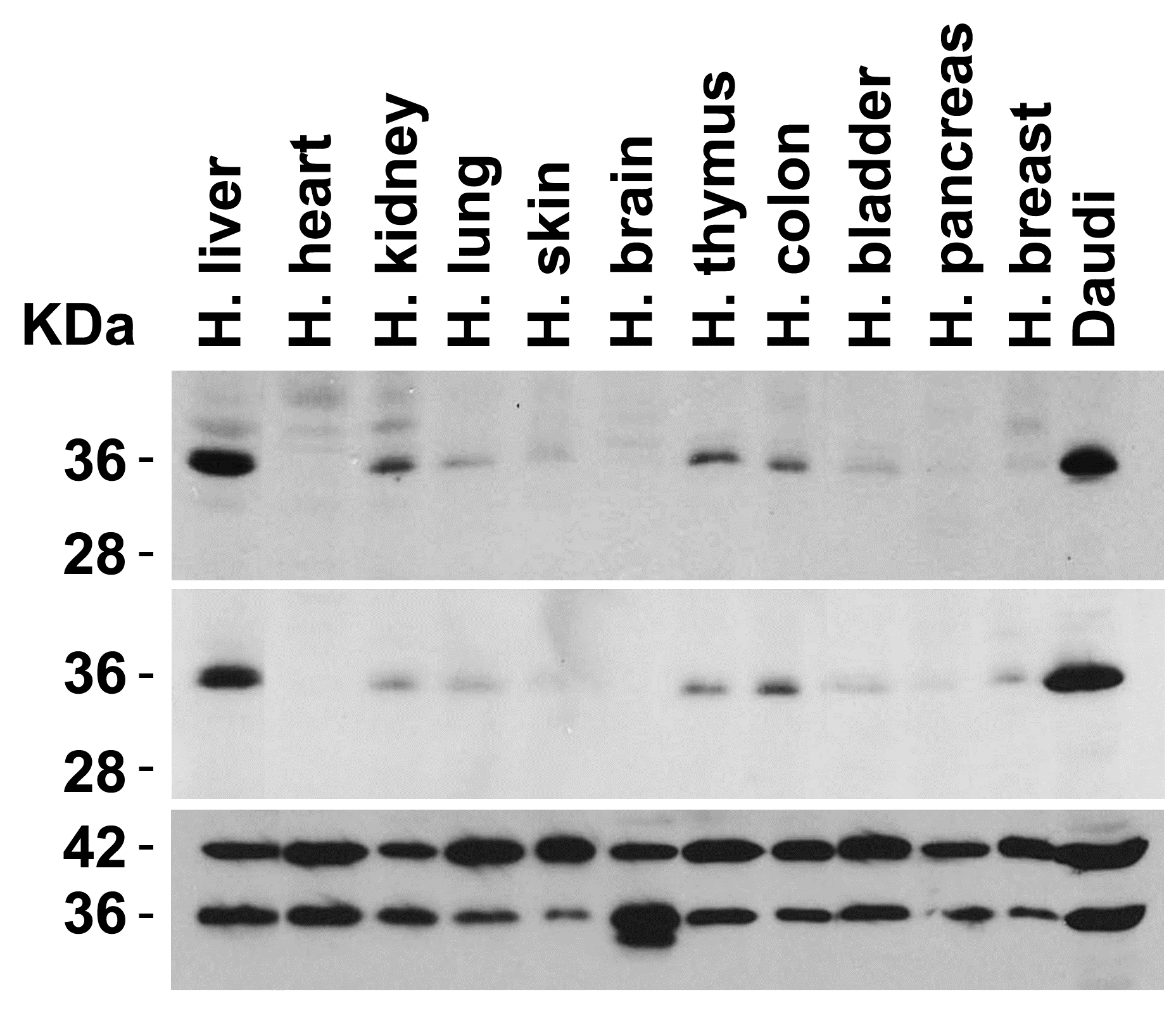
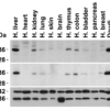
Description Independent Antibody Validation (IAV) via Protein Expression Profile in Human Tissues
Loading: 15 ug of lysates per lane. Antibodies: MyD88 2125 (2 ug/mL), MyD88 2127 (2 ug/mL), beta-actin (1 ug/mL), and GAPDH (0.02 ug/mL), 1 h incubation at RT in 5% NFDM/TBST.Secondary: Goat anti-rabbit IgG HRP conjugate at 1:10000 dilution.
Loading: 15 ug of lysates per lane. Antibodies: MyD88 2125 (2 ug/mL), MyD88 2127 (2 ug/mL), beta-actin (1 ug/mL), and GAPDH (0.02 ug/mL), 1 h incubation at RT in 5% NFDM/TBST.Secondary: Goat anti-rabbit IgG HRP conjugate at 1:10000 dilution.

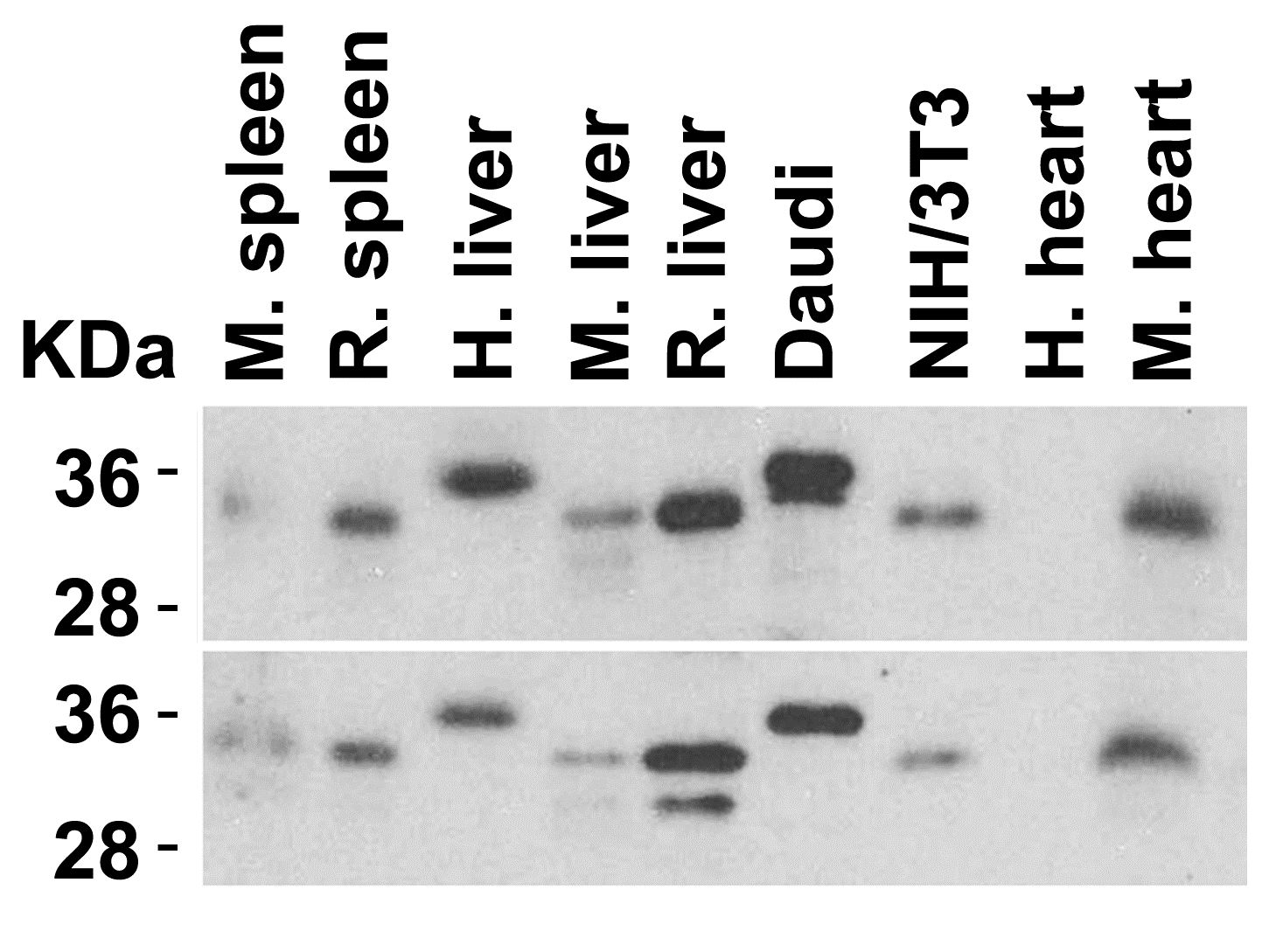
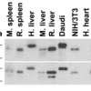
Description Animal Species Reactivity
Loading: Lysates/proteins at 15 ug per lane. Antibodies: 2125 (2 ug/mL) or 2127 (2 ug/mL). 1 h incubation at RT in 5% NFDM/TBST.Secondary: Goat anti-rabbit IgG HRP conjugate at 1:10000 dilution.
Loading: Lysates/proteins at 15 ug per lane. Antibodies: 2125 (2 ug/mL) or 2127 (2 ug/mL). 1 h incubation at RT in 5% NFDM/TBST.Secondary: Goat anti-rabbit IgG HRP conjugate at 1:10000 dilution.

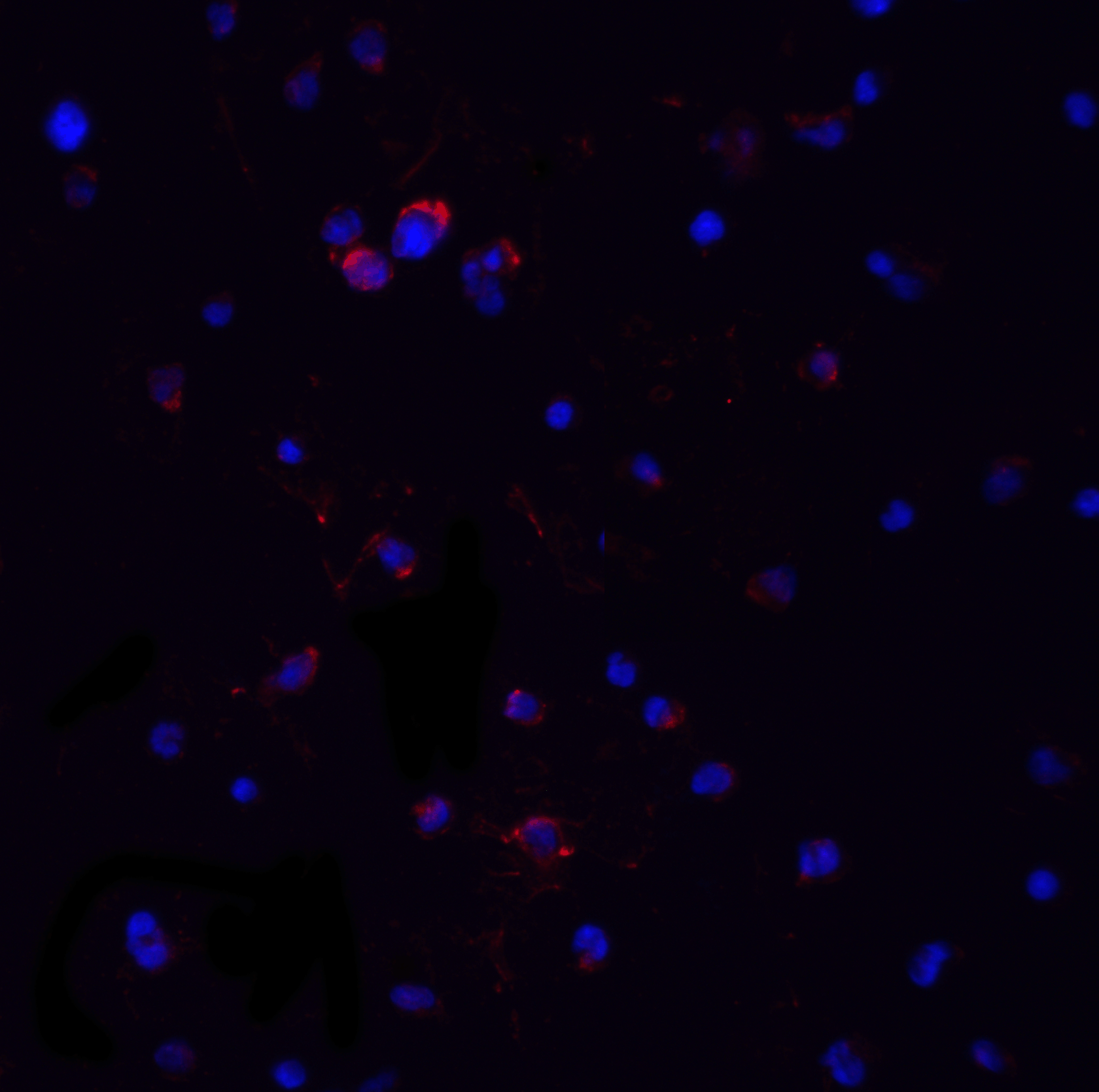
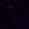
Description Immunofluorescence Validation of MyD88 in Jurkat Cells
Immunofluorescent analysis of 4% paraformaldehyde-fixed Jurkat cells labeling MyD88 with 2127 at 20 ug/mL, followed by goat anti-rabbit IgG secondary antibody at 1/500 dilution (red) and DAPI staining (blue).
Immunofluorescent analysis of 4% paraformaldehyde-fixed Jurkat cells labeling MyD88 with 2127 at 20 ug/mL, followed by goat anti-rabbit IgG secondary antibody at 1/500 dilution (red) and DAPI staining (blue).

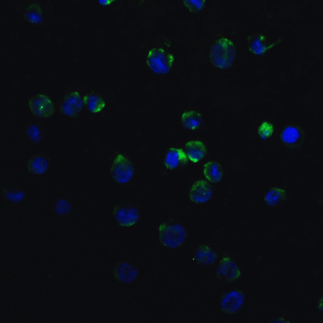
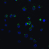
Description Immunofluorescence Validation of MyD88 in K562 Cells
Immunofluorescent analysis of 4% paraformaldehyde-fixed K562 cells labeling MyD88 with 2127 at 20 ug/mL, followed by goat anti-rabbit IgG secondary antibody at 1/500 dilution (green) and DAPI staining (blue).
Immunofluorescent analysis of 4% paraformaldehyde-fixed K562 cells labeling MyD88 with 2127 at 20 ug/mL, followed by goat anti-rabbit IgG secondary antibody at 1/500 dilution (green) and DAPI staining (blue).

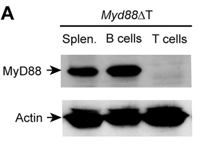
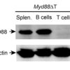
Description KO Validation of MyD88 in Mouse T cells (Rahman et al., 2011)
Splenocytes were isolated from a ;Myd88ΔT ;mouse in which MyD88 was specifically disrupted in T cells. T and B cells were FACS purified, and MyD88 expression was examined by Western blot with anti-MyD88 antibodies (2127). MyD88 expression was detected in Splenocytes and B cells, but not in T cells.
Splenocytes were isolated from a ;Myd88ΔT ;mouse in which MyD88 was specifically disrupted in T cells. T and B cells were FACS purified, and MyD88 expression was examined by Western blot with anti-MyD88 antibodies (2127). MyD88 expression was detected in Splenocytes and B cells, but not in T cells.

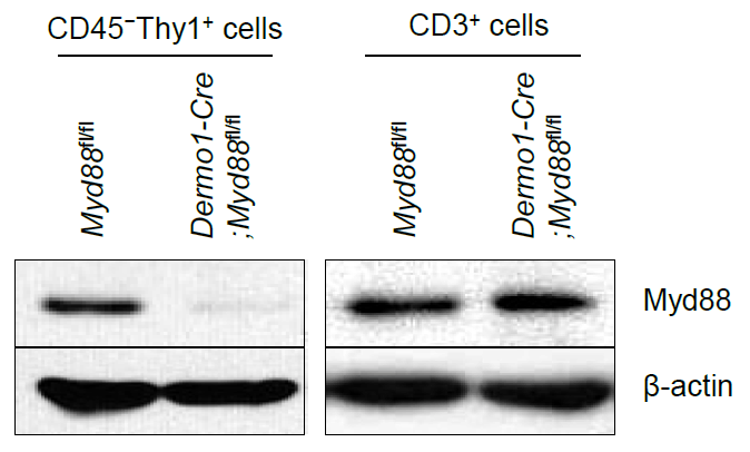
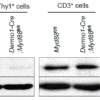
Description KO Validation in CD45 - Thy1 + cells (Kim et al., 2017)
The expression of Myd88 protein was analyzed in CD45 - Thy1 + intestinal mesenchymal cells and CD3 + intestinalT cells, by immunoblotting with anti-Myd88 antibodies (2127). MyD88 expression was not detected in knockout cells.
The expression of Myd88 protein was analyzed in CD45 - Thy1 + intestinal mesenchymal cells and CD3 + intestinalT cells, by immunoblotting with anti-Myd88 antibodies (2127). MyD88 expression was not detected in knockout cells.

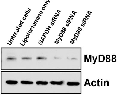
Description KD Validation in Raw 264.7 cells (Altimeier et al., 2416)
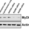
The Transfection of RAW 264.7 cells with MyD88-specific siRNAresulted in attenuation of MyD88 protein by Western blot analysis with anti-Myd88 antibodies (2127).
The Transfection of RAW 264.7 cells with MyD88-specific siRNAresulted in attenuation of MyD88 protein by Western blot analysis with anti-Myd88 antibodies (2127).

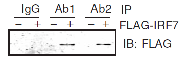
Description Immunoprecipitation Validation in HEK293 cells (Kawai et al., 2004)
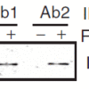
HEK293 cells were transiently transfected with FLAG-IRF7. Cell lysates were immunoprecipitated with control rabbit anti-mouse immunoglobulin serum (IgG) or anti-MyD88 (Ab1 and Ab2), followed by immunoblotting with anti-FLAG.
HEK293 cells were transiently transfected with FLAG-IRF7. Cell lysates were immunoprecipitated with control rabbit anti-mouse immunoglobulin serum (IgG) or anti-MyD88 (Ab1 and Ab2), followed by immunoblotting with anti-FLAG.
Handling
Storage This antibody is stable for at least one (1) year at -20°C. Avoid multiple freeze- thaw cycles.
Dilution Instructions Dilute in PBS or medium which is identical to that used in the assay system.
Application Instructions Immunoblotting: use at 1:500-1:1,000 dilution.
Positive control: Whole cell lysate from Jurkat cells.
Positive control: Whole cell lysate from Jurkat cells.
References & Data Sheet
Data Sheet  Download PDF Data Sheet
 Download PDF Data Sheet

