Anti-Livin Antibody (11006)
$445.00
SKU: 11006
Categories: Antibody Products, Apoptosis Antibodies, Products
Overview
Product Name Anti-Livin Antibody (11006)
Description Anti-Livin Rabbit Polyclonal Antibody
Target Livin
Species Reactivity Human
Applications ELISA,WB,IHC-P,IF
Host Rabbit
Clonality Polyclonal
Isotype IgG
Immunogen Peptide corresponding to aa 264- 280 of the short form and aa 281-298 of the long form of human Livin (accession no.
Properties
Form Liquid
Concentration Lot Specific
Formulation PBS, pH 7.4.
Buffer Formulation Phosphate Buffered Saline
Buffer pH pH 7.4
Format Purified
Purification Purified by peptide immuno-affinity chromatography
Specificity Information
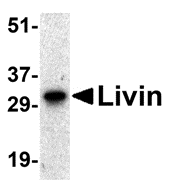
Specificity This antibody recognizes human Livin (33kDa).
Target Name Baculoviral IAP repeat-containing protein 7
Target ID Livin
Uniprot ID Q96CA5
Alternative Names EC 2.3.2.27, Kidney inhibitor of apoptosis protein, KIAP, Livin, Melanoma inhibitor of apoptosis protein, ML-IAP, RING finger protein 50, RING-type E3 ubiquitin transferase BIRC7 [Cleaved into: Baculoviral IAP repeat-containing protein 7 30kDa subunit
Gene Name BIRC7
Gene ID 79444
Accession Number NP_071444
Sequence Location Nucleus, Cytoplasm, Golgi apparatus
Biological Function Apoptotic regulator capable of exerting proapoptotic and anti-apoptotic activities and plays crucial roles in apoptosis, cell proliferation, and cell cycle control (PubMed:11162435, PubMed:11024045, PubMed:11084335, PubMed:16729033, PubMed:17294084). Its anti-apoptotic activity is mediated through the inhibition of CASP3, CASP7 and CASP9, as well as by its E3 ubiquitin-protein ligase activity (PubMed:11024045, PubMed:16729033). As it is a weak caspase inhibitor, its anti-apoptotic activity is thought to be due to its ability to ubiquitinate DIABLO/SMAC targeting it for degradation thereby promoting cell survival (PubMed:16729033). May contribute to caspase inhibition, by blocking the ability of DIABLO/SMAC to disrupt XIAP/BIRC4-caspase interactions (PubMed:16729033). Protects against apoptosis induced by TNF or by chemical agents such as adriamycin, etoposide or staurosporine (PubMed:11162435, PubMed:11084335, PubMed:11865055). Suppression of apoptosis is mediated by activation of MAPK8/JNK1, and possibly also of MAPK9/JNK2 (PubMed:11865055). This activation depends on TAB1 and MAP3K7/TAK1 (PubMed:11865055). In vitro, inhibits CASP3 and proteolytic activation of pro-CASP9 (PubMed:11024045). {PubMed:11024045, PubMed:11084335, PubMed:11162435, PubMed:11865055, PubMed:16729033, PubMed:17294084}.; [Isoform 1]: Blocks staurosporine-induced apoptosis (PubMed:11322947). Promotes natural killer (NK) cell-mediated killing (PubMed:18034418). {PubMed:11322947, PubMed:18034418}.; [Isoform 2]: Blocks etoposide-induced apoptosis (PubMed:11162435, PubMed:11322947). Protects against natural killer (NK) cell-mediated killing (PubMed:18034418). {PubMed:11162435, PubMed:11322947, PubMed:18034418}.
Research Areas Apoptosis
Background Apoptosis is prevented by the inhibitor of apoptosis (IAP) proteins. A novel member in the IAP protein family has been identified and designated Livin and KIAP for kidney IAP. Livin/KIAP contains a single baculoviral IAP repeat (BIR) domain and a RING finger domain and has two isoforms termed Livin-a and Livin-b. Transfection of Livin in cells results in protection from apoptosis induced by FADD, BAX, RIP, RIP3 and DR6. Livin has direct interaction with several caspases including caspase-3, -7, and -9. Livin inhibits the activation of caspase-9 induced by Apaf- 1, cytochrome c, and dATP. The two isoforms of Livin appear to have different functions and tissue distributions.
Application Images


Description Western blot analysis of Livin expression in human Raji cell lysate with Livin antibody at 0.5 ug/mL.

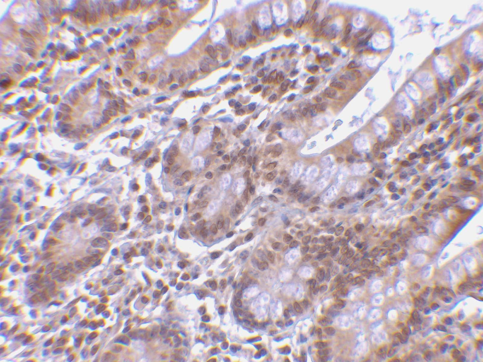
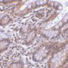
Description Immunohistochemistry of Livin in human small intestine tissue with Livin antibody at 5 ug/mL.

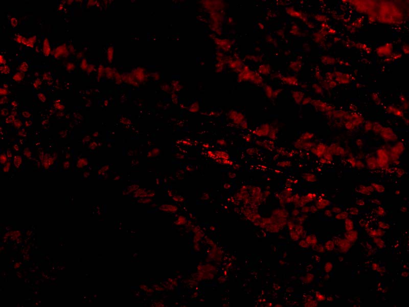
Description Immunofluorescence of Livin in Human Small Intestine cells with Livin antibody at 20 ug/mL.
Handling
Storage This antibody is stable for at least one (1) year at -20°C. Avoid multiple freeze-thaw cycles.
Dilution Instructions Dilute in PBS or medium which is identical to that used in the assay system.
Application Instructions Immunoblotting: use at 0.5-1.0ug/mL.
Positive control: Raji cell lysate.
Immunohistochemistry: use at 5ug/mL.
Positive control: Raji cell lysate.
Immunohistochemistry: use at 5ug/mL.
References & Data Sheet
Data Sheet  Download PDF Data Sheet
 Download PDF Data Sheet

