Anti-LAMP1 Antibody (56270)
$466.00
SKU: 56270
Categories: Antibody Products, Cell Adhesion Molecule Antibodies, Products
Overview
Product Name Anti-LAMP1 Antibody (56270)
Description Anti-LAMP1 Antibody Mouse Monoclonal Antibody
Target LAMP1
Species Reactivity Rat, Hamster
Applications WB,ICC/IF,IP
Host Mouse
Clonality Monoclonal
Clone ID Ly1C6
Isotype IgG1
Immunogen Rat liver lysosome membranes.
Properties
Form Liquid
Concentration Lot Specific
Formulation PBS, pH 7.4.
Buffer Formulation Phosphate Buffered Saline
Buffer pH pH 7.4
Format Purified
Purification Purified by Protein G affinity chromatography
Specificity Information
Specificity This antibody recognizes human, rat, and hamster LAMP1.Accession no.: NP_036989.1 Gene ID: 25328
Target Name Lysosome-associated membrane glycoprotein 1
Target ID LAMP1
Uniprot ID P14562
Alternative Names LAMP-1, Lysosome-associated membrane protein 1, 120 kDa lysosomal membrane glycoprotein, LGP-120, CD107 antigen-like family member A, CD antigen CD107a
Gene Name Lamp1
Sequence Location Cell membrane, Endosome membrane, Lysosome membrane, Late endosome membrane, Cytolytic granule membrane
Biological Function Lysosomal membrane glycoprotein which plays an important role in lysosome biogenesis, autophagy, and cholesterol homeostasis (By similarity). Plays also an important role in NK-cells cytotoxicity. Mechanistically, participates in cytotoxic granule movement to the cell surface and perforin trafficking to the lytic granule. In addition, protects NK-cells from degranulation-associated damage induced by their own cytotoxic granule content. Presents carbohydrate ligands to selectins. Also implicated in tumor cell metastasis (By similarity). {UniProtKB:P11279, UniProtKB:P11438}.
Research Areas Cell adhesion
Background Lysosome-associated membrane proteins (LAMP1 and LAMP2) are major constituents of the lysosomal membrane. The structures of these two proteins are closely related with 37% sequence homology. Both are transmembrane glycoproteins localized primarily in lysosomes and late endosomes. Cell surface LAMP1 and LAMP2 promote adhesion of human peripheral blood mononuclear cells (PBMC) to vascular endothelium which suggests that they are involved in adhesion of PBMC at sites of inflammation.
Application Images


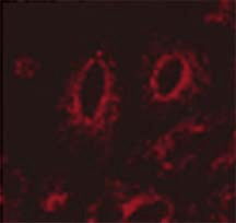
Description Immunocytochemistry/Immunofluorescence analysis using Mouse Anti-LAMP1 Monoclonal Antibody, Clone Ly1C6 (56270). Tissue: transfected HeLa cells. Species: Human. Primary Antibody: Mouse Anti-LAMP1 Monoclonal Antibody (56270) at 1:1000. Secondary Antibody: APC Goat Anti-Mouse (red). Courtesy of: Robert H Edwards, U. of Cali, San Fran School of Medicine.

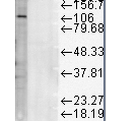
Description Western Blot analysis of Rat liver microsome lysate showing detection of LAMP1 protein using Mouse Anti-LAMP1 Monoclonal Antibody, Clone Ly1C6 (56270). Load: 15 µg. Block: 1.5% BSA for 30 minutes at RT. Primary Antibody: Mouse Anti-LAMP1 Monoclonal Antibody (56270) at 1:1000 for 2 hours at RT. Secondary Antibody: Sheep Anti-Mouse IgG: HRP for 1 hour at RT.
Handling
Storage This antibody is stable for at least one (1) year at -20°C.
Dilution Instructions Dilute in PBS or medium which is identical to that used in the assay system.
Application Instructions Immunoblotting: Use at 1ug/mL. A band of ~120kDa is detected.
Immunocytochemistry: Use at 5-10ug/mL.
These are recommended concentrations.
Enduser should determine optimal concentrations for their application.
Immunocytochemistry: Use at 5-10ug/mL.
These are recommended concentrations.
Enduser should determine optimal concentrations for their application.
References & Data Sheet
References Lewis V et al. 1985. J Cell Biol 100: 1839-1847.
PMID 3922993
Data Sheet  Download PDF Data Sheet
 Download PDF Data Sheet


