Anti-IKKε (CT) Antibody (2329)
$445.00
SKU: 2329
Categories: Antibody Products, Neuroscience and Signal Transduction Antibodies, Products
Overview
Product Name Anti-IKKε (CT) Antibody (2329)
Description Anti-IKKε (CT) Rabbit Polyclonal Antibody
Target IKKε (CT)
Species Reactivity Human
Applications ELISA,WB,IHC-P,IF
Host Rabbit
Clonality Polyclonal
Isotype IgG
Immunogen Peptide corresponding to aa 705-716 of human IKKepsilon.
Properties
Form Liquid
Concentration Lot Specific
Formulation PBS, pH 7.4.
Buffer Formulation Phosphate Buffered Saline
Buffer pH pH 7.4
Format Purified
Purification Purified by peptide immuno-affinity chromatography
Specificity Information
Specificity This antibody recognizes human IKKepsilon (80 kD). No cross-reactivity with IKKalpha, IKKb or IKKg.
Target Name Inhibitor of nuclear factor κ-B kinase subunitε
Target ID IKKε (CT)
Uniprot ID Q14164
Alternative Names I-κ-B kinaseε, IKK-E, IKK-ε, IkBKE, EC 2.7.11.10, Inducible I κ-B kinase, IKK-i
Gene Name IKBKE
Gene ID 9641
Accession Number NP_054721
Sequence Location Cytoplasm, Nucleus. Nucleus, PML body
Biological Function Serine/threonine kinase that plays an essential role in regulating inflammatory responses to viral infection, through the activation of the type I IFN, NF-kappa-B and STAT signaling. Also involved in TNFA and inflammatory cytokines, like Interleukin-1, signaling. Following activation of viral RNA sensors, such as RIG-I-like receptors, associates with DDX3X and phosphorylates interferon regulatory factors (IRFs), IRF3 and IRF7, as well as DDX3X. This activity allows subsequent homodimerization and nuclear translocation of the IRF3 leading to transcriptional activation of pro-inflammatory and antiviral genes including IFNB. In order to establish such an antiviral state, IKBKE forms several different complexes whose composition depends on the type of cell and cellular stimuli. Thus, several scaffolding molecules including IPS1/MAVS, TANK, AZI2/NAP1 or TBKBP1/SINTBAD can be recruited to the IKBKE-containing-complexes. Activated by polyubiquitination in response to TNFA and interleukin-1, regulates the NF-kappa-B signaling pathway through, at least, the phosphorylation of CYLD. Phosphorylates inhibitors of NF-kappa-B thus leading to the dissociation of the inhibitor/NF-kappa-B complex and ultimately the degradation of the inhibitor. In addition, is also required for the induction of a subset of ISGs which displays antiviral activity, may be through the phosphorylation of STAT1 at 'Ser-708'. Phosphorylation of STAT1 at 'Ser-708' seems also to promote the assembly and DNA binding of ISGF3 (STAT1:STAT2:IRF9) complexes compared to GAF (STAT1:STAT1) complexes, in this way regulating the balance between type I and type II IFN responses. Protects cells against DNA damage-induced cell death. Also plays an important role in energy balance regulation by sustaining a state of chronic, low-grade inflammation in obesity, wich leads to a negative impact on insulin sensitivity. Phosphorylates AKT1. {PubMed:17568778, PubMed:18583960, PubMed:19153231, PubMed:20188669, PubMed:21138416, PubMed:21464307, PubMed:22532683, PubMed:23453969, PubMed:23478265}.
Research Areas Neuroscience
Background Nuclear factor kappa B (NF-kappaB) is a ubiquitous transcription factor and key mediator of gene expression during immune and inflammatory responses. NF-kappaB activates numerous genes in response to extracellular stimuli, such as IL-1, TNFalpha, and LPS. NF-kappaB is associated with IkappaB in cytoplasm, which inhibits NF-kappaB activity. IkappaB is phos-phorylated by the IkappaB kinase (IKK) complex which contains IKKalpha, IKKbeta, and IKKg. A novel molecule in the IKK complex, designated IKKepsilon, is required for the activation of NF-kappaB by PMA and T cell receptors but not TNFalpha and IL-1. IKKepsilon is expressed in a variety of tissues and is inducible by TNFalpha, IL-1, and LPS.
Application Images


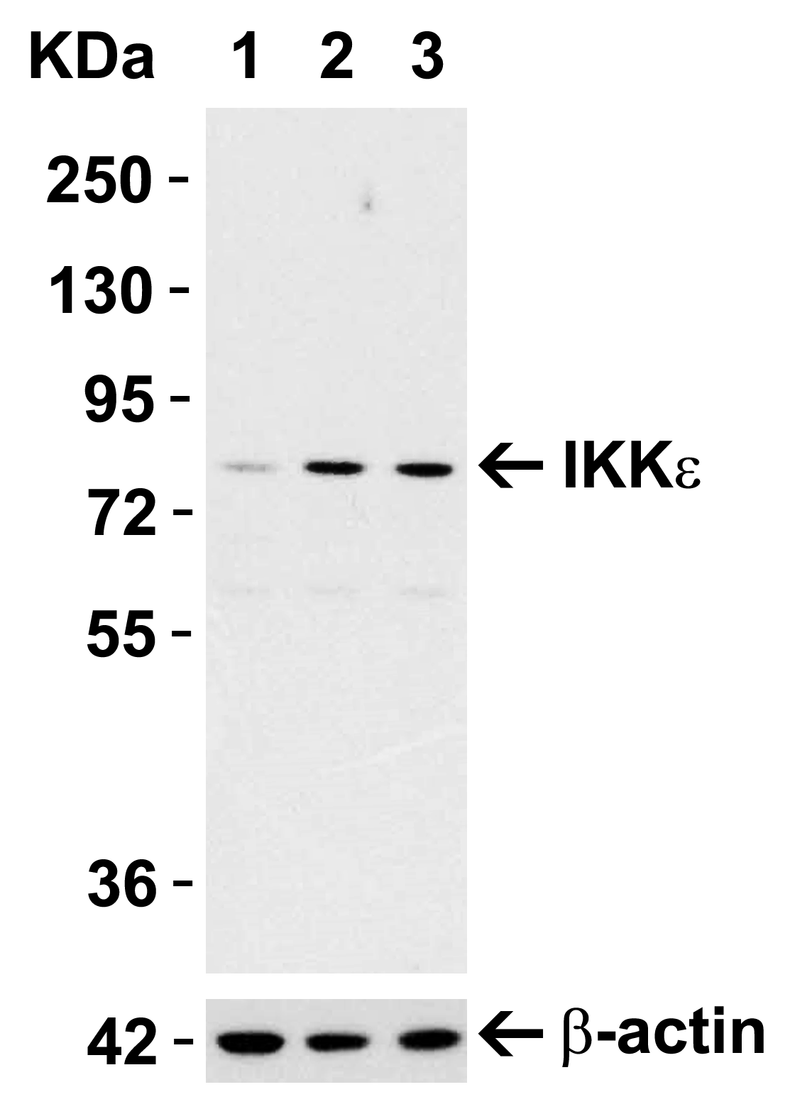
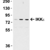
Description Induction Validation in Mouse Cell Line
Loading: 15 ug of Raw264.7 cell lysates per lane. Antibodies: IKK epsilon 2329 (1 ug/mL), 1h incubation at RT in 5% NFDM/TBST.Secondary: Goat anti-rabbit IgG HRP conjugate at 1:10000 dilution.Cells were treated with LPS (0.3 ug/mL) for 3 hrs (lane 2) and 6 hrs (lane 3) or with control (lane 1).
Loading: 15 ug of Raw264.7 cell lysates per lane. Antibodies: IKK epsilon 2329 (1 ug/mL), 1h incubation at RT in 5% NFDM/TBST.Secondary: Goat anti-rabbit IgG HRP conjugate at 1:10000 dilution.Cells were treated with LPS (0.3 ug/mL) for 3 hrs (lane 2) and 6 hrs (lane 3) or with control (lane 1).

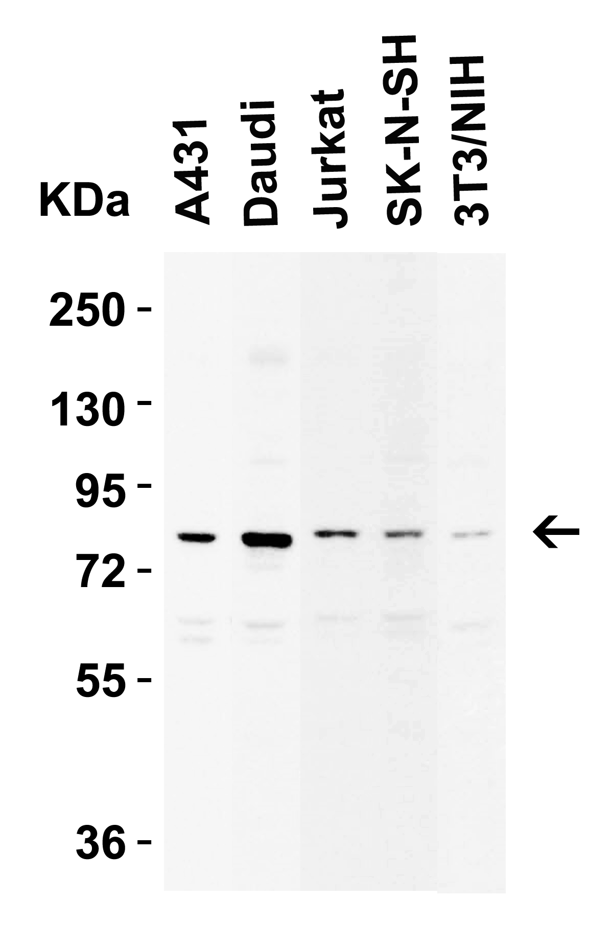
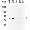
Description Western Blot Validation in Human and Mouse Cell Lines
Loading: 15 ug of lysates per lane. Antibodies: IKK epsilon 2329 (1 ug/mL), 1h incubation at RT in 5% NFDM/TBST.Secondary: Goat anti-rabbit IgG HRP conjugate at 1:10000 dilution.
Loading: 15 ug of lysates per lane. Antibodies: IKK epsilon 2329 (1 ug/mL), 1h incubation at RT in 5% NFDM/TBST.Secondary: Goat anti-rabbit IgG HRP conjugate at 1:10000 dilution.

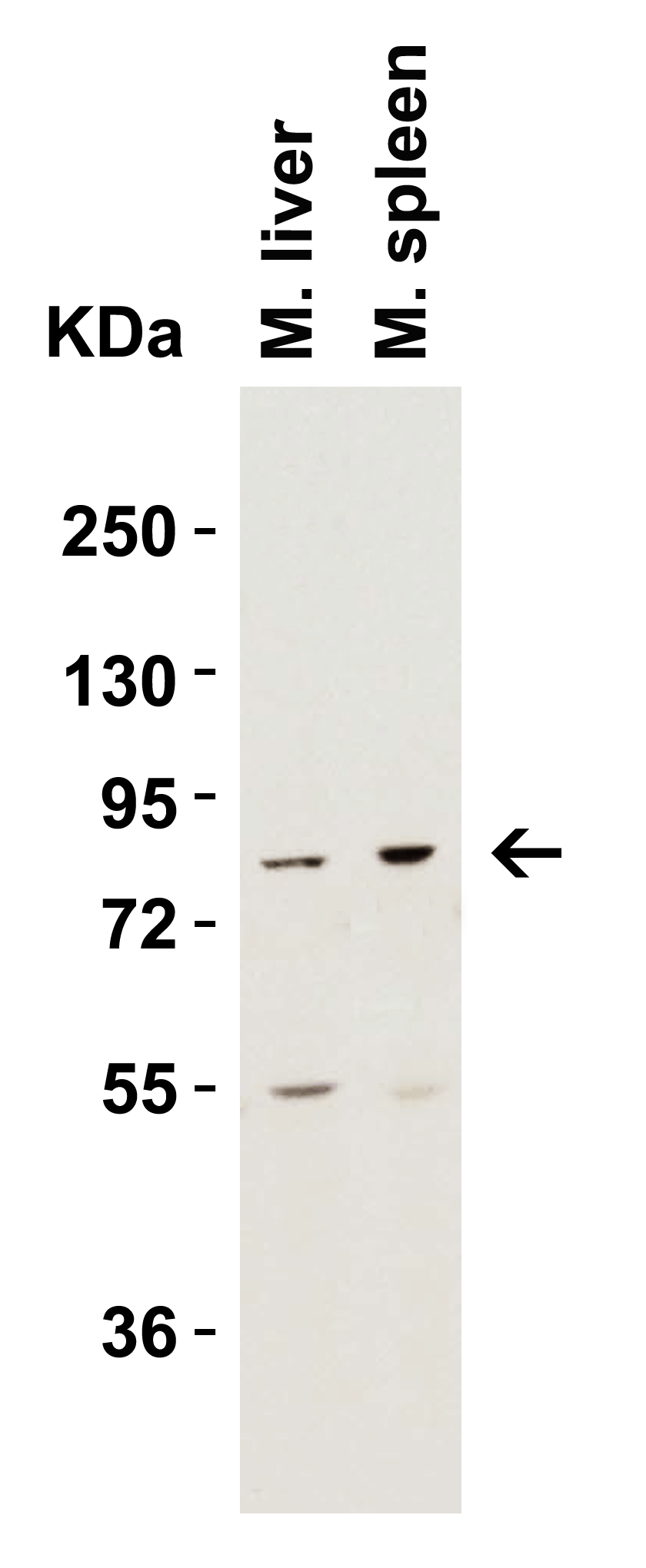
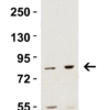
Description Western Blot Validation in Mouse Tissue Lysates
Loading: 15 ug of lysates per lane. Antibodies: IKK epsilon 2329 (2 ug/mL), 1h incubation at RT in 5% NFDM/TBST.Secondary: Goat anti-rabbit IgG HRP conjugate at 1:10000 dilution.
Loading: 15 ug of lysates per lane. Antibodies: IKK epsilon 2329 (2 ug/mL), 1h incubation at RT in 5% NFDM/TBST.Secondary: Goat anti-rabbit IgG HRP conjugate at 1:10000 dilution.

Description Western Blot Validation in Jurkat Cell Lysate
Loading: 15 ug of lysates per lane. Antibodies: IKK epsilon 2329 (2 ug/mL), 1h incubation at RT in 5% NFDM/TBST.Secondary: Goat anti-rabbit IgG HRP conjugate at 1:10000 dilution.
Loading: 15 ug of lysates per lane. Antibodies: IKK epsilon 2329 (2 ug/mL), 1h incubation at RT in 5% NFDM/TBST.Secondary: Goat anti-rabbit IgG HRP conjugate at 1:10000 dilution.

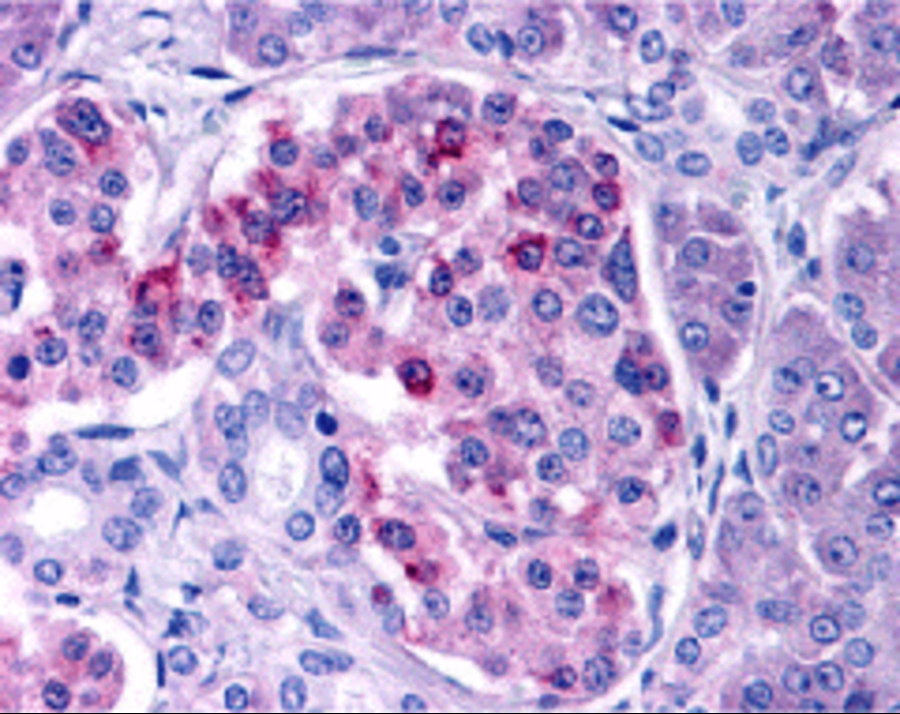
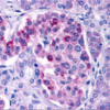
Description Immunohistochemistry Validation of IKK epsilon in Human Pancreas Tissue
Immunohistochemical analysis of paraffin-embedded human pancreas tissue using anti-IKK epsilon antibody (2329) at 10 ug/ml. Tissue was fixed with formaldehyde and blocked with 10% serum for 1 h at RT; antigen retrieval was by heat mediation with a citrate buffer (pH6). Samples were incubated with primary antibody overnight at 4C. A goat anti-rabbit IgG H&L (HRP) at 1/250 was used as secondary. Counter stained with Hematoxylin.
Immunohistochemical analysis of paraffin-embedded human pancreas tissue using anti-IKK epsilon antibody (2329) at 10 ug/ml. Tissue was fixed with formaldehyde and blocked with 10% serum for 1 h at RT; antigen retrieval was by heat mediation with a citrate buffer (pH6). Samples were incubated with primary antibody overnight at 4C. A goat anti-rabbit IgG H&L (HRP) at 1/250 was used as secondary. Counter stained with Hematoxylin.

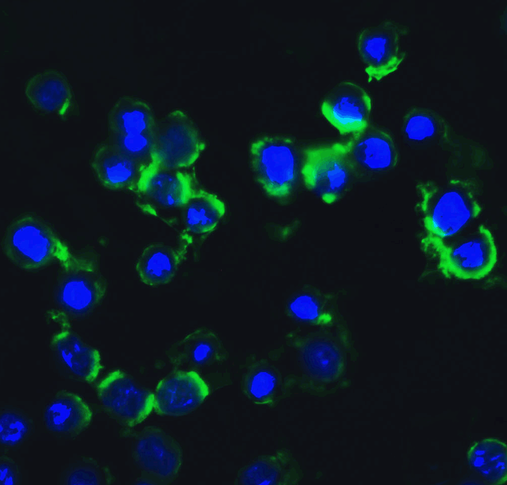
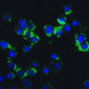
Description Immunofluorescence Validation of IKK epsilon in HeLa Cells
Immunofluorescent analysis of 4% paraformaldehyde-fixed HeLa cells labeling IKK epsilon with 2329 at 20 ug/mL, followed by goat anti-rabbit IgG secondary antibody at 1/500 dilution (green) and DAPI staining (blue).
Immunofluorescent analysis of 4% paraformaldehyde-fixed HeLa cells labeling IKK epsilon with 2329 at 20 ug/mL, followed by goat anti-rabbit IgG secondary antibody at 1/500 dilution (green) and DAPI staining (blue).
Handling
Storage This antibody is stable for at least one (1) year at -20°C. Avoid multiple freeze-thaw cycles.
Dilution Instructions Dilute in PBS or medium which is identical to that used in the assay system.
Application Instructions Immunoblotting: use at 1:500-1:1,000 dilution.
Positive control: Whole cell lysate from Jurkat cells.
Positive control: Whole cell lysate from Jurkat cells.
References & Data Sheet
Data Sheet  Download PDF Data Sheet
 Download PDF Data Sheet

