Anti-GABARAPL1 Antibody (56570)
$466.00
SKU: 56570
Categories: Antibody Products, Neuroscience and Signal Transduction Antibodies, Products
Overview
Product Name Anti-GABARAPL1 Antibody (56570)
Description Anti-GABARAPL1 Rabbit Polyclonal Antibody
Target GABARAPL1
Species Reactivity Human
Applications WB,ICC/IF
Host Rabbit
Clonality Polyclonal
Immunogen Synthetic peptide corresponding to the C-terminus of human GABARAPL1
Properties
Form Liquid
Concentration 1.0 mg/mL
Formulation PBS, pH 7.4, 50% glycerol, 0.09% sodium azide.
Buffer Formulation Phosphate Buffered Saline
Buffer pH pH 7.4
Buffer Anti-Microbial 0.09% Sodium Azide
Buffer Cryopreservative 50% Glycerol
Format Purified
Purification Purified by peptide immuno-affinity chromatography
Specificity Information
Specificity This antibody recognizes human GABARAPL1.
Target Name γ-aminobutyric acid receptor-associated protein-like 1
Target ID GABARAPL1
Uniprot ID Q9H0R8
Alternative Names Early estrogen-regulated protein, GABA(A receptor-associated protein-like 1, Glandular epithelial cell protein 1, GEC-1
Gene Name GABARAPL1
Sequence Location Cytoplasmic vesicle, autophagosome, Cytoplasmic vesicle membrane, Cytoplasm, cytoskeleton, Endoplasmic reticulum, Golgi apparatus
Biological Function Ubiquitin-like modifier that increases cell-surface expression of kappa-type opioid receptor through facilitating anterograde intracellular trafficking of the receptor (PubMed:16431922). Involved in formation of autophagosomal vacuoles (PubMed:20404487). While LC3s are involved in elongation of the phagophore membrane, the GABARAP/GATE-16 subfamily is essential for a later stage in autophagosome maturation (PubMed:20404487). Through its interaction with the reticulophagy receptor TEX264, participates in the remodeling of subdomains of the endoplasmic reticulum into autophagosomes upon nutrient stress, which then fuse with lysosomes for endoplasmic reticulum turnover (PubMed:31006538, PubMed:31006537). {PubMed:16431922, PubMed:20404487, PubMed:31006537, PubMed:31006538}.
Research Areas Neuroscience
Background GABARAP, GABARAPL1 and GABARAPL2 genes are differentially expressed in normal and pathological tissues. The GABARAP gene is highly expressed in endocrine tissues while the GABARAPL1 gene is predominantly expressed in the central nervous system, but they both are underexpressed in a large variety of cancer cell lines. However, the analysis of GABARAPL1 expression in a cohort of 256 breast adenocarcinoma revealed that a low GABARAPL1 expression was correlated with a high risk of metastasis, in particular for lymph node-positive patients.
Application Images


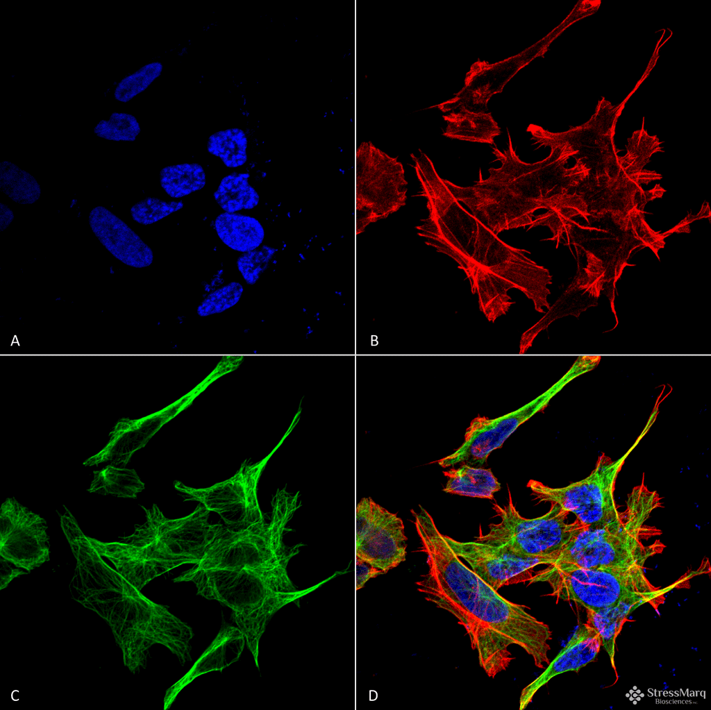
Description Immunocytochemistry/Immunofluorescence analysis using Rabbit Anti-GABARAPL1 Polyclonal Antibody (56570). Tissue: Neuroblastoma cell line (SK-N-BE). Species: Human. Fixation: 4% Formaldehyde for 15 min at RT. Primary Antibody: Rabbit Anti-GABARAPL1 Polyclonal Antibody (56570) at 1:100 for 60 min at RT. Secondary Antibody: Goat Anti-Rabbit ATTO 488 at 1:100 for 60 min at RT. Counterstain: Phalloidin Texas Red F-Actin stain; DAPI (blue) nuclear stain at 1:1000, 1:5000 for 60min RT, 5min RT. Localization: Cytoplasm, Cytoskeleton. Magnification: 60X. (A) DAPI (blue) nuclear stain (B) Phalloidin Texas Red F-Actin stain (C) GABARAPL1 Antibody (D) Composite.

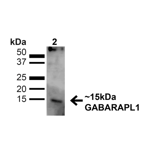
Description Western blot analysis of Human Cervical cancer cell line (HeLa) lysate showing detection of ~14kDa GABARAPL1 protein using Rabbit Anti-GABARAPL1 Polyclonal Antibody (56570). Lane 1: MW Ladder. Lane 2: Human HeLa (20 µg). Load: 20 µg. Block: 5% milk + TBST for 1 hour at RT. Primary Antibody: Rabbit Anti-GABARAPL1 Polyclonal Antibody (56570) at 1:1000 for 1 hour at RT. Secondary Antibody: Goat Anti-Rabbit: HRP at 1:2000 for 1 hour at RT. Color Development: TMB solution for 12 min at RT. Predicted/Observed Size: ~14kDa.
Handling
Storage This antibody is stable for at least one (1) year at -20°C.
Dilution Instructions Dilute in PBS or medium that is identical to that used in the assay system.
Application Instructions Immunoblotting: use at dilution of 1:1,000. A band of ~14-16kDa is detected.
Immunofluorescence: use at dilution of 1:100
These are recommended working dilutions.
Endusers should determine optimal dilutions for their applications.
Immunofluorescence: use at dilution of 1:100
These are recommended working dilutions.
Endusers should determine optimal dilutions for their applications.
References & Data Sheet
Data Sheet  Download PDF Data Sheet
 Download PDF Data Sheet


