Anti-DR5 Antibody (2421)
$445.00
SKU: 2421
Categories: Antibody Products, Apoptosis Antibodies, Products
Overview
Product Name Anti-DR5 Antibody (2421)
Description Anti-DR5 Rabbit Polyclonal Antibody
Target DR5
Species Reactivity Human
Applications ELISA,WB,IHC-P,IF
Host Rabbit
Clonality Polyclonal
Isotype IgG
Immunogen Peptide corresponding to aa 388- 407 of human DR5 precursor (accession no. AF012535).
Properties
Form Liquid
Concentration Lot Specific
Formulation PBS, pH 7.4.
Buffer Formulation Phosphate Buffered Saline
Buffer pH pH 7.4
Format Purified
Purification Purified by peptide immuno-affinity chromatography
Specificity Information
Specificity This antibody recognizes full-length human and mouse DR5 (57kDa).
Target Name Tumor necrosis factor receptor superfamily member 10B
Target ID DR5
Uniprot ID O14763
Alternative Names Death receptor 5, TNF-related apoptosis-inducing ligand receptor 2, TRAIL receptor 2, TRAIL-R2, CD antigen CD262
Gene Name TNFRSF10B
Gene ID 8795
Accession Number NP_003833
Sequence Location Membrane; Single-pass type I membrane protein.
Biological Function Receptor for the cytotoxic ligand TNFSF10/TRAIL (PubMed:10549288). The adapter molecule FADD recruits caspase-8 to the activated receptor. The resulting death-inducing signaling complex (DISC) performs caspase-8 proteolytic activation which initiates the subsequent cascade of caspases (aspartate-specific cysteine proteases) mediating apoptosis. Promotes the activation of NF-kappa-B. Essential for ER stress-induced apoptosis. {PubMed:10542098, PubMed:10549288, PubMed:15322075}.
Research Areas Apoptosis
Background Apoptosis is induced by certain cytokines including TNF and Fas ligand in the TNF family through their death domain-containing receptors. TRAIL/Apo2L is a new member of the TNF family. A novel death domain- containing receptor for TRAIL was recently identified and designated DR5, Apo2, TRAIL-R2, TRICK2, or KILLER by several independent laboratories. Like DR4, DR5 transcript is widely expressed in normal tissues and in many types of tumor cells. DR5 binds to TRAIL and mediates TRAIL- induced cell death. Overexpression of DR5 induces apoptosis and activates NF- B.
Application Images


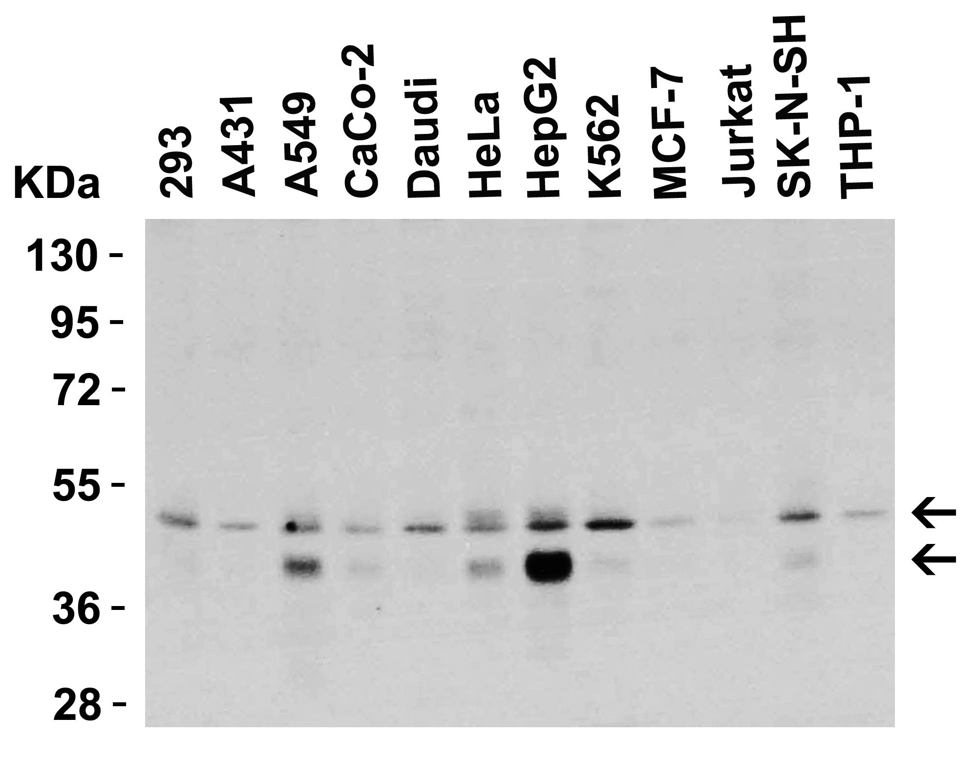
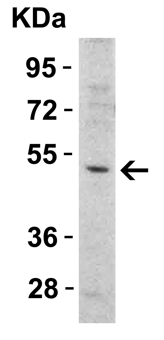
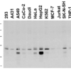
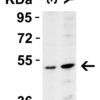
Description Western Blot Validation in Human Cell Lines
Loading: 15 ug of lysates per lane. Antibodies: DR5 2421, (0.5 ug/mL), 1h incubation at RT in 5% NFDM/TBST.Secondary: Goat anti-rabbit IgG HRP conjugate at 1:10000 dilution.
Loading: 15 ug of lysates per lane. Antibodies: DR5 2421, (0.5 ug/mL), 1h incubation at RT in 5% NFDM/TBST.Secondary: Goat anti-rabbit IgG HRP conjugate at 1:10000 dilution.

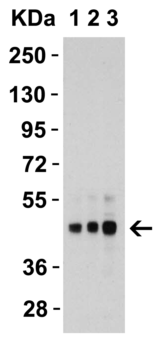
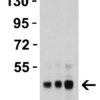
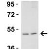
Description Western Blot Validation in Human HepG2 Cells
Loading: 15 ug of lysates per lane. Antibodies: DR5 2421, 1h incubation at RT in 5% NFDM/TBST.Secondary: Goat anti-rabbit IgG HRP conjugate at 1:10000 dilution.Lane 1: 1 ug/mLLane 2: 2 ug/mLLane 3: 4 ug/mL
Loading: 15 ug of lysates per lane. Antibodies: DR5 2421, 1h incubation at RT in 5% NFDM/TBST.Secondary: Goat anti-rabbit IgG HRP conjugate at 1:10000 dilution.Lane 1: 1 ug/mLLane 2: 2 ug/mLLane 3: 4 ug/mL

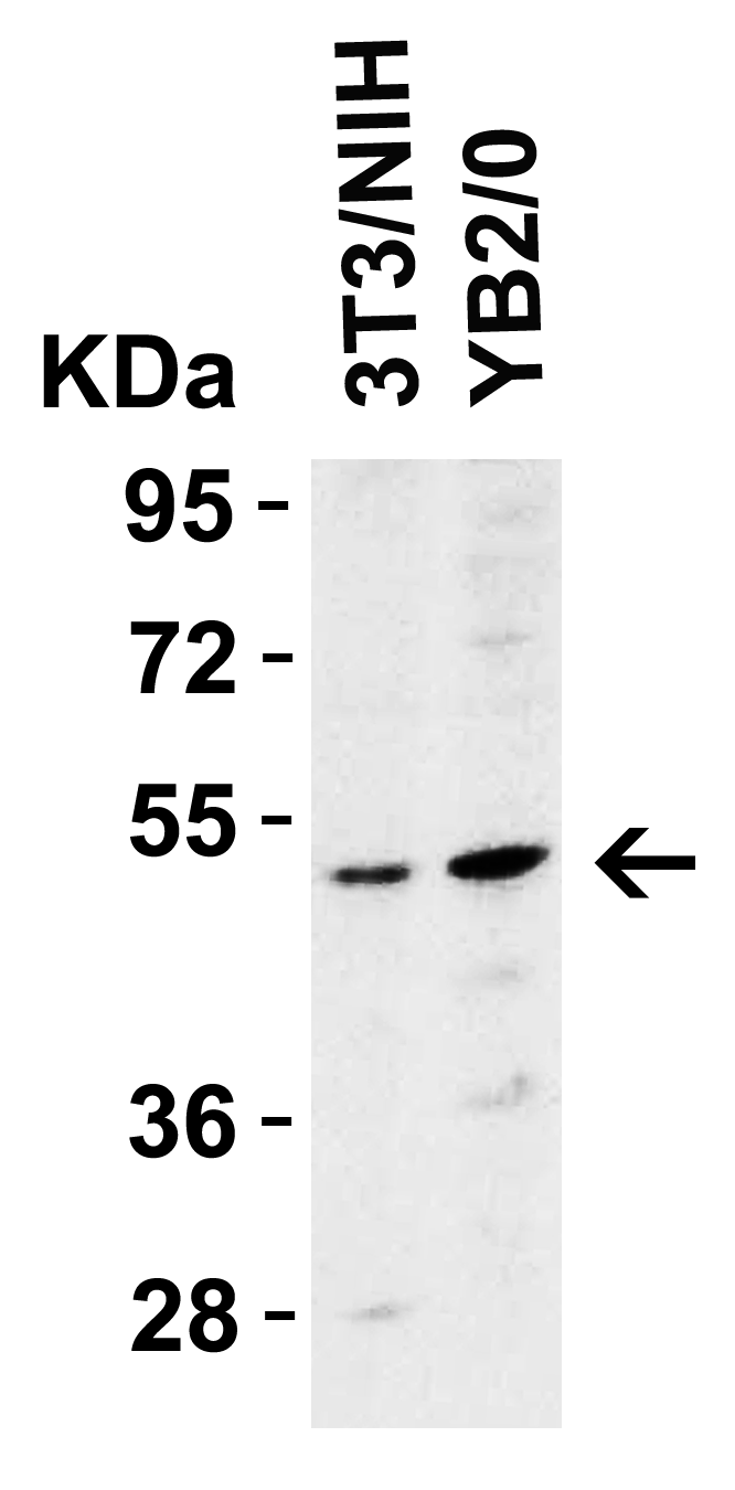
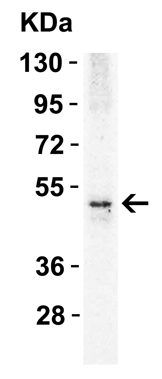
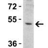
Description Western Blot Validation in Mouse and Rat Cell Lines
Loading: 15 ug of lysates per lane. Antibodies: DR5 2421, (2 ug/mL), 1h incubation at RT in 5% NFDM/TBST.Secondary: Goat anti-rabbit IgG HRP
Loading: 15 ug of lysates per lane. Antibodies: DR5 2421, (2 ug/mL), 1h incubation at RT in 5% NFDM/TBST.Secondary: Goat anti-rabbit IgG HRP

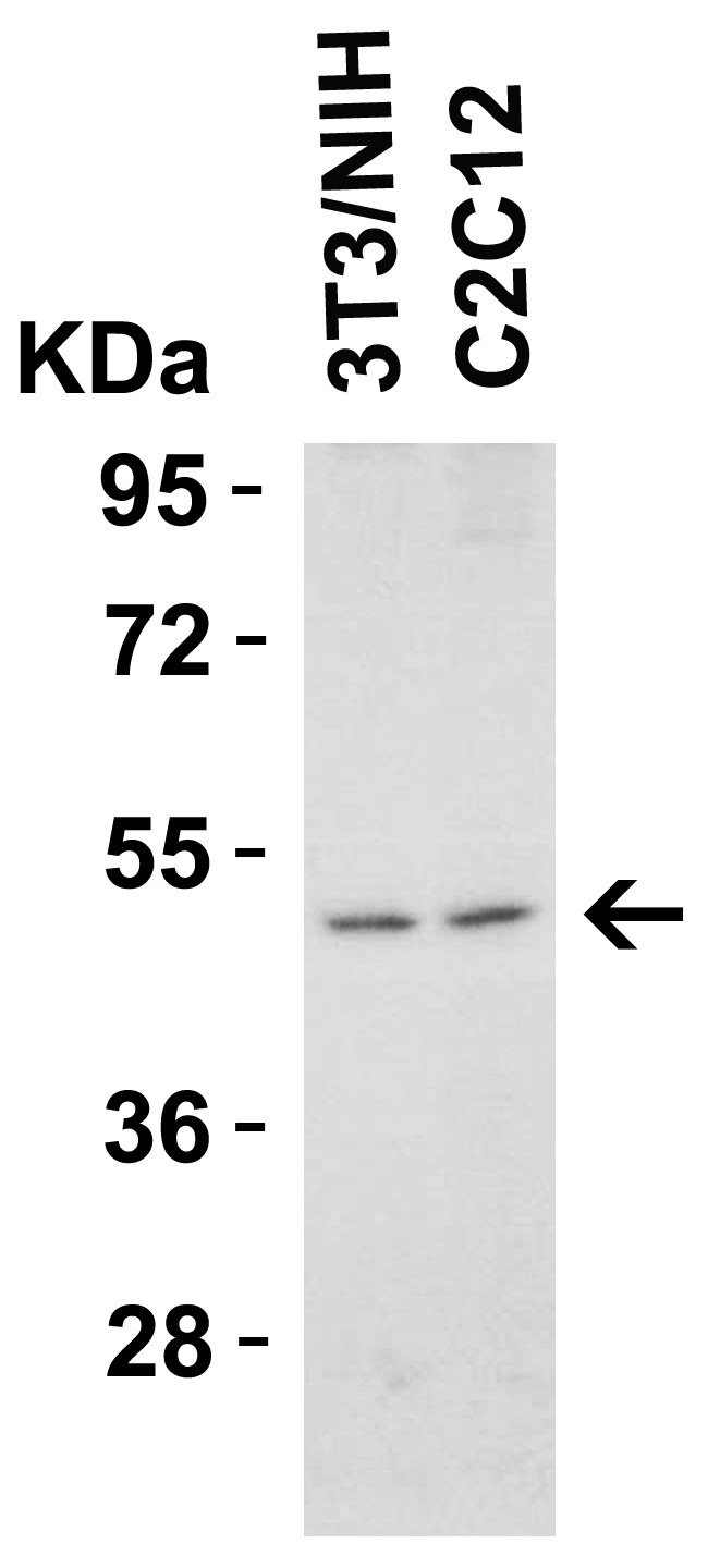
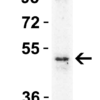
Description Western Blot Validation in Mouse Cell Lines
Loading: 15 ug of lysates per lane. Antibodies: DR5 2421, (1 ug/mL), 1h incubation at RT in 5% NFDM/TBST.Secondary: Goat anti-rabbit IgG HRP conjugate at 1:10000 dilution.
Loading: 15 ug of lysates per lane. Antibodies: DR5 2421, (1 ug/mL), 1h incubation at RT in 5% NFDM/TBST.Secondary: Goat anti-rabbit IgG HRP conjugate at 1:10000 dilution.

Description Western Blot Validation in Mouse Heart
Loading: 15 ug of lysatesper lane. Antibodies: DR5 2421, (1 ug/mL), 1h incubation at RT in 5% NFDM/TBST.Secondary: Goat anti-rabbit IgG HRP conjugate at 1:10000 dilution.
Loading: 15 ug of lysatesper lane. Antibodies: DR5 2421, (1 ug/mL), 1h incubation at RT in 5% NFDM/TBST.Secondary: Goat anti-rabbit IgG HRP conjugate at 1:10000 dilution.

Description Western Blot Validation in Rat Skeletal Muscle
Loading: 15 ug of lysate per lane. Antibodies: DR5 2421, (1 ug/mL), 1h incubation at RT in 5% NFDM/TBST.Secondary: Goat anti-rabbit IgG HRP conjugate at 1:10000 dilution.
Loading: 15 ug of lysate per lane. Antibodies: DR5 2421, (1 ug/mL), 1h incubation at RT in 5% NFDM/TBST.Secondary: Goat anti-rabbit IgG HRP conjugate at 1:10000 dilution.

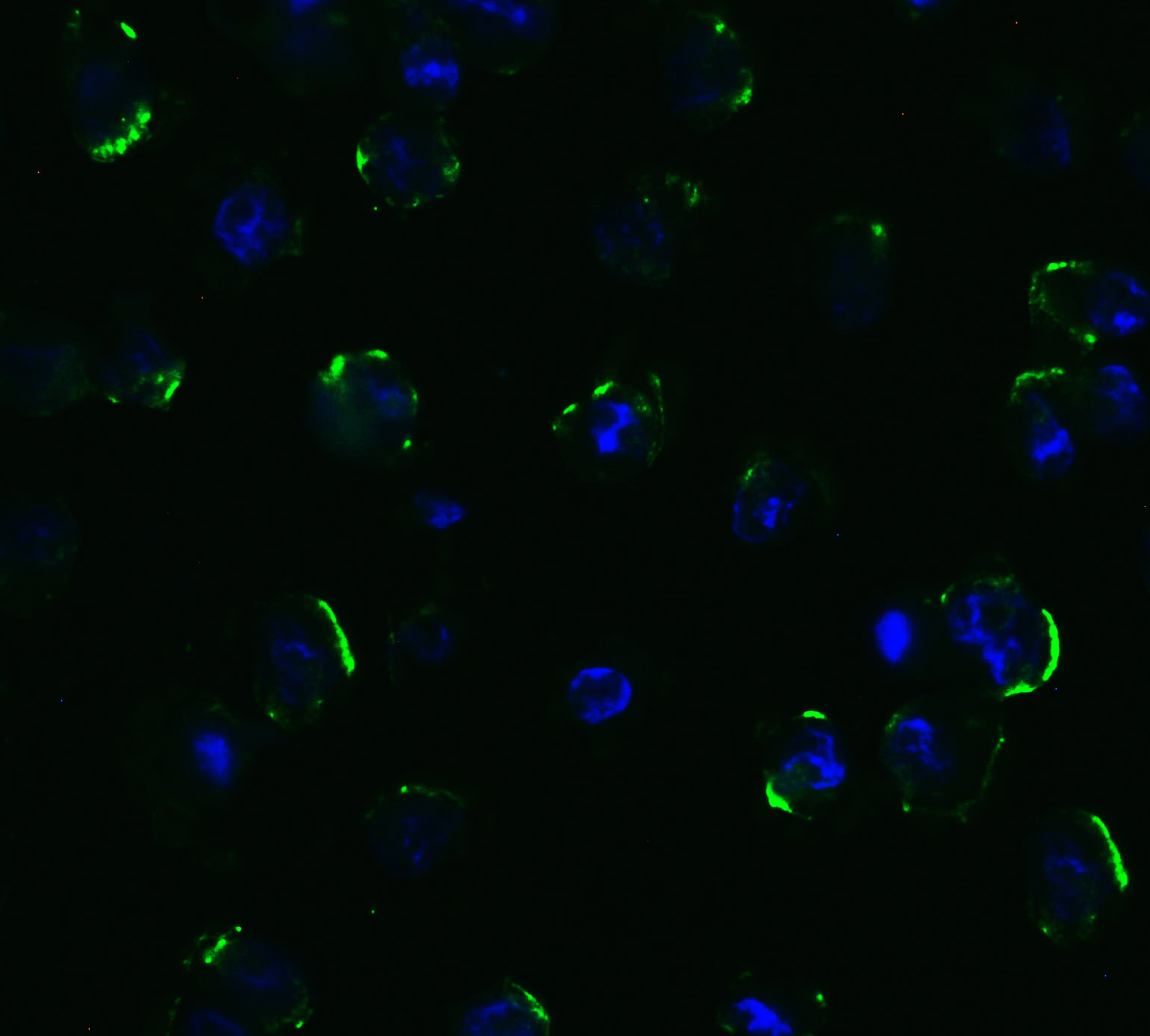
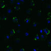
Description Immunofluorescence Validation of DR5 in Human HepG2 Cells
Immunofluorescent analysis of 4% paraformaldehyde-fixed human HepG2 cells labeling DR5 with 2421 at 5 ug/mL, followed by goat anti-rabbit IgG secondary antibody at 1/500 dilution (green) and DAPI (blue).
Immunofluorescent analysis of 4% paraformaldehyde-fixed human HepG2 cells labeling DR5 with 2421 at 5 ug/mL, followed by goat anti-rabbit IgG secondary antibody at 1/500 dilution (green) and DAPI (blue).

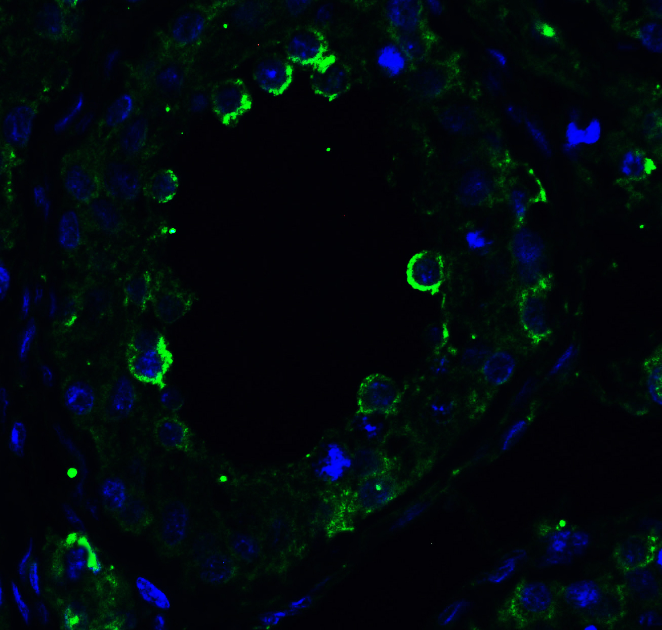
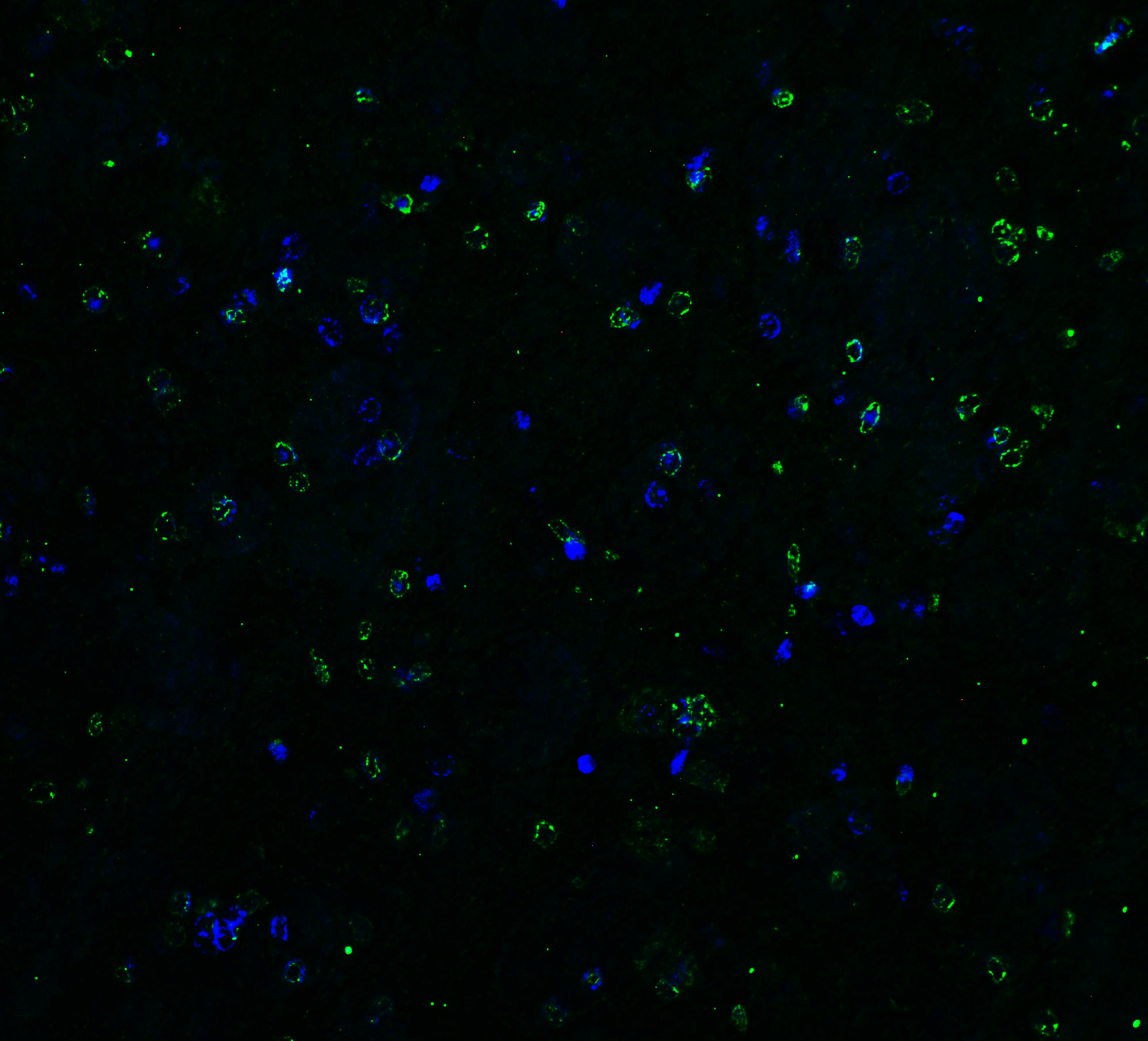
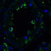
Description Immunofluorescence Validation of DR5 in Human Testis
Immunofluorescent analysis of 4% paraformaldehyde-fixed human testis tissue labeling DR5 with 2421 at 10 ug/mL, followed by goat anti-rabbit IgG secondary antibody at 1/500 dilution (green) and DAPI (blue).
Immunofluorescent analysis of 4% paraformaldehyde-fixed human testis tissue labeling DR5 with 2421 at 10 ug/mL, followed by goat anti-rabbit IgG secondary antibody at 1/500 dilution (green) and DAPI (blue).

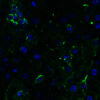
Description Immunofluorescence Validation of DR5 in Mouse Pancreas
Immunofluorescent analysis of 4% paraformaldehyde-fixed mouse pancreas tissue labeling DR5 with 2421 at 10 ug/mL, followed by goat anti-rabbit IgG secondary antibody at 1/500 dilution (green) and DAPI (blue).
Immunofluorescent analysis of 4% paraformaldehyde-fixed mouse pancreas tissue labeling DR5 with 2421 at 10 ug/mL, followed by goat anti-rabbit IgG secondary antibody at 1/500 dilution (green) and DAPI (blue).

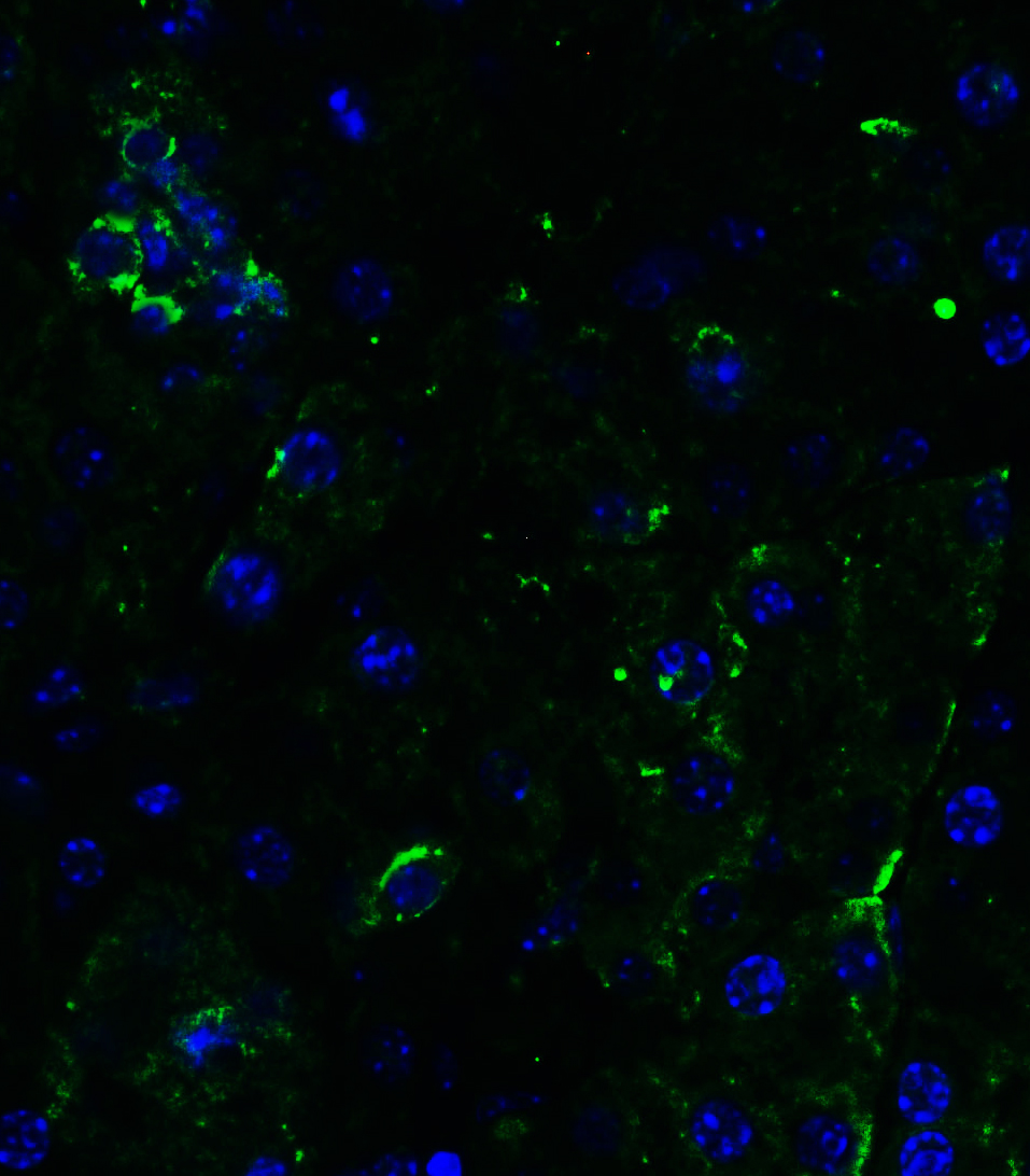
Description Immunofluorescence Validation of DR5 in Rat Brain
Immunofluorescent analysis of 4% paraformaldehyde-fixed rat brain tissue labeling DR5 with 2421 at 5 ug/mL, followed by goat anti-rabbit IgG secondary antibody at 1/500 dilution (green) and DAPI (blue).
Immunofluorescent analysis of 4% paraformaldehyde-fixed rat brain tissue labeling DR5 with 2421 at 5 ug/mL, followed by goat anti-rabbit IgG secondary antibody at 1/500 dilution (green) and DAPI (blue).

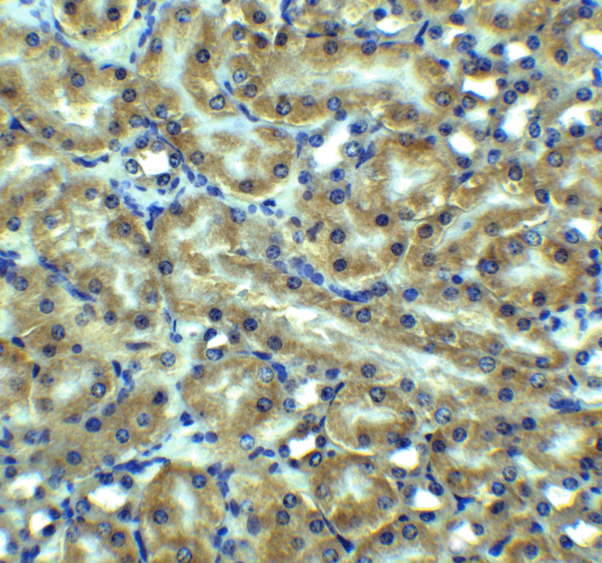
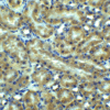
Description Immunohistochemistry Validation of DR5 in Mouse kidney tissue
Immunohistochemical analysis of paraffin-embedded mouse kidney tissue using anti-DR5 antibody (2421) at 5ug/ml. Tissue was fixed with formaldehyde and blocked with 10% serum for 1 h at RT; antigen retrieval was by heat mediation with a citrate buffer (pH6). Samples were incubated with primary antibody overnight at 4C. A goat anti-rabbit IgG H&L (HRP) at 1/250 was used as secondary. Counter stained with Hematoxylin.
Immunohistochemical analysis of paraffin-embedded mouse kidney tissue using anti-DR5 antibody (2421) at 5ug/ml. Tissue was fixed with formaldehyde and blocked with 10% serum for 1 h at RT; antigen retrieval was by heat mediation with a citrate buffer (pH6). Samples were incubated with primary antibody overnight at 4C. A goat anti-rabbit IgG H&L (HRP) at 1/250 was used as secondary. Counter stained with Hematoxylin.

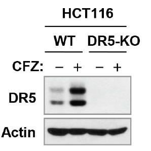
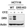
Description KO Validation of DR5 in HCT116 Cells (Han et al., 2015)
Anti-cancer drug, Carfilzomib (CFZ), induced up-regulation of DR5 and the expression of DR5 was not detected in DR5-KO HCT 116 cell line with anti-DR5 antibodies (2421).
Anti-cancer drug, Carfilzomib (CFZ), induced up-regulation of DR5 and the expression of DR5 was not detected in DR5-KO HCT 116 cell line with anti-DR5 antibodies (2421).
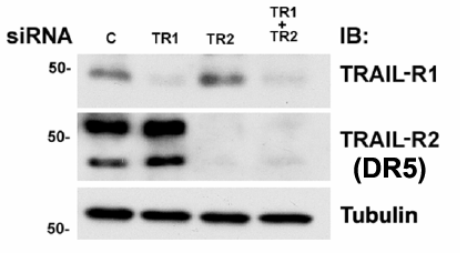
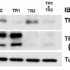
Description KD Validation of DR5 in MB231 Cells (Rahman et al., 2009)
Western blot analysis with anti-DR5 antibodies was performed for DR5 in MB231 cells transfected with control siRNA or DR5 siRNA. DR5 expression was disrupted after DR5 siRNA knockdown.
Western blot analysis with anti-DR5 antibodies was performed for DR5 in MB231 cells transfected with control siRNA or DR5 siRNA. DR5 expression was disrupted after DR5 siRNA knockdown.

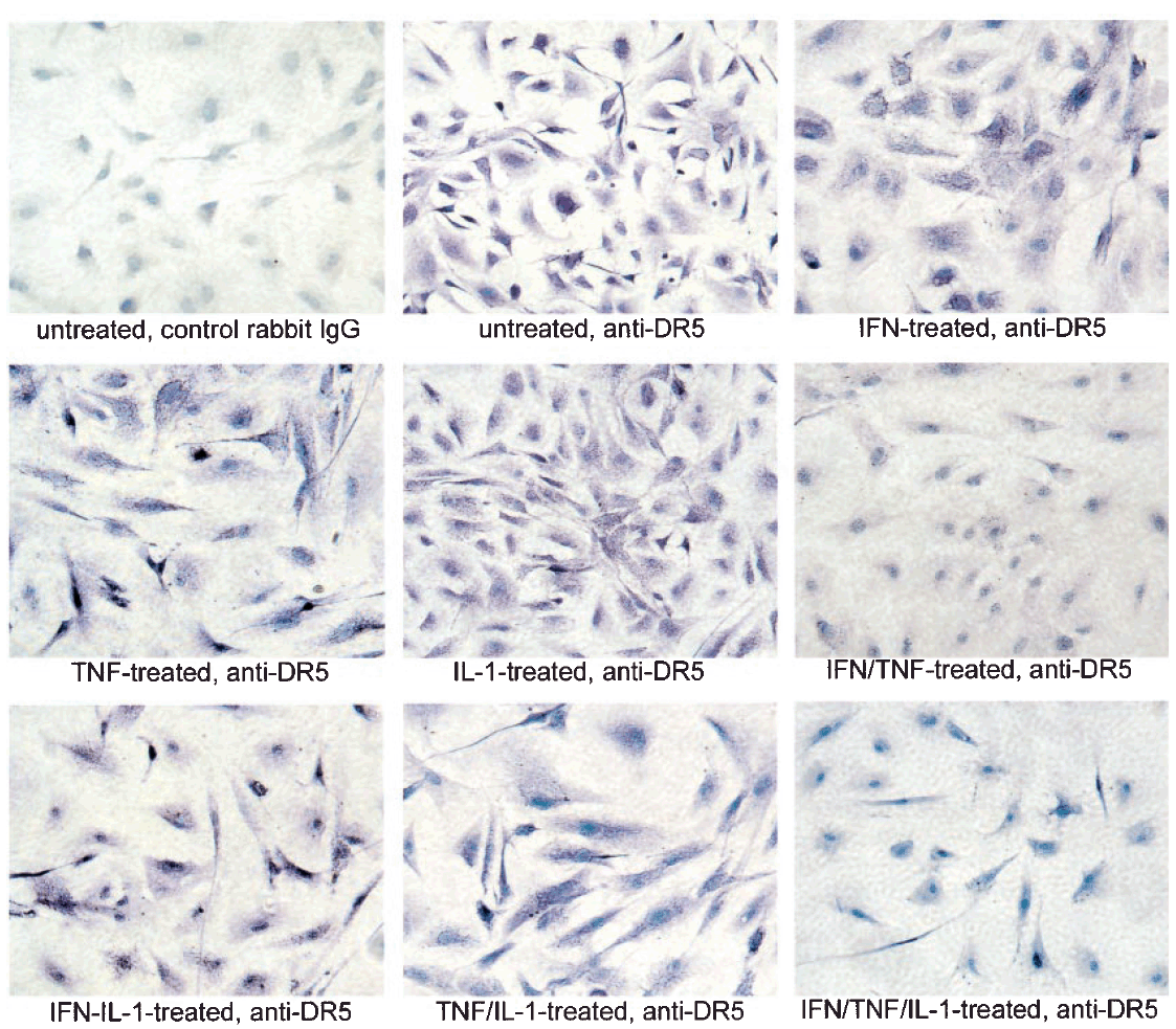
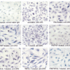
Description Regulated Expression Validation of DR5 in Thyroid Epithelial Cells (Bretz et al., 2002)
Immunostaining with anti-DR5 antibodies shows high levels of DR5 expression in untreated cells and cells treated with each of the three cytokines alone or TNFalpha combined with IL-1b. In contrast, treatment with both IFNg and TNFalpha or all three cytokines greatly reduces DR5 staining. The reduction in staining appears most significant in cytoplasmic regions while some staining is maintained in or around the nucleus.
Immunostaining with anti-DR5 antibodies shows high levels of DR5 expression in untreated cells and cells treated with each of the three cytokines alone or TNFalpha combined with IL-1b. In contrast, treatment with both IFNg and TNFalpha or all three cytokines greatly reduces DR5 staining. The reduction in staining appears most significant in cytoplasmic regions while some staining is maintained in or around the nucleus.
Handling
Storage This antibody is stable for at least one (1) year at -20°C. Avoid multiple freeze-thaw cycles.
Dilution Instructions Dilute in PBS or medium which is identical to that used in the assay system.
Application Instructions Immunoblotting : use at 2ug/mL.
Immunocytochemistry: use at 5ug/mL.
Positive control: Whole cell lysate from HeLa cells or K562 cells.
These are recommended concentrations.
Enduser should determine optimal concentrations for their applications.
Immunocytochemistry: use at 5ug/mL.
Positive control: Whole cell lysate from HeLa cells or K562 cells.
These are recommended concentrations.
Enduser should determine optimal concentrations for their applications.
References & Data Sheet
Data Sheet  Download PDF Data Sheet
 Download PDF Data Sheet

