Anti-DR4 (CT) Antibody (2404)
$445.00
SKU: 2404
Categories: Antibody Products, Apoptosis Antibodies, Products
Overview
Product Name Anti-DR4 (CT) Antibody (2404)
Description Anti-DR4 (CT) Rabbit Polyclonal Antibody
Target DR4 (CT)
Species Reactivity Human
Applications ELISA,WB,IHC-P,ICC,IF
Host Rabbit
Clonality Polyclonal
Isotype IgG
Immunogen Peptide corresponding to aa 427-445 at the C-terminus of human DR4 (accession no. ACC51226).
Properties
Form Liquid
Concentration Lot Specific
Formulation PBS, pH 7.4.
Buffer Formulation Phosphate Buffered Saline
Buffer pH pH 7.4
Format Purified
Purification Purified by peptide immuno-affinity chromatography
Specificity Information
Specificity This antibody recognizes full-length human DR4 (57kDa).
Target Name Tumor necrosis factor receptor superfamily member 10A
Target ID DR4 (CT)
Uniprot ID O00220
Alternative Names Death receptor 4, TNF-related apoptosis-inducing ligand receptor 1, TRAIL receptor 1, TRAIL-R1, CD antigen CD261
Gene Name TNFRSF10A
Gene ID 8797
Accession Number NP_003835.3
Sequence Location Cell membrane, Membrane raft, Cytoplasm, cytosol
Biological Function Receptor for the cytotoxic ligand TNFSF10/TRAIL (PubMed:26457518). The adapter molecule FADD recruits caspase-8 to the activated receptor. The resulting death-inducing signaling complex (DISC) performs caspase-8 proteolytic activation which initiates the subsequent cascade of caspases (aspartate-specific cysteine proteases) mediating apoptosis (PubMed:19090789). Promotes the activation of NF-kappa-B (PubMed:9430227). {PubMed:19090789, PubMed:9430227}.
Research Areas Apoptosis
Background Apoptosis is induced by certain cytokines including TNF and Fas ligand of the TNF family through their death domain-containing receptors, TNFR1 and Fas. Another cell death receptor was recently identified and designated DR4. The ligand for DR4 has been designated TRAIL and is a new member of the TNF family. Therefore, DR4 is also called TRAIL receptor-1. DR4 is expressed in most human tissues including spleen, peripheral blood, small intestine, and thymus. Like TNFR1, Fas and DR3, DR4 mediates TRAIL-induced apoptosis and NF- B activation in many tissues and cells.
Application Images


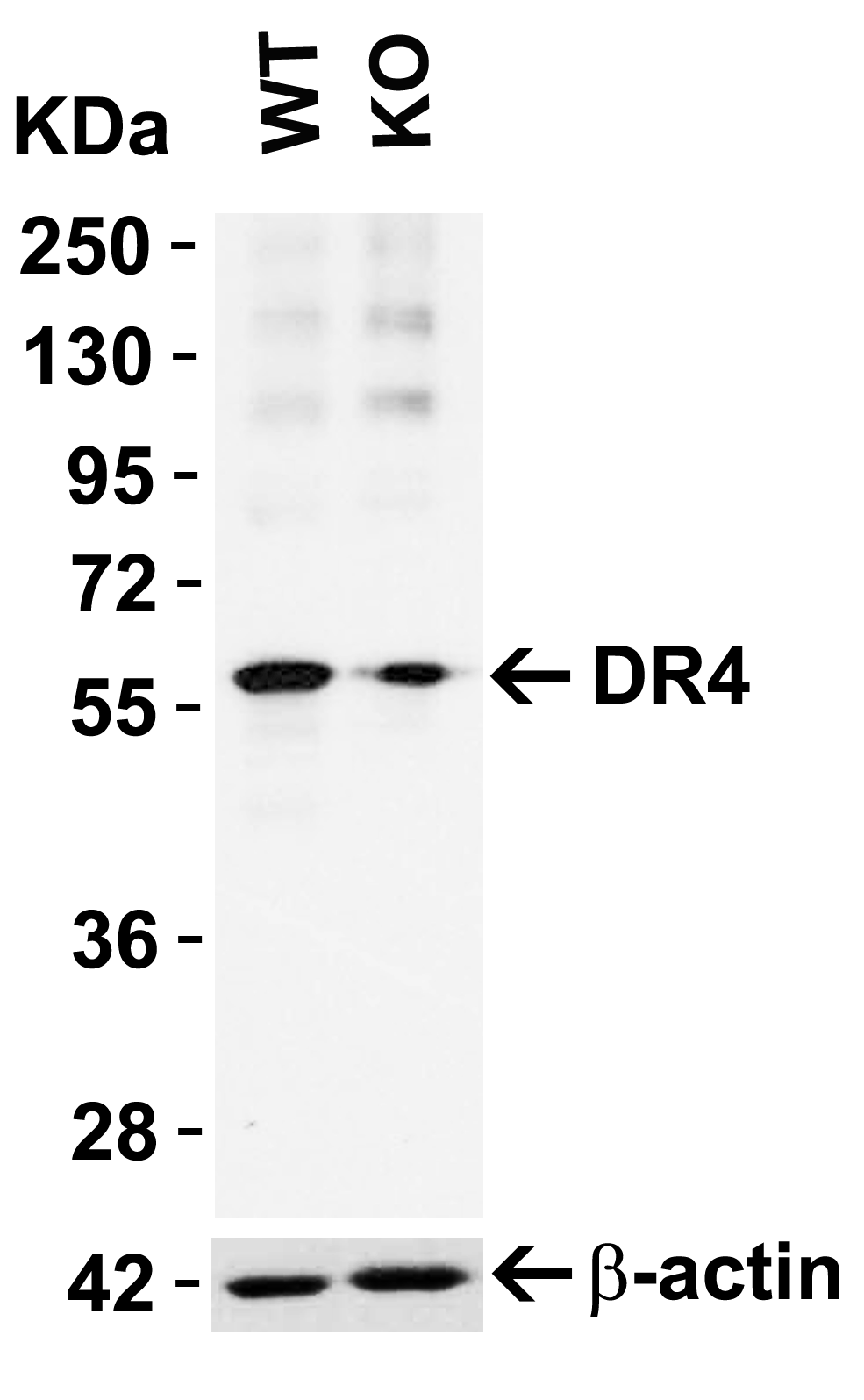
Description KO Validation in HeLa Cells
Loading: 10 ug of HeLa WT cell lysates or DR4 KO cell lysates. Antibodies: DR4 2404 (1 ug/mL) and beta-actin 3779 (1 ug/mL), 1 h incubation at RT in 5% NFDM/TBST.Secondary: Goat Anti-Rabbit IgG HRP conjugate at 1:10000 dilution.
Loading: 10 ug of HeLa WT cell lysates or DR4 KO cell lysates. Antibodies: DR4 2404 (1 ug/mL) and beta-actin 3779 (1 ug/mL), 1 h incubation at RT in 5% NFDM/TBST.Secondary: Goat Anti-Rabbit IgG HRP conjugate at 1:10000 dilution.

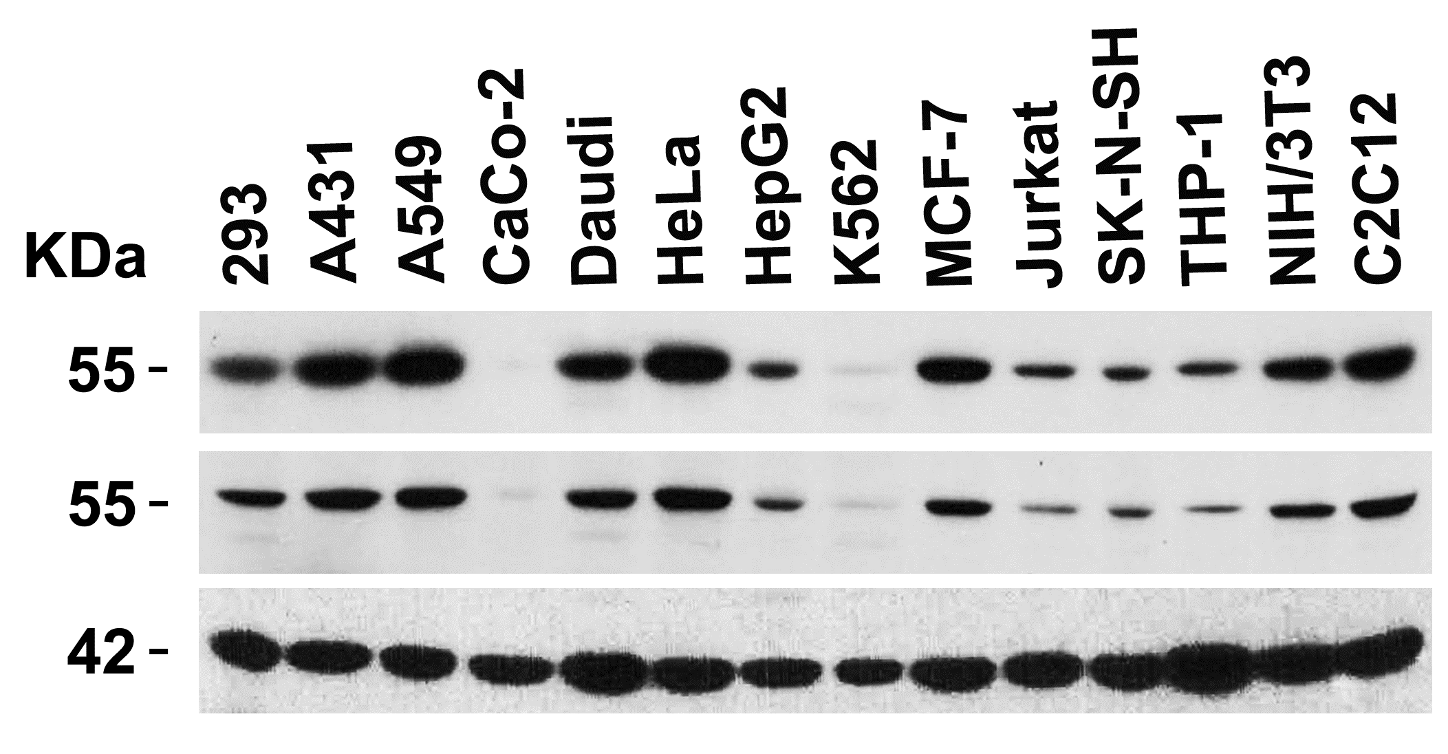
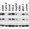
Description Independent Antibody Validation (IAV) via Protein Expression Profile in Cell Lines
Loading: 15 ug of lysates per lane. Antibodies: DR4 2404 (1 ug/mL), DR4 1167 ( 4 ug/mL), and beta-actin (1 ug/mL), 1h incubation at RT in 5% NFDM/TBST.Secondary: Goat anti-rabbit IgG HRP conjugate at 1:10000 dilution.
Loading: 15 ug of lysates per lane. Antibodies: DR4 2404 (1 ug/mL), DR4 1167 ( 4 ug/mL), and beta-actin (1 ug/mL), 1h incubation at RT in 5% NFDM/TBST.Secondary: Goat anti-rabbit IgG HRP conjugate at 1:10000 dilution.

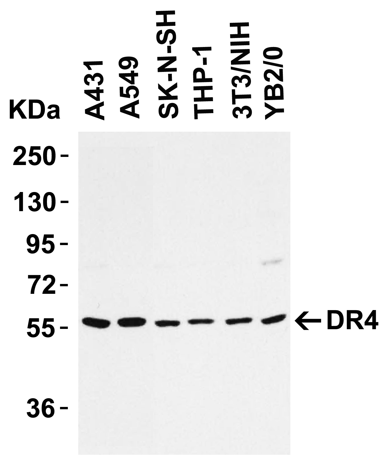
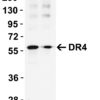
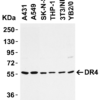
Description Western Blot Validation in Cell Lines
Loading: 15 ug of cell lysates per lane. Antibodies: DR4 2404 (1 ug/mL), 1h incubation at RT in 5% NFDM/TBST.Secondary: Goat anti-rabbit IgG HRP conjugate at 1:10000 dilution.
Loading: 15 ug of cell lysates per lane. Antibodies: DR4 2404 (1 ug/mL), 1h incubation at RT in 5% NFDM/TBST.Secondary: Goat anti-rabbit IgG HRP conjugate at 1:10000 dilution.

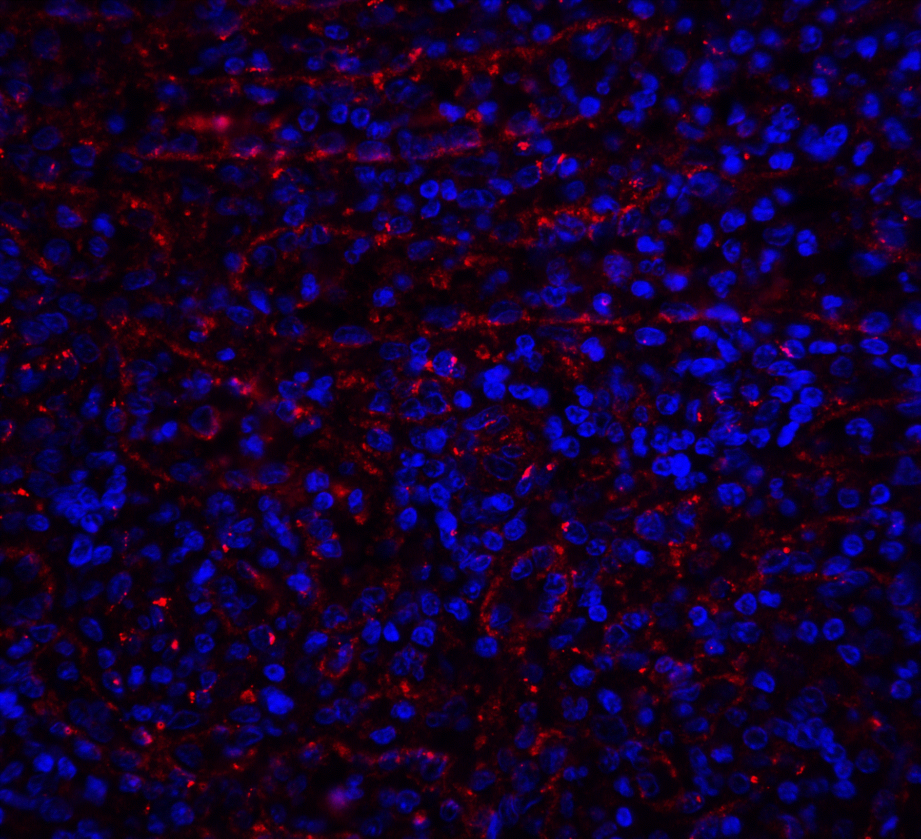
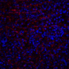
Description Immunofluorescence Validation of DR4
Immunofluorescent analysis of 4% paraformaldehyde-fixed human spleen tissue labeling DR4 with 2404 at 20 ug/mL, followed by goat anti-rabbit IgG secondary antibody at 1/500 dilution (red) and DAPI staining (blue). Image showing membrane staining on human spleen cells.
Immunofluorescent analysis of 4% paraformaldehyde-fixed human spleen tissue labeling DR4 with 2404 at 20 ug/mL, followed by goat anti-rabbit IgG secondary antibody at 1/500 dilution (red) and DAPI staining (blue). Image showing membrane staining on human spleen cells.

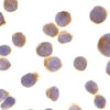
Description Immunocytochemistry Validation of DR4 in HeLa Cells
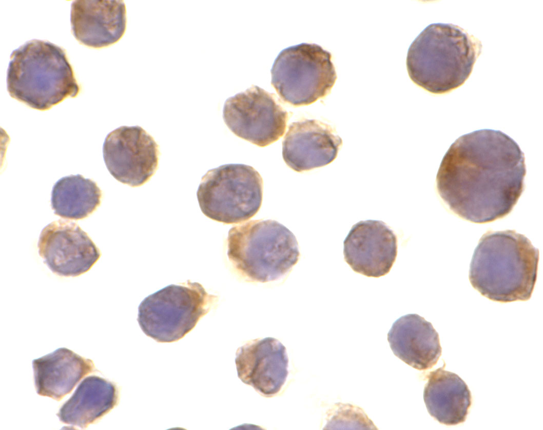
Immunocytochemical analysis of HeLa cells using anti-DR4 antibody (2404) at 10 ug/ml. Cells was fixed with formaldehyde and blocked with 10% serum for 1 h at RT; antigen retrieval was by heat mediation with a citrate buffer (pH6). Samples were incubated with primary antibody overnight at 4 C. A goat anti-rabbit IgG H&L (HRP) at 1/250 was used as secondary. Counter stained with Hematoxylin
Immunocytochemical analysis of HeLa cells using anti-DR4 antibody (2404) at 10 ug/ml. Cells was fixed with formaldehyde and blocked with 10% serum for 1 h at RT; antigen retrieval was by heat mediation with a citrate buffer (pH6). Samples were incubated with primary antibody overnight at 4 C. A goat anti-rabbit IgG H&L (HRP) at 1/250 was used as secondary. Counter stained with Hematoxylin

Description Immunohistochemistry Validation of DR4
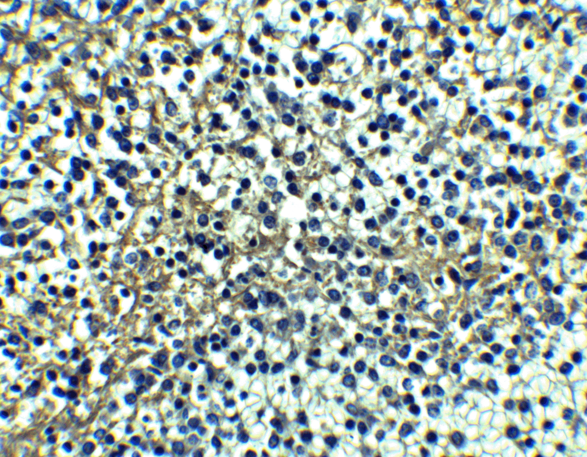
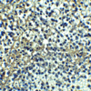
Immunohistochemical analysis of paraffin-embedded human spleen tissue using anti-DR4 antibody (2404) at 10 ug/ml. Tissue was fixed with formaldehyde and blocked with 10% serum for 1 h at RT; antigen retrieval was by heat mediation with a citrate buffer (pH6). Samples were incubated with primary antibody overnight at 4uC. A goat anti-rabbit IgG H&L (HRP) at 1/250 was used as secondary. Counter stained with Hematoxylin.
Immunohistochemical analysis of paraffin-embedded human spleen tissue using anti-DR4 antibody (2404) at 10 ug/ml. Tissue was fixed with formaldehyde and blocked with 10% serum for 1 h at RT; antigen retrieval was by heat mediation with a citrate buffer (pH6). Samples were incubated with primary antibody overnight at 4uC. A goat anti-rabbit IgG H&L (HRP) at 1/250 was used as secondary. Counter stained with Hematoxylin.

Description KD Validation in SW480 cells (Goda et al., 2008)
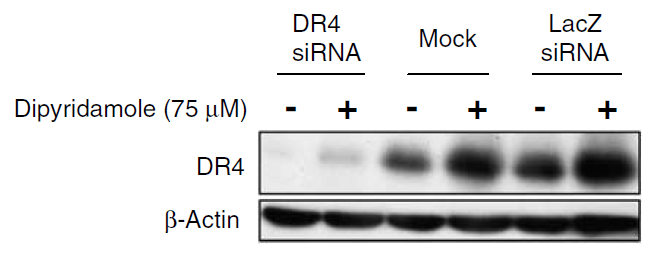
The expression of DR4 was knocked down via DR4 siRNA, 24 h latercells were treated with dipyridamole for 24 h. DR4 protein expression detected by anti-DR4 antibodies (2404) was disrupted. Dipyridamole up-regulated the expression of DR4.
The expression of DR4 was knocked down via DR4 siRNA, 24 h latercells were treated with dipyridamole for 24 h. DR4 protein expression detected by anti-DR4 antibodies (2404) was disrupted. Dipyridamole up-regulated the expression of DR4.

Description Immunofluorescence Validation of DR4 in rat brain (Cantarella et al., 2014)
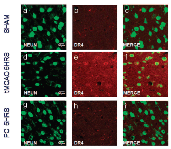
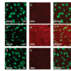
DR4 protein expression detected by anti-DR4 antibodies (2404) was increased after transient brain ischemia (tMCAO) and decreased after pre-conditioning stimulus. Confocal microscopic images displaying NeuN (a,d, g) (green), DR4 (b, e, h) (red), and Merge (c, f, i) (yellow) in the brain peri-ischemic region of rats after 5 h.
DR4 protein expression detected by anti-DR4 antibodies (2404) was increased after transient brain ischemia (tMCAO) and decreased after pre-conditioning stimulus. Confocal microscopic images displaying NeuN (a,d, g) (green), DR4 (b, e, h) (red), and Merge (c, f, i) (yellow) in the brain peri-ischemic region of rats after 5 h.

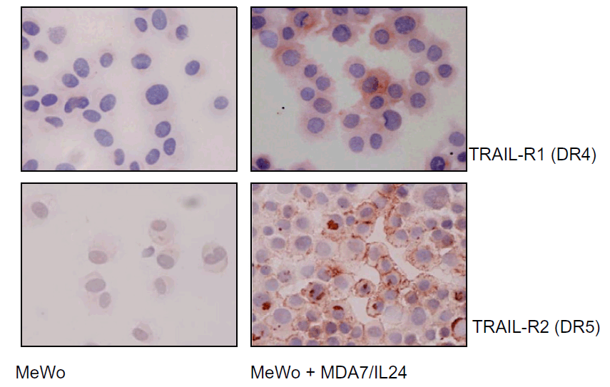
Description Immunocytochemistry Validation of DR4 in human melanoma cells (Ekmekcioglu et al., 2008)
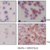
MeWo melanoma cells were exposed to affinity-purified MDA7/IL-24. After 48 h of treatment, cells were collected and cytospins prepared for cytochemical assessment of their TRAIL receptor (R1 and R2) expression (anti-DR4 (2404) or anti-DR5, AEC, hematoxylin). Both DR4 and DR5 expression were upregulated in MeWo cells after treatment.
MeWo melanoma cells were exposed to affinity-purified MDA7/IL-24. After 48 h of treatment, cells were collected and cytospins prepared for cytochemical assessment of their TRAIL receptor (R1 and R2) expression (anti-DR4 (2404) or anti-DR5, AEC, hematoxylin). Both DR4 and DR5 expression were upregulated in MeWo cells after treatment.

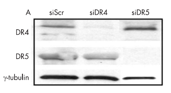
Description KD Validation in Huh7 cells (Malhi et al., 2416)
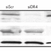
Western blot analysis with anti-DR4 antibodies (2404) was performed for DR5 and DR4 expression using whole cell lysates from Huh 7 cells transfected with respective siRNAs. In cells treated with siDR4, a decrease in DR4 level was observed, DR5 levels were unchanged. Scrambled siRNA was used as control.
Western blot analysis with anti-DR4 antibodies (2404) was performed for DR5 and DR4 expression using whole cell lysates from Huh 7 cells transfected with respective siRNAs. In cells treated with siDR4, a decrease in DR4 level was observed, DR5 levels were unchanged. Scrambled siRNA was used as control.

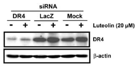
Description KD Validation in HeLa cells (Horinaka et al., 2005)
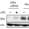
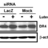
HeLa cells were transfected with DR4siRNA or LacZ control siRNA. At 24 h after transfection, the cells were treated with or without 20 uM luteolin for 24 h. Western blot analysis was carried out with anti-DR4 antibodies (2404). DR4 expression was markedly reduced after DR4 knockdown.
HeLa cells were transfected with DR4siRNA or LacZ control siRNA. At 24 h after transfection, the cells were treated with or without 20 uM luteolin for 24 h. Western blot analysis was carried out with anti-DR4 antibodies (2404). DR4 expression was markedly reduced after DR4 knockdown.
Handling
Storage This antibody is stable for at least one (1) year at -20°C. Avoid multiple freeze-thaw cycles.
Dilution Instructions Dilute in PBS or medium which is identical to that used in the assay system.
Application Instructions Immunoblotting : use at 1ug/mL.
Immunocytochemistry: use at 10ug/mL.
Positive control: Whole cell lysate from HeLa cells.
These are recommended concentrations.
Enduser should determine optimal concentrations for their applications.
Immunocytochemistry: use at 10ug/mL.
Positive control: Whole cell lysate from HeLa cells.
These are recommended concentrations.
Enduser should determine optimal concentrations for their applications.
References & Data Sheet
Data Sheet  Download PDF Data Sheet
 Download PDF Data Sheet

