Anti-Dicer Antibody (12532)
$466.00
SKU: 12532
Categories: Antibody Products, DNA/RNA/Oxidative Stress Antibodies, Products
Overview
Product Name Anti-Dicer Antibody (12532)
Description Anti-Dicer Mouse Monoclonal Antibody
Target Dicer
Species Reactivity Human, Mouse, Rat
Applications WB,ICC/IF
Host Mouse
Clonality Monoclonal
Clone ID S167-7
Isotype IgG1
Immunogen Fusion protein corresponding to aa 1638-1899 of mouse Endoribonuclease Dicer (accession no.Q8R418).
Properties
Form Liquid
Concentration 1.0 mg/mL
Formulation PBS, pH 7.4, 50% glycerol, 0.09% sodium azide.Purified by Protein G affinity chromatography.
Buffer Formulation Phosphate Buffered Saline
Buffer pH pH 7.4
Buffer Anti-Microbial 0.09% Sodium Azide
Buffer Cryopreservative 50% Glycerol
Format Purified
Purification Purified by Protein G affinity chromatography
Specificity Information
Specificity This antibody recognizes human, mouse and rat Dicer.
Target Name Endoribonuclease Dicer
Target ID Dicer
Uniprot ID Q8R418
Alternative Names EC 3.1.26.3, Double-strand-specific ribonuclease mDCR-1
Gene Name Dicer1
Accession Number NP_683750.2
Sequence Location Cytoplasm
Biological Function Double-stranded RNA (dsRNA) endoribonuclease playing a central role in short dsRNA-mediated post-transcriptional gene silencing. Cleaves naturally occurring long dsRNAs and short hairpin pre-microRNAs (miRNA) into fragments of twenty-one to twenty-three nucleotides with 3' overhang of two nucleotides, producing respectively short interfering RNAs (siRNA) and mature microRNAs. SiRNAs and miRNAs serve as guide to direct the RNA-induced silencing complex (RISC) to complementary RNAs to degrade them or prevent their translation. Gene silencing mediated by siRNAs, also called RNA interference, controls the elimination of transcripts from mobile and repetitive DNA elements of the genome but also the degradation of exogenous RNA of viral origin for instance. The miRNA pathway on the other side is a mean to specifically regulate the expression of target genes (By similarity). {UniProtKB:Q9UPY3}.; [Isoform 2]: More active than isoform 1 to process long double-stranded RNA into siRNAs. Responsible for the accumulation of endogenous siRNAs observed in mouse oocytes compared to somatic cells and it regulates meiotic spindle organization in female germline.
Research Areas DNA
Background Dicer is a member of the RNase III family that specifically cleaves double-stranded RNAs to generate microRNAs (miRNAs). After long primary transcript pri-miRNAs are processed to stem-looped pre- miRNAs by Drosha, pre-miRNAs are transported to the cytoplasm and further processed by Dicer to produce 22- nucleotide mature miRNAs . The mature miRNA then becomes a part of the RNA- Induced Silencing Complex (RISC) and can bind to the 3′ UTR of the target mRNA.
Application Images


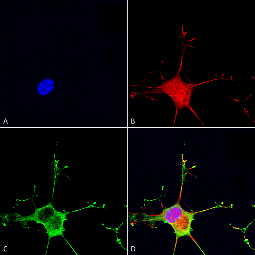
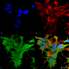
Description Immunocytochemistry/Immunofluorescence analysis using Mouse Anti-Dicer Monoclonal Antibody, Clone S167-7 (12532). Tissue: Neuroblastoma cells (SH-SY5Y). Species: Human. Fixation: 4% PFA for 15 min. Primary Antibody: Mouse Anti-Dicer Monoclonal Antibody (12532) at 1:50 for overnight at 4°C with slow rocking. Secondary Antibody: AlexaFluor 488 at 1:1000 for 1 hour at RT. Counterstain: Phalloidin-iFluor 647 (red) F-Actin stain; Hoechst (blue) nuclear stain at 1:800, 1.6mM for 20 min at RT. (A) Hoechst (blue) nuclear stain. (B) Phalloidin-iFluor 647 (red) F-Actin stain. (C) Dicer Antibody (D) Composite.

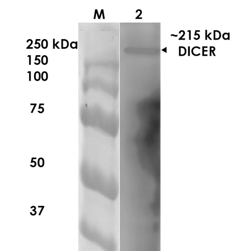
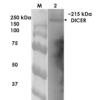
Description Western Blot analysis of Rat Brain Membrane showing detection of ~215 kDa Dicer protein using Mouse Anti-Dicer Monoclonal Antibody, Clone S167-7 (12532). Lane 1: MW Ladder. Lane 2: Rat Brain Membrane. Load: 10 µg. Block: 5% milk. Primary Antibody: Mouse Anti-Dicer Monoclonal Antibody (12532) at 1:1000 for 1 hour at RT. Secondary Antibody: Goat Anti-Mouse IgG: HRP at 1:200 for 1 hour at RT. Color Development: TMB solution for 10 min at RT. Predicted/Observed Size: ~215 kDa.

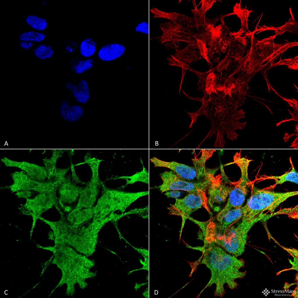
Description Immunocytochemistry/Immunofluorescence analysis using Mouse Anti-Dicer Monoclonal Antibody, Clone S167-7 (12532). Tissue: Neuroblastoma cell line (SK-N-BE). Species: Human. Fixation: 4% Formaldehyde for 15 min at RT. Primary Antibody: Mouse Anti-Dicer Monoclonal Antibody (12532) at 1:100 for 60 min at RT. Secondary Antibody: Goat Anti-Mouse ATTO 488 at 1:100 for 60 min at RT. Counterstain: Phalloidin Texas Red F-Actin stain; DAPI (blue) nuclear stain at 1:1000; 1:5000 for 60 min RT, 5 min RT. Localization: Cytoplasm. Magnification: 60X. (A) DAPI (blue) nuclear stain. (B) Phalloidin Texas Red F-Actin stain. (C) Dicer Antibody. (D) Composite.
Handling
Storage This antibody is stable for at least one (1) year at -20°C.
Dilution Instructions Dilute in PBS or medium which is identical to that used in the assay system.
Application Instructions Immunoblotting: use at 1ug/mL. Predicted molecular weight is ~215kDa.
Positive control: rat brain lysate.
These are recommended concentrations.
Endusers should determine optimal concentrations for their applications.
Positive control: rat brain lysate.
These are recommended concentrations.
Endusers should determine optimal concentrations for their applications.
References & Data Sheet
Data Sheet  Download PDF Data Sheet
 Download PDF Data Sheet

