Anti-DEDAF Antibody (2227)
$445.00
| Host | Quantity | Applications | Species Reactivity | Data Sheet | |
|---|---|---|---|---|---|
| Rabbit | 100ug | ELISA,WB,IHC-P,IF | Human, Mouse, Rat |  |
SKU: 2227
Categories: Antibody Products, Apoptosis Antibodies, Products
Overview
Product Name Anti-DEDAF Antibody (2227)
Description Anti-DEDAF Rabbit Polyclonal Antibody
Target DEDAF
Species Reactivity Human, Mouse, Rat
Applications ELISA,WB,IHC-P,IF
Host Rabbit
Clonality Polyclonal
Isotype IgG
Immunogen Peptide corresponding to aa 215-229 of human DEDAF (accession no. AF179286). This sequence is identical to that of mouse.
Properties
Form Liquid
Concentration Lot Specific
Formulation PBS, pH 7.4.
Buffer Formulation Phosphate Buffered Saline
Buffer pH pH 7.4
Format Purified
Purification Purified by peptide immuno-affinity chromatography
Specificity Information
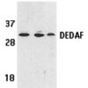
Specificity This antibody recognizes human, mouse, and rat DEDAF (32kDa).
Target Name RING1 and YY1-binding protein
Target ID DEDAF
Uniprot ID Q8N488
Alternative Names Apoptin-associating protein 1, APAP-1, Death effector domain-associated factor, DED-associated factor, YY1 and E4TF1-associated factor 1
Gene Name RYBP
Gene ID 23429
Accession Number NP_036366
Sequence Location Nucleus, Cytoplasm, Nucleus, nucleoplasm
Biological Function Component of a Polycomb group (PcG) multiprotein PRC1-like complex, a complex class required to maintain the transcriptionally repressive state of many genes, including Hox genes, throughout development. PcG PRC1-like complex acts via chromatin remodeling and modification of histones; it mediates monoubiquitination of histone H2A 'Lys-119', rendering chromatin heritably changed in its expressibility (PubMed:25519132). Component of a PRC1-like complex that mediates monoubiquitination of histone H2A 'Lys-119' on the X chromosome and is required for normal silencing of one copy of the X chromosome in XX females. May stimulate ubiquitination of histone H2A 'Lys-119' by recruiting the complex to target sites (By similarity). Inhibits ubiquitination and subsequent degradation of TP53, and thereby plays a role in regulating transcription of TP53 target genes (PubMed:19098711). May also regulate the ubiquitin-mediated proteasomal degradation of other proteins like FANK1 to regulate apoptosis (PubMed:14765135, PubMed:27060496). May be implicated in the regulation of the transcription as a repressor of the transcriptional activity of E4TF1 (PubMed:11953439). May bind to DNA (By similarity). May play a role in the repression of tumor growth and metastasis in breast cancer by down-regulating SRRM3 (PubMed:27748911). {UniProtKB:Q8CCI5, PubMed:11953439, PubMed:14765135, PubMed:19098711, PubMed:27060496, PubMed:27748911}.
Research Areas Apoptosis
Background Cell death signals are transduced by death domain-containing adapter molecules and members of the caspase family of proteases. A novel protein that interacts with the death effector domain of capase-8 and caspase-10 has been designated DEDAF and is dedentical to the transcriptional repressor RYBP. DEDAF is expressed in multiple tissues and cell lines and appears to be involved in the regulation of both cytoplasmic and nuclear events of apoptosis.
Application Images


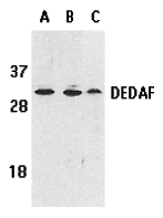
Description Western blot analysis of DEDAF expression in human A549 (lane A), HepG2 (lane B), and mouse 3T3 (lane C) cell lysates with DEDAF antibody at 1 ug/ml.

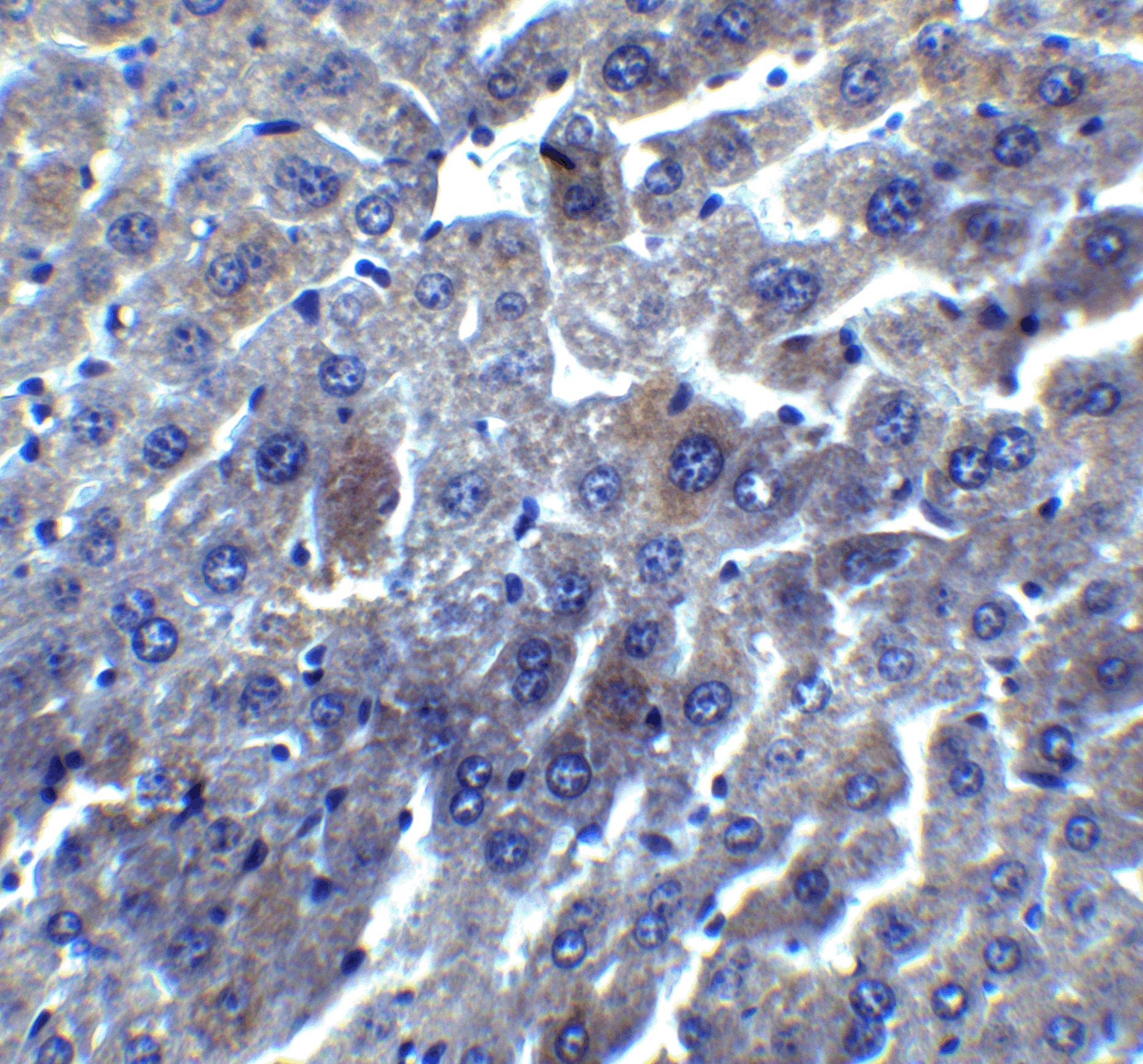
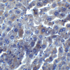
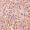
Description Immunohistochemistry of DEDAF in mouse liver tissue with DEDAF antibody at 5 ug/mL.

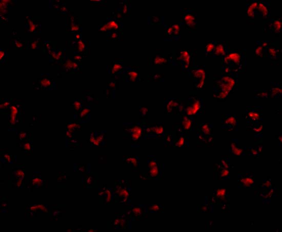
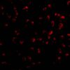
Description Immunofluorescence of DEDAF in A549 cells with DEDAF antibody at 20 ug/mL.

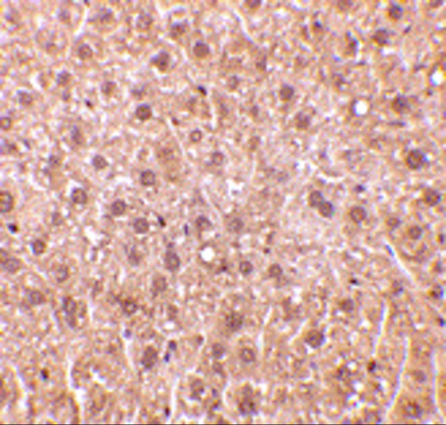
Description Immunohistochemistry of DEDAF in mouse liver tissue with DEDAF antibody at 10 ug/mL.

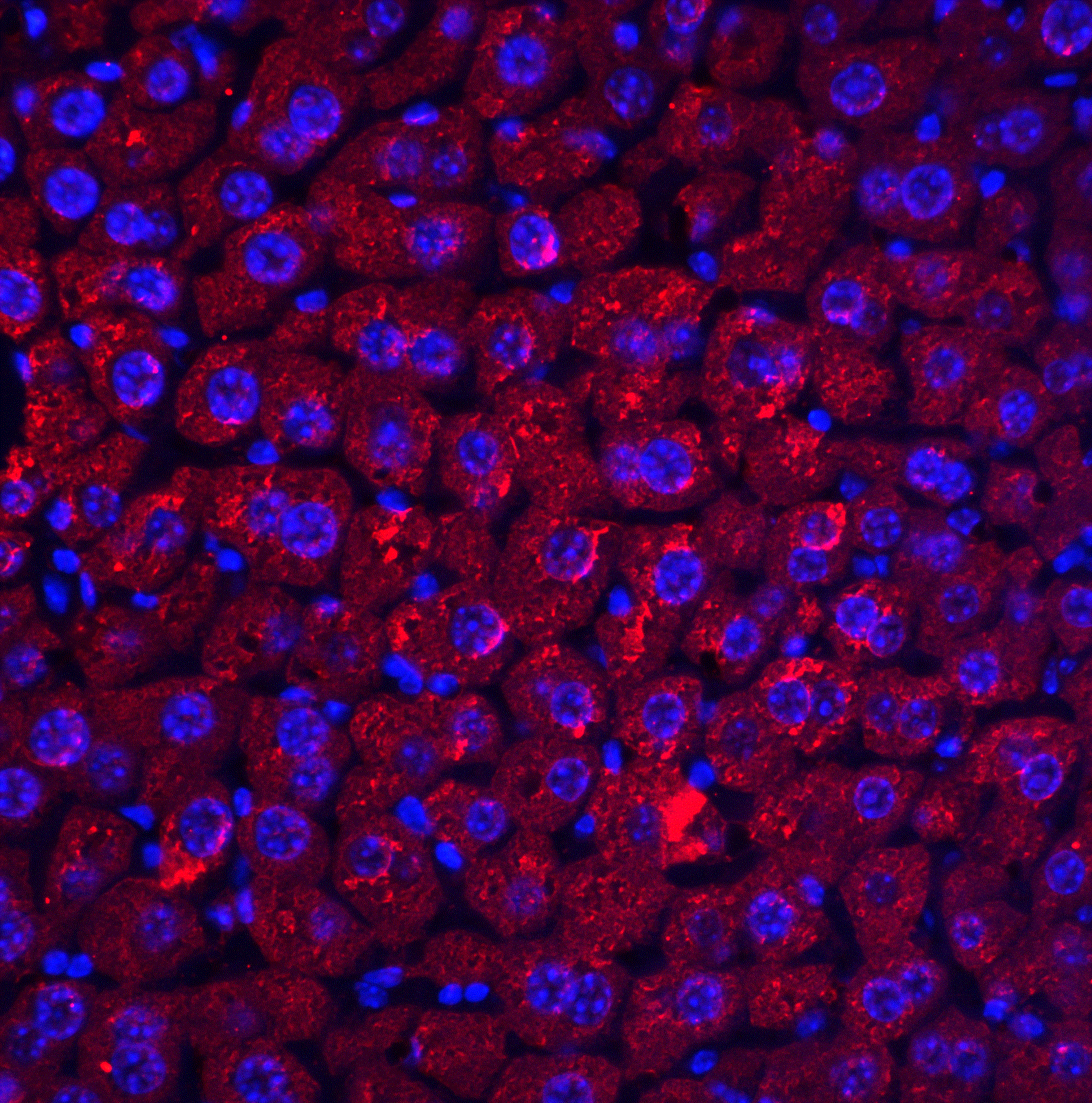
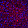
Description Immunofluorescence of DEDAF in mouse liver tissue with DEDAF antibody at 20 ug/ml.
Handling
Storage This antibody is stable for at least one (1) year at -20°C. Avoid multiple freeze-thaw cycles.
Dilution Instructions Dilute in PBS or medium which is identical to that used in the assay system.
Application Instructions Immunoblotting: use at 1ug/mL.
Immunohistochemistry: use at 10ug/mL.
These are recommended concentrations.
Enduser should determine optimal concentrations for their applications.
Positive control: Whole cell lysate from A549 or HepG2 cells.
Immunohistochemistry: use at 10ug/mL.
These are recommended concentrations.
Enduser should determine optimal concentrations for their applications.
Positive control: Whole cell lysate from A549 or HepG2 cells.
References & Data Sheet
Data Sheet  Download PDF Data Sheet
 Download PDF Data Sheet

