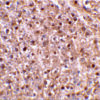
Anti-Caspase-12 (IN) Antibody (2327)
$445.00
| Host | Quantity | Applications | Species Reactivity | Data Sheet | |
|---|---|---|---|---|---|
| Rabbit | 100ug | ELISA,WB,IHC-P,IF | Human, Mouse, Rat |  |
SKU: 2327
Categories: Antibody Products, Apoptosis Antibodies, Products
Overview
Product Name Anti-Caspase-12 (IN) Antibody (2327)
Description Anti-Caspase-12 (IN) Rabbit Polyclonal Antibody
Target Caspase-12 (IN)
Species Reactivity Human, Mouse, Rat
Applications ELISA,WB,IHC-P,IF
Host Rabbit
Clonality Polyclonal
Isotype IgG
Immunogen Peptide corresponding to aa 100-116 of mouse caspase-12 (accession no. CAA73532).
Properties
Form Liquid
Concentration Lot Specific
Formulation PBS, pH 7.4.
Buffer Formulation Phosphate Buffered Saline
Buffer pH pH 7.4
Format Purified
Purification Purified by peptide immuno-affinity chromatography
Specificity Information
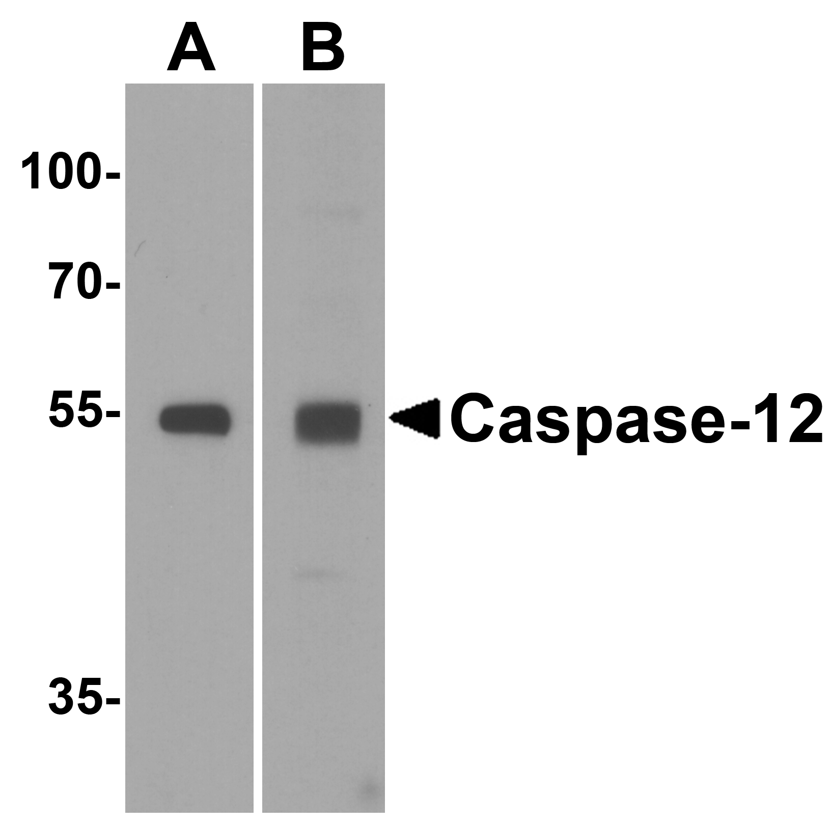
Specificity This antibody recognizes human, mouse, and rat caspase-12 (approx. 53kDa).
Target Name Caspase-12
Target ID Caspase-12 (IN)
Uniprot ID O08736
Alternative Names CASP-12, EC 3.4.22.-
Gene Name Casp12
Gene ID 12364
Accession Number NP_033938.3
Biological Function Involved in the activation cascade of caspases responsible for apoptosis execution. {ECO:0000250}.
Research Areas Apoptosis
Background Three distinct signaling pathways lead to programmed cell death. The death receptor and mitochondrial pathways are the main pathways in which the proteases caspase-8 and caspase- 9, respectively, are involved. Endoplasmic reticulum (ER) stress is the third pathway, and caspase-12 is involved. Caspase-12, localized in the ER, is activated by ER stress and mediates ER stress-induced apoptosis. Several proteins that are associated with Alzheimer's disease, including -secretase, presenilin, and – amyloid precursor protein (APP), co-localize in the ER with caspase-12. Caspase-12 is ubiquitously expressed in mouse tissues.
Application Images


Description Western blot analysis of Caspase-12 in (A) human spleen and (B) mouse spleen tissue lysate with Caspase-12 antibody at 1 ug/mL.

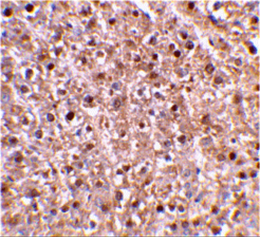
Description Immunohistochemical staining of mouse liver tissue using caspase-12 antibody at 2 ug/mL.

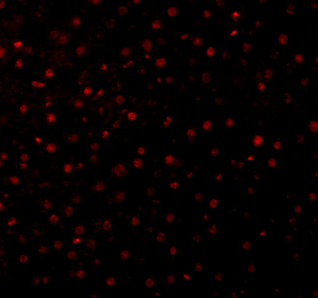
Description Immunofluorescence of Caspase-12 in Mouse Liver cells with Caspase-12 antibody at 10 ug/mL.
Handling
Storage This antibody is stable for at least one (1) year at -20°C. Avoid multiple freeze-thaw cycles.
Dilution Instructions Dilute in PBS or medium which is identical to that used in the assay system.
Application Instructions Immunoblotting: use at 1ug/mL.
Immunohistochemistry: use at 2ug/mL.
These are recommended concentrations.
Enduser should determine optimal concentrations for their applications.
Positive control: Tissue lysate of mouse brain.
Immunohistochemistry: use at 2ug/mL.
These are recommended concentrations.
Enduser should determine optimal concentrations for their applications.
Positive control: Tissue lysate of mouse brain.
References & Data Sheet
Data Sheet  Download PDF Data Sheet
 Download PDF Data Sheet

