Anti-BAFF (CT) Antibody (2221)
$445.00
| Host | Quantity | Applications | Species Reactivity | Data Sheet | |
|---|---|---|---|---|---|
| Rabbit | 100ug | ELISA,WB,ICC,IF,IHC | Human, Mouse, Rat |  |
SKU: 2221
Categories: Antibody Products, Apoptosis Antibodies, Products
Overview
Product Name Anti-BAFF (CT) Antibody (2221)
Description Anti-BAFF (CT) Rabbit Polyclonal Antibody
Target BAFF (CT)
Species Reactivity Human, Mouse, Rat
Applications ELISA,WB,ICC,IF,IHC
Host Rabbit
Clonality Polyclonal
Isotype IgG
Immunogen Peptide corresponding to aa 254-269 at the C-terminus of human BAFF.
Properties
Form Liquid
Concentration Lot Specific
Formulation PBS, pH 7.4.
Buffer Formulation Phosphate Buffered Saline
Buffer pH pH 7.4
Format Purified
Purification Purified by peptide immuno-affinity chromatography
Specificity Information
Specificity This antibody recognizes human, mouse, and rat BAFF (approx. 38kDa).
Target Name Tumor necrosis factor ligand superfamily member 13B
Target ID BAFF (CT)
Uniprot ID Q9Y275
Alternative Names B lymphocyte stimulator, BLyS, B-cell-activating factor, BAFF, Dendritic cell-derived TNF-like molecule, TNF- and APOL-related leukocyte expressed ligand 1, TALL-1, CD antigen CD257 [Cleaved into: Tumor necrosis factor ligand superfamily member 13b, membrane form; Tumor necrosis factor ligand superfamily member 13b, soluble form]
Gene Name TNFSF13B
Gene ID 10673
Accession Number NP_006564
Sequence Location Cell membrane; Single-pass type II membrane protein.; [Tumor necrosis factor ligand superfamily member 13b, soluble form]: Secreted.
Biological Function Cytokine that binds to TNFRSF13B/TACI and TNFRSF17/BCMA. TNFSF13/APRIL binds to the same 2 receptors. Together, they form a 2 ligands -2 receptors pathway involved in the stimulation of B- and T-cell function and the regulation of humoral immunity. A third B-cell specific BAFF-receptor (BAFFR/BR3) promotes the survival of mature B-cells and the B-cell response. {PubMed:10973284}.; Isoform 2 seems to inhibit isoform 1 secretion and bioactivity. {PubMed:10973284}.
Research Areas Apoptosis
Background A novel member of the TNF superfamily was recently identified and designated BAFF (B Cell Activating Factor of the TNF Family) or BlyS (B Lymphocyte Stimulator), TALL-1 (TNF- and ApoL-Related Leukocyte-Expressed Ligand), and THANK (TNF Homologue that Activates Apoptosis, NF- B and c-jun N-terminal Kinase). BAFF is designated a B cell stimulator since it induces B cell proliferation and immunoglobulin secretion. BAFF and its receptors are involved in the development of systemic lupus erythematosus and other B cell-associated autoimmune diseases.
Application Images


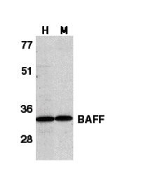
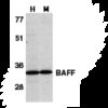
Description Western Blot Validation in Human HL60 Cell Lysate (H) and Mouse Spleen Lysate (M)
Loading: 15 ug of lysates per lane. Antibodies: BAFF 2221 (1 ug/mL), 1h incubation at RT in 5% NFDM/TBST.Secondary: Goat anti-rabbit IgG HRP conjugate at 1:10000 dilution.
Loading: 15 ug of lysates per lane. Antibodies: BAFF 2221 (1 ug/mL), 1h incubation at RT in 5% NFDM/TBST.Secondary: Goat anti-rabbit IgG HRP conjugate at 1:10000 dilution.

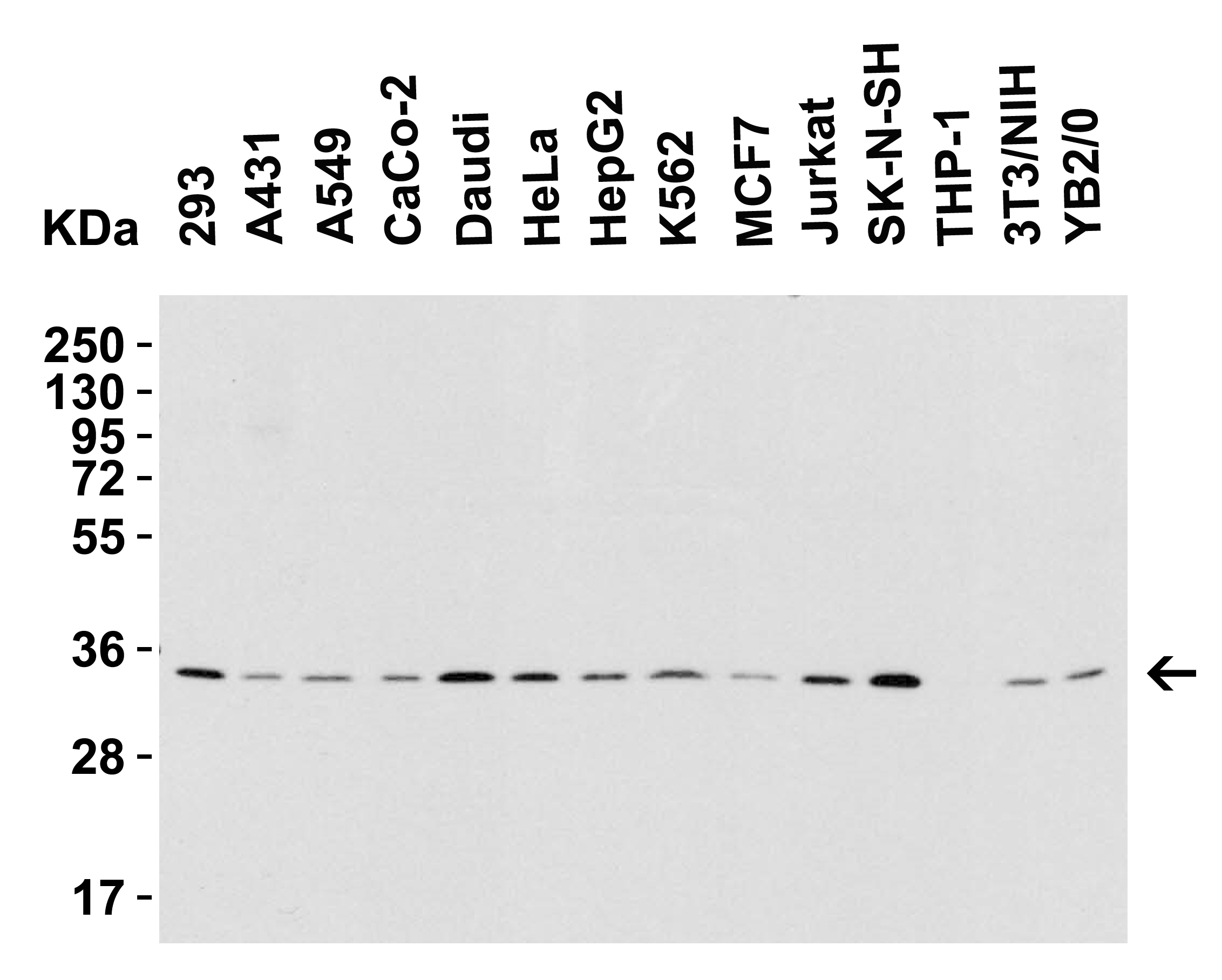
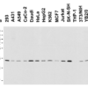
Description Western Blot Validation in Human, Mouse and Rat Cell Lines
Loading: 15 ug of lysates per lane. Antibodies: BAFF 2221 (1 ug/mL), 1h incubation at RT in 5% NFDM/TBST.Secondary: Goat anti-rabbit IgG HRP conjugate at 1:10000 dilution.
Loading: 15 ug of lysates per lane. Antibodies: BAFF 2221 (1 ug/mL), 1h incubation at RT in 5% NFDM/TBST.Secondary: Goat anti-rabbit IgG HRP conjugate at 1:10000 dilution.

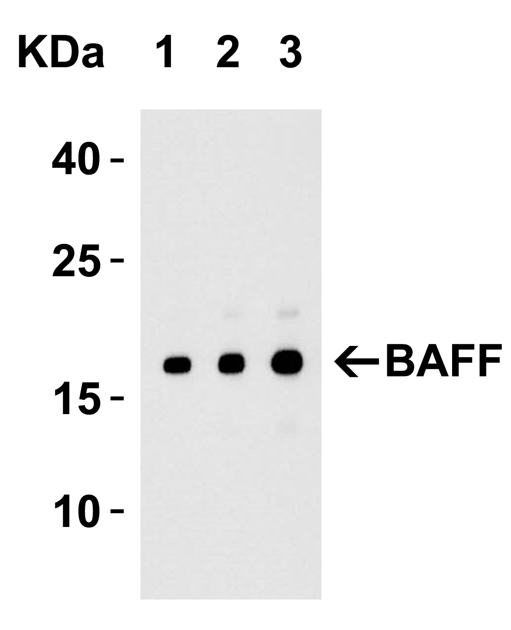
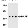
Description Western Blot Validation with Recombinant Protein
Loading: 30 ng of human BAFF recombinant protein per lane. Antibodies: BAFF 2221 (Lane 1: 0.25 ug/mL; Lane 2: 0.5 ug/mL and Lane 3: 1 ug/mL), 1h incubation at RT in 5% NFDM/TBST.Secondary: Goat anti-rabbit IgG HRP conjugate at 1:10000 dilution.Observed at around 18kD.
Loading: 30 ng of human BAFF recombinant protein per lane. Antibodies: BAFF 2221 (Lane 1: 0.25 ug/mL; Lane 2: 0.5 ug/mL and Lane 3: 1 ug/mL), 1h incubation at RT in 5% NFDM/TBST.Secondary: Goat anti-rabbit IgG HRP conjugate at 1:10000 dilution.Observed at around 18kD.

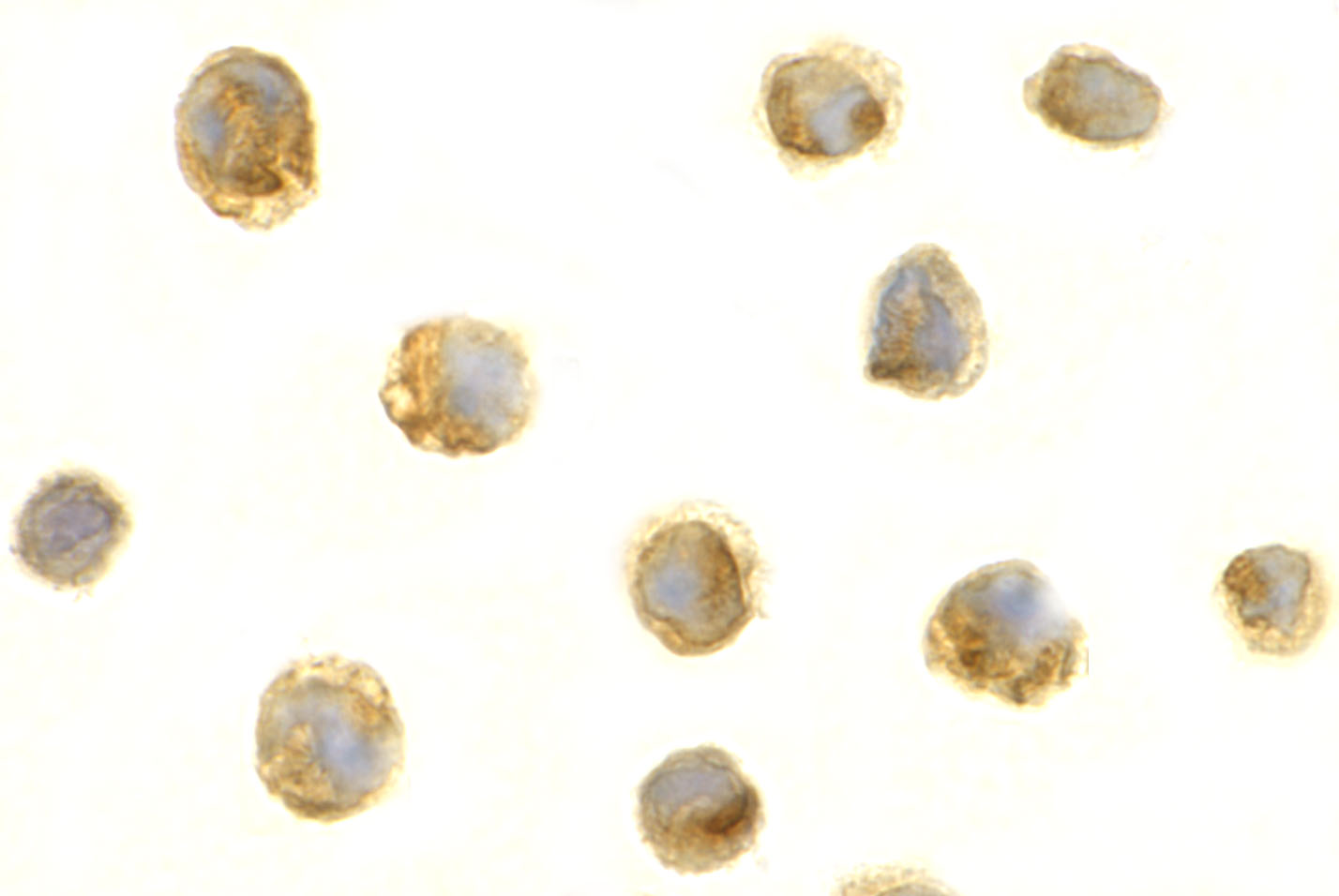
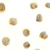
Description Immunocytochemistry Validation of BAFF in HL60 Cells
Immunocytochemical analysis of HL60 cells using anti-BAFF antibody (2221) at 1 ug/ml. Cells was fixed with formaldehyde and blocked with 10% serum for 1 h at RT; antigen retrieval was by heat mediation with a citrate buffer (pH6). Samples were incubated with primary antibody overnight at 4C. A goat anti-rabbit IgG H&L (HRP) at 1/250 was used as secondary. Counter stained with Hematoxylin.
Immunocytochemical analysis of HL60 cells using anti-BAFF antibody (2221) at 1 ug/ml. Cells was fixed with formaldehyde and blocked with 10% serum for 1 h at RT; antigen retrieval was by heat mediation with a citrate buffer (pH6). Samples were incubated with primary antibody overnight at 4C. A goat anti-rabbit IgG H&L (HRP) at 1/250 was used as secondary. Counter stained with Hematoxylin.

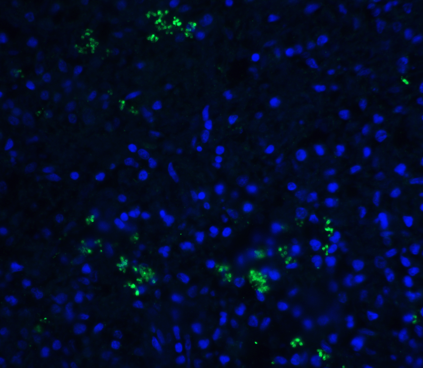
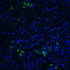
Description Immunofluorescence Validation of BAFF in Human Spleen Tissue
Immunofluorescent analysis of 4% paraformaldehyde-fixed human spleen tissue labeling BAFF with 2221 at 20 ug/mL, followed by goat anti-rabbit IgG secondary antibody at 1/500 dilution (green) and DAPI staining (blue).
Immunofluorescent analysis of 4% paraformaldehyde-fixed human spleen tissue labeling BAFF with 2221 at 20 ug/mL, followed by goat anti-rabbit IgG secondary antibody at 1/500 dilution (green) and DAPI staining (blue).

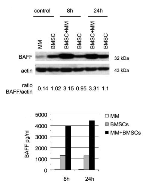
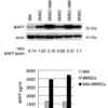
Description Regulated Expression Validation of BAFF in Myeloma Patients (Tai et al., 2006)
Immunoblot analysis was performed to monitor protein expression of BAFF with anti-BAFF antibodies in multiple myeloma cells with or without BMSCs. BAFF expression in cocultures at 8hr or 24hr was up-regulated by ~3.5-fold relative to BMSCs alone.
Immunoblot analysis was performed to monitor protein expression of BAFF with anti-BAFF antibodies in multiple myeloma cells with or without BMSCs. BAFF expression in cocultures at 8hr or 24hr was up-regulated by ~3.5-fold relative to BMSCs alone.

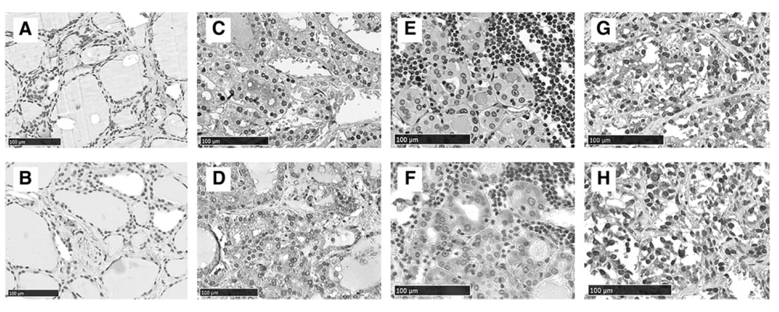
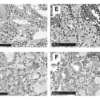
Description Immunohistochemistry Validation of BAFF in Thyroid of Patients with Graves’ Diseases (Campi et al., 2015)
BAFF expression detected by anti-BAFF antibodies (2221) was remarkably increased in thyrocytes from multinodular goiter (C) compared with either Hashimoto's thyroiditis (E) or Graves' disease (G) while no staining was found in normal thyroid tissue (A).
BAFF expression detected by anti-BAFF antibodies (2221) was remarkably increased in thyrocytes from multinodular goiter (C) compared with either Hashimoto's thyroiditis (E) or Graves' disease (G) while no staining was found in normal thyroid tissue (A).

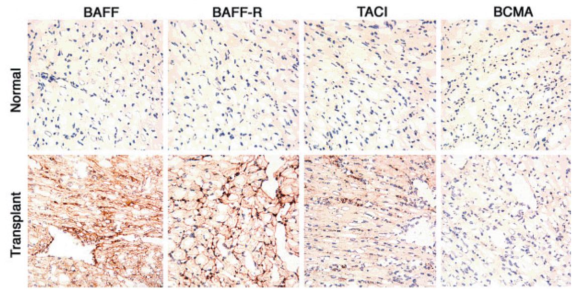
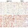
Description Immunohistochemistry Validation of BAFF in Murine Cardiac Transplants at Rejection (Ye et al., 2004)
BAFF expression detected by anti-BAFF antibodies (2221) was upregulated in intragraft leukocytes due to rejection at 7 days after heart transplant.
BAFF expression detected by anti-BAFF antibodies (2221) was upregulated in intragraft leukocytes due to rejection at 7 days after heart transplant.
Handling
Storage This antibody is stable for at least one (1) year at -20°C. Avoid multiple freeze-thaw cycles.
Dilution Instructions Dilute in PBS or medium which is identical to that used in the assay system.
Application Instructions Immunoblotting: use at 1ug/mL.
Immunocytochemistry: use at 1ug/mL.
These are recommended concentrations.
Enduser should determine optimal concentration for their application.
Positive control: Whole cell lysate from HL60 cells.
Immunocytochemistry: use at 1ug/mL.
These are recommended concentrations.
Enduser should determine optimal concentration for their application.
Positive control: Whole cell lysate from HL60 cells.
References & Data Sheet
Data Sheet  Download PDF Data Sheet
 Download PDF Data Sheet

