Anti-Ataxin-1 Antibody (56550)
$466.00
| Host | Quantity | Applications | Species Reactivity | Data Sheet | |
|---|---|---|---|---|---|
| Mouse | 100ug | WB,IHC,ICC/IF,IP | Human, Mouse, Rat |  |
SKU: 56550
Categories: Antibody Products, Neuroscience and Signal Transduction Antibodies, Products
Overview
Product Name Anti-Ataxin-1 Antibody (56550)
Description Anti-Ataxin-1 Mouse Monoclonal Antibody
Target Ataxin-1
Species Reactivity Human, Mouse, Rat
Applications WB,IHC,ICC/IF,IP
Host Mouse
Clonality Monoclonal
Clone ID S76-8
Isotype IgG2b
Immunogen Synthetic peptide corresponding to aa 164-197 (ATTPSQRSQLEAYSTLLAN- MGSLSQAPGHKVEPP) of mouse Ataxin-1. This sequence is 100
Properties
Form Liquid
Concentration 1.0 mg/mL
Formulation PBS, pH 7.4, 0.1% sodium azide, 50% glycerol.
Buffer Formulation Phosphate Buffered Saline
Buffer pH pH 7.4
Buffer Anti-Microbial 0.1% Sodium Azide
Buffer Cryopreservative 50% Glycerol
Format Purified
Purification Purified by Protein G affinity chromatography
Specificity Information
Specificity This antibody recognizes human, mouse, and rat Ataxin-1.
Target Name Ataxin-1
Target ID Ataxin-1
Uniprot ID P54254
Alternative Names Spinocerebellar ataxia type 1 protein homolog
Gene Name Atxn1
Sequence Location Cytoplasm, Nucleus
Biological Function Chromatin-binding factor that repress Notch signaling in the absence of Notch intracellular domain by acting as a CBF1 corepressor. Binds to the HEY promoter and might assist, along with NCOR2, RBPJ-mediated repression (By similarity). May be involved in RNA metabolism (By similarity). In concert with CIC and ATXN1L, involved in brain development (PubMed:28288114). {UniProtKB:P54253, PubMed:28288114}.
Research Areas Neuroscience
Background Ataxin-1 is a member of the ATXN1 protein family. It is a neurodegenerative disorder protein that is believed to play a role in metabolism of RNA. A mutation of Ataxin-1 causes spinocerebellar ataxia type-1 (SCA1), a progressive neurodegenerative disease that primarily affects Purjinke cells in the brain stem and cerebellum. Ataxin-1 is ubiquitously expressed except in brain where it is limited to populations of neurons.
Application Images


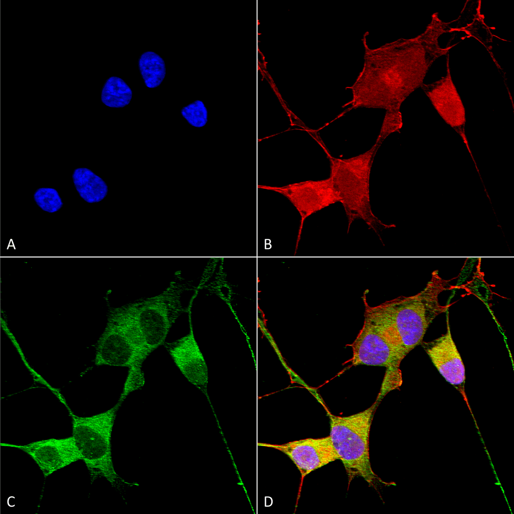
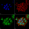
Description Immunocytochemistry/Immunofluorescence analysis using Mouse Anti-Ataxin 1 Monoclonal Antibody, Clone S76-8 (56550). Tissue: Neuroblastoma cells (SH-SY5Y). Species: Human. Fixation: 4% PFA for 15 min. Primary Antibody: Mouse Anti-Ataxin 1 Monoclonal Antibody (56550) at 1:100 for overnight at 4°C with slow rocking. Secondary Antibody: AlexaFluor 488 at 1:1000 for 1 hour at RT. Counterstain: Phalloidin-iFluor 647 (red) F-Actin stain; Hoechst (blue) nuclear stain at 1:800, 1.6mM for 20 min at RT. (A) Hoechst (blue) nuclear stain. (B) Phalloidin-iFluor 647 (red) F-Actin stain. (C) Ataxin 1 Antibody (D) Composite.

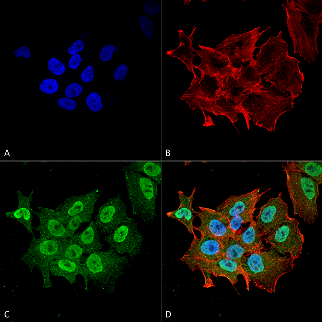
Description Immunocytochemistry/Immunofluorescence analysis using Mouse Anti-Ataxin 1 Monoclonal Antibody, Clone S76-8 (56550). Tissue: Neuroblastoma cell line (SK-N-BE). Species: Human. Fixation: 4% Formaldehyde for 15 min at RT. Primary Antibody: Mouse Anti-Ataxin 1 Monoclonal Antibody (56550) at 1:100 for 60 min at RT. Secondary Antibody: Goat Anti-Mouse ATTO 488 at 1:200 for 60 min at RT. Counterstain: Phalloidin Texas Red F-Actin stain; DAPI (blue) nuclear stain at 1:1000, 1:5000 for 60 min at RT, 5 min at RT. Localization: Cytoplasm, Nucleus. Magnification: 60X. (A) DAPI (blue) nuclear stain. (B) Phalloidin Texas Red F-Actin stain. (C) Ataxin 1 Antibody. (D) Composite.

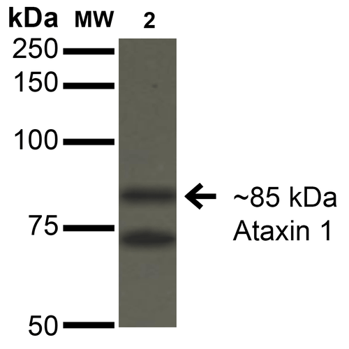
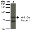
Description Western Blot analysis of Monkey COS-1 cells transfected with Ataxin- 1 showing detection of ~85 kDa Ataxin 1 protein using Mouse Anti-Ataxin 1 Monoclonal Antibody, Clone S76-8 (56550). Lane 1: Molecular Weight Ladder. Lane 2: Monkey COS-1 cells transfected with Ataxin- 1. Load: 15 µg. Block: 2% BSA and 2% Skim Milk in 1X TBST. Primary Antibody: Mouse Anti-Ataxin 1 Monoclonal Antibody (56550) at 1:200 for 16 hours at 4°C. Secondary Antibody: Goat Anti-Mouse IgG: HRP at 1:1000 for 1 hour RT. Color Development: ECL solution for 6 min in RT. Predicted/Observed Size: ~85 kDa.
Handling
Storage This product is stable for at least one (1) year at -20°C.
Dilution Instructions Dilute in PBS or medium that is identical to that used in the assay system.
Application Instructions Immunoblotting: use at 1-5ug/mL. A band of ~85kDa is detected.
Immunofluorescence: use at 10ug/mL.
These are recommended concentrations.
Enduser should determine optimal concentrations for their application.
Immunofluorescence: use at 10ug/mL.
These are recommended concentrations.
Enduser should determine optimal concentrations for their application.
References & Data Sheet
Data Sheet  Download PDF Data Sheet
 Download PDF Data Sheet

