Anti-ASC Antibody (2287)
$445.00
SKU: 2287
Categories: Antibody Products, Apoptosis Antibodies, Products
Overview
Product Name Anti-ASC Antibody (2287)
Description Anti-ASC Rabbit Polyclonal Antibody
Target ASC
Species Reactivity Human
Applications ELISA,WB,ICC,IHC-P
Host Rabbit
Clonality Polyclonal
Isotype IgG
Immunogen Peptide corresponding to aa 182-195 of human ASC.
Properties
Form Liquid
Concentration Lot Specific
Formulation PBS, pH 7.4.
Buffer Formulation Phosphate Buffered Saline
Buffer pH pH 7.4
Format Purified
Purification Purified by peptide immuno-affinity chromatography
Specificity Information
Specificity This antibody recognizes human ASC (25kDa).
Target Name Apoptosis-associated speck-like protein containing a CARD
Target ID ASC
Uniprot ID Q9ULZ3
Alternative Names hASC, Caspase recruitment domain-containing protein 5, PYD and CARD domain-containing protein, Target of methylation-induced silencing 1
Gene Name PYCARD
Gene ID 29108
Accession Number NP_037390
Sequence Location Cytoplasm, Inflammasome, Endoplasmic reticulum, Mitochondrion, Nucleus
Biological Function Functions as key mediator in apoptosis and inflammation. Promotes caspase-mediated apoptosis involving predominantly caspase-8 and also caspase-9 in a probable cell type-specific manner. Involved in activation of the mitochondrial apoptotic pathway, promotes caspase-8-dependent proteolytic maturation of BID independently of FADD in certain cell types and also mediates mitochondrial translocation of BAX and activates BAX-dependent apoptosis coupled to activation of caspase-9, -2 and -3. Involved in macrophage pyroptosis, a caspase-1-dependent inflammatory form of cell death and is the major constituent of the ASC pyroptosome which forms upon potassium depletion and rapidly recruits and activates caspase-1. In innate immune response believed to act as an integral adapter in the assembly of the inflammasome which activates caspase-1 leading to processing and secretion of proinflammatory cytokines. The function as activating adapter in different types of inflammasomes is mediated by the pyrin and CARD domains and their homotypic interactions. Required for recruitment of caspase-1 to inflammasomes containing certain pattern rPubMed:11103777, PubMed:12486103, PubMed:12646168, PubMed:14499617, PubMed:14730312, PubMed:15030775, PubMed:16585594, PubMed:16964285, PubMed:16982856, PubMed:17349957, PubMed:17599095, PubMed:19158675, PubMed:19158676, PubMed:19234215, PubMed:19494289, PubMed:21487011, PubMed:22732093, PubMed:28314590, PubMed:30674671, PubMed:34678144}.; [Isoform 2]: May have a regulating effect on the function as inflammasome adapter. {PubMed:19759850, PubMed:20482797}.; [Isoform 3]: Seems to inhibit inflammasome-mediated maturation of interleukin-1 beta. {PubMed:20482797}.
Research Areas Apoptosis
Background A novel CARD-containing protein was recently identified and designated ASC (or TMS1). ASC induces apoptosis by activating caspase-9 and inhibits survival of human breast cancer cells . ASC is widely expressed in human and mouse tissues.
Application Images


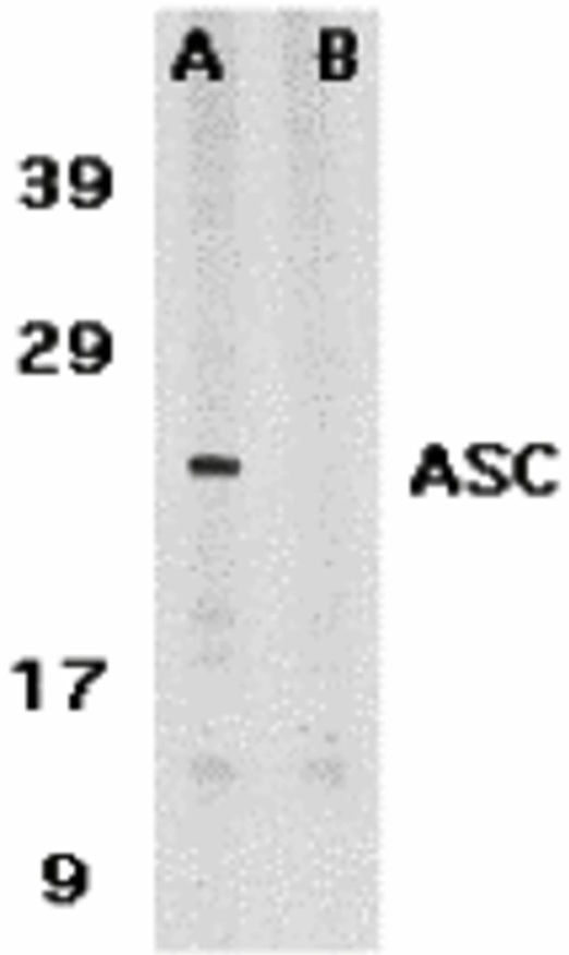
Description Western Blot Validation in Human HL60 Cells
Loading: 15 ug of lysates per lane. Antibodies: ASC 2287, (1 ug/mL) in the absence (A) or presence (B) of blocking peptide, 1h incubation at RT in 5% NFDM/TBST.Secondary: Goat anti-rabbit IgG HRP conjugate at 1:10000 dilution.
Loading: 15 ug of lysates per lane. Antibodies: ASC 2287, (1 ug/mL) in the absence (A) or presence (B) of blocking peptide, 1h incubation at RT in 5% NFDM/TBST.Secondary: Goat anti-rabbit IgG HRP conjugate at 1:10000 dilution.

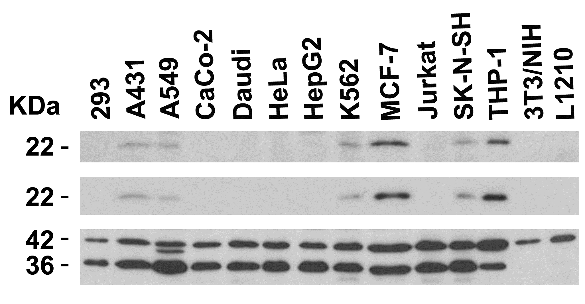
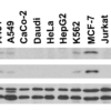
Description Independent Antibody Validation (IAV) via Protein Expression Profile in Cell Lines
Loading: 15 ug of lysates per lane. Antibodies: ASC 2287, (2 ug/mL), ASC 39-001, (2 ug/mL), beta-actin (1 ug/mL) and GAPDH (0.02 ug/mL), 1h incubation at RT in 5% NFDM/TBST.Secondary: Goat anti-rabbit or goat anti-mouse (for ASC 39001) IgG HRP conjugate at 1:10000 dilution.
Loading: 15 ug of lysates per lane. Antibodies: ASC 2287, (2 ug/mL), ASC 39-001, (2 ug/mL), beta-actin (1 ug/mL) and GAPDH (0.02 ug/mL), 1h incubation at RT in 5% NFDM/TBST.Secondary: Goat anti-rabbit or goat anti-mouse (for ASC 39001) IgG HRP conjugate at 1:10000 dilution.

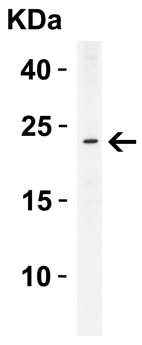
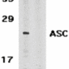
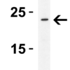
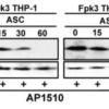
Description Western Blot Validation in Human THP-1 Cells
Loading: 15 ug of lysate per lane. Antibodies: ASC 2287, (2 ug/mL), 1h incubation at RT in 5% NFDM/TBST.Secondary: Goat anti-rabbit IgG HRP conjugate at 1:10000 dilution.
Loading: 15 ug of lysate per lane. Antibodies: ASC 2287, (2 ug/mL), 1h incubation at RT in 5% NFDM/TBST.Secondary: Goat anti-rabbit IgG HRP conjugate at 1:10000 dilution.

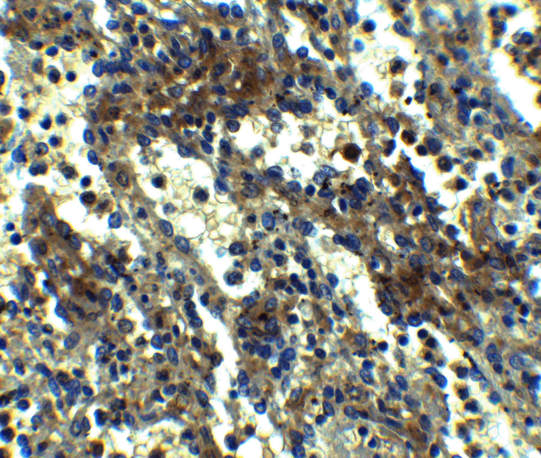
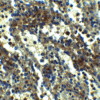
Description Immunohistochemistry Validation of ASC in Human Spleen Tissue
Immunohistochemical analysis of paraffin-embedded human spleen tissue using anti-ASC antibody (2287) at 2.5 ug/ml. Tissue was fixed with formaldehyde and blocked with 10% serum for 1 h at RT; antigen retrieval was by heat mediation with a citrate buffer (pH6). Samples were incubated with primary antibody overnight at 4C. A goat anti-rabbit IgG H&L (HRP) at 1/250 was used as secondary. Counter stained with Hematoxylin.
Immunohistochemical analysis of paraffin-embedded human spleen tissue using anti-ASC antibody (2287) at 2.5 ug/ml. Tissue was fixed with formaldehyde and blocked with 10% serum for 1 h at RT; antigen retrieval was by heat mediation with a citrate buffer (pH6). Samples were incubated with primary antibody overnight at 4C. A goat anti-rabbit IgG H&L (HRP) at 1/250 was used as secondary. Counter stained with Hematoxylin.

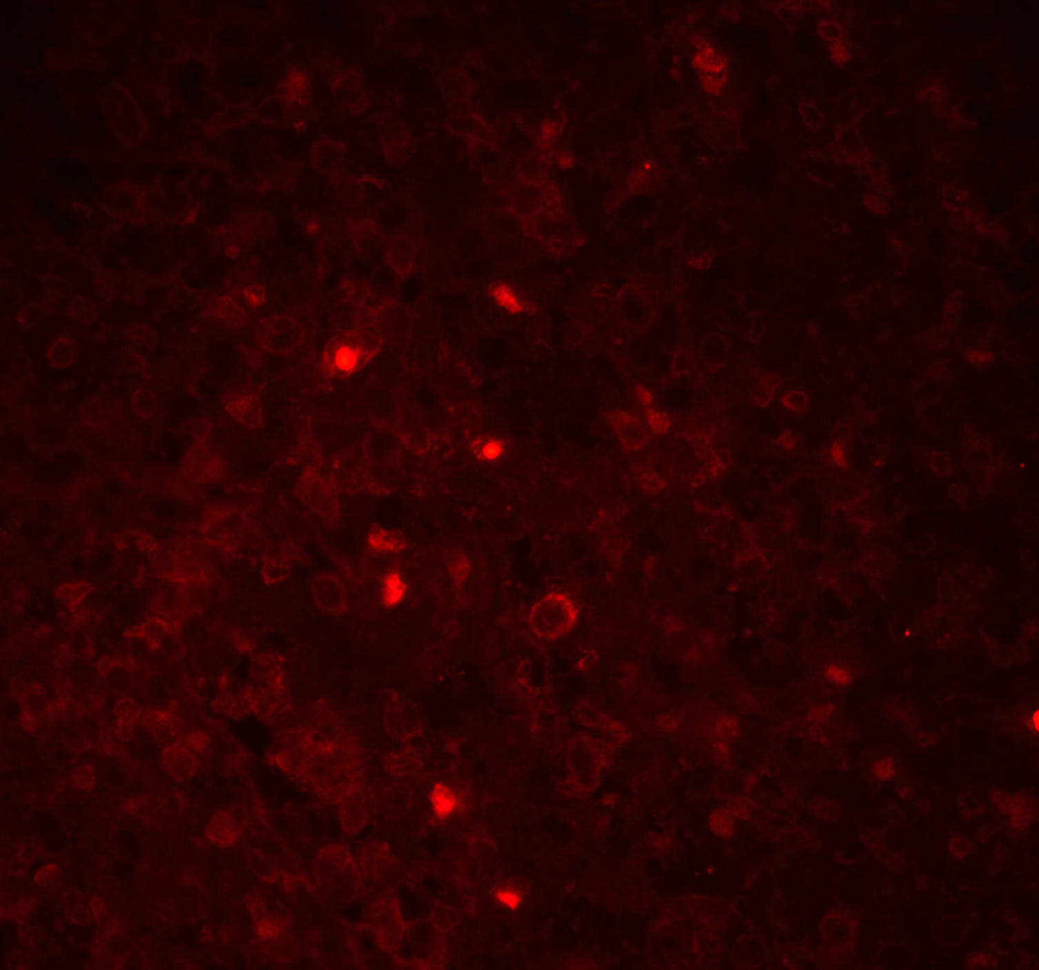
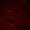
Description Immunofluorescence Validation of ASC in Human Spleen Tissue
Immunofluorescent analysis of 4% paraformaldehyde-fixed Human Spleen Tissue labeling ASC with 2287 at 20 ug/mL, followed by goat anti-rabbit IgG secondary antibody at 1/500 dilution (red).
Immunofluorescent analysis of 4% paraformaldehyde-fixed Human Spleen Tissue labeling ASC with 2287 at 20 ug/mL, followed by goat anti-rabbit IgG secondary antibody at 1/500 dilution (red).

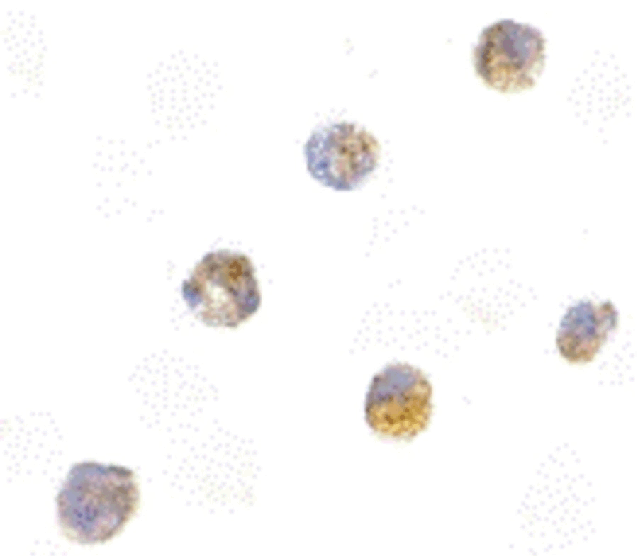
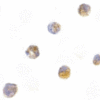
Description Immunocytochemistry Validation of ASC in HL60 Cells
Immunocytochemical analysis of HL60 cells using anti-ASC antibody (2287) at 5 ug/ml. Cells was fixed with formaldehyde and blocked with 10% serum for 1 h at RT; antigen retrieval was by heat mediation with a citrate buffer (pH6). Samples were incubated with primary antibody overnight at 4C. A goat anti-rabbit IgG H&L (HRP) at 1/250 was used as secondary. Counter stained with Hematoxylin.
Immunocytochemical analysis of HL60 cells using anti-ASC antibody (2287) at 5 ug/ml. Cells was fixed with formaldehyde and blocked with 10% serum for 1 h at RT; antigen retrieval was by heat mediation with a citrate buffer (pH6). Samples were incubated with primary antibody overnight at 4C. A goat anti-rabbit IgG H&L (HRP) at 1/250 was used as secondary. Counter stained with Hematoxylin.

Description KD Validation of ASC in THP-1 Cells (Dowds et al., 2004)
Immunofluorescence analysis with anti-ASC antibodies (2287) was performed for BIM in 293 cells transfected with GFP siRNA or ASC siRNA. ASC expression was disrupted after ASC siRNA knockdown.
Immunofluorescence analysis with anti-ASC antibodies (2287) was performed for BIM in 293 cells transfected with GFP siRNA or ASC siRNA. ASC expression was disrupted after ASC siRNA knockdown.

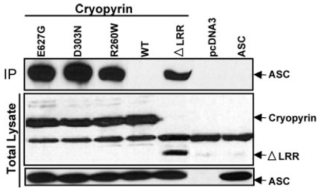
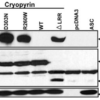
Description Overexpression Validation of ASC in HEK293T Cells (Dowds et al., 2004)
Western blot analysis with anti-ASC antibodies (2287) was performed for ASC in HEK293T cells transfected with pcDNA3-ASC.
Western blot analysis with anti-ASC antibodies (2287) was performed for ASC in HEK293T cells transfected with pcDNA3-ASC.
Handling
Storage This antibody is stable for at least one (1) year at -20°C. Avoid multiple freeze-thaw cycles.
Dilution Instructions Dilute in PBS or medium which is identical to that used in the assay system.
Application Instructions Immunoblotting: use at 1ug/mL.
Immunocytochemistry: use at 5ug/mL.
These are recommended concentrations.
Enduser should determine optimal concentrations for their applications.
Positive control: Whole cell lysate of HL60 cells.
Immunocytochemistry: use at 5ug/mL.
These are recommended concentrations.
Enduser should determine optimal concentrations for their applications.
Positive control: Whole cell lysate of HL60 cells.
References & Data Sheet
Data Sheet  Download PDF Data Sheet
 Download PDF Data Sheet

