Anti-Ankyrin-B Antibody (56518)
$466.00
| Host | Quantity | Applications | Species Reactivity | Data Sheet | |
|---|---|---|---|---|---|
| Mouse | 100ug | WB,IHC,ICC/IF | Human, Mouse, Rat |  |
SKU: 56518
Categories: Antibody Products, Neuroscience and Signal Transduction Antibodies, Products
Overview
Product Name Anti-Ankyrin-B Antibody (56518)
Description Anti-Ankyrin-B Mouse Monoclonal Antibody
Target Ankyrin-B
Species Reactivity Human, Mouse, Rat
Applications WB,IHC,ICC/IF
Host Mouse
Clonality Monoclonal
Clone ID S105-13
Isotype IgG1
Immunogen Synthetic peptide correspond- ing to aa 203-496 of human Ankyrin-B (accession no. NP_001120965.1)
Properties
Form Liquid
Concentration 1.0 mg/mL
Formulation PBS, pH 7.4, 50% glycerol, 0.09% sodium azide.Purified by Protein G affinity chromatography.
Buffer Formulation Phosphate Buffered Saline
Buffer pH pH 7.4
Buffer Anti-Microbial 0.09% Sodium Azide
Buffer Cryopreservative 50% Glycerol
Format Purified
Purification Purified by Protein G affinity chromatography
Specificity Information
Specificity This antibody recognizes human, mouse, and rat Ankyrin-B.
Target Name Ankyrin-2
Target ID Ankyrin-B
Uniprot ID Q01484
Alternative Names ANK-2, Ankyrin-B, Brain ankyrin, Non-erythroid ankyrin
Gene Name ANK2
Sequence Location Cytoplasm, cytoskeleton, Membrane, Cytoplasm, myofibril, sarcomere, M line, Apical cell membrane, Cell membrane, Cell junction, synapse, postsynaptic cell membrane, Early endosome, Recycling endosome, Lysosome, Mitochondrion, Cytoplasm, myofibril, sarcomere, Z line, Cell membrane, sarcolemma, T-tubule
Biological Function Plays an essential role in the localization and membrane stabilization of ion transporters and ion channels in several cell types, including cardiomyocytes, as well as in striated muscle cells. In skeletal muscle, required for proper localization of DMD and DCTN4 and for the formation and/or stability of a special subset of microtubules associated with costameres and neuromuscular junctions. In cardiomyocytes, required for coordinate assembly of Na/Ca exchanger, SLC8A1/NCX1, Na/K ATPases ATP1A1 and ATP1A2 and inositol 1,4,5-trisphosphate (InsP3) receptors at sarcoplasmic reticulum/sarcolemma sites. Required for expression and targeting of SPTBN1 in neonatal cardiomyocytes and for the regulation of neonatal cardiomyocyte contraction rate (PubMed:12571597). In the inner segment of rod photoreceptors, required for the coordinated expression of the Na/K ATPase, Na/Ca exchanger and beta-2-spectrin (SPTBN1) (By similarity). Plays a role in endocytosis and intracellular protein transport. Associates with phosphatidylinositol 3-phosphate (PI3P)-positive organelles and binds dynactin to promote long-range motility of cells. Recruits RABGAP1L to (PI3P)-positive early endosomes, where RABGAP1L inactivates RAB22A, and promotes polarized trafficking to the leading edge of the migrating cells. Part of the ANK2/RABGAP1L complex which is required for the polarized recycling of fibronectin receptor ITGA5 ITGB1 to the plasma membrane that enables continuous directional cell migration (By similarity). {UniProtKB:Q8C8R3, PubMed:12571597}.
Research Areas Neuroscience
Background Ankyrins are a family of adaptor proteins that mediate the attachment of integral membrane proteins to the spectrin-actin based membrane skeleton. Ankyrins have binding sites for the beta subunit of spectrin and for at least 12 families of integral membrane proteins. This linkage is required to maintain the integrity of the plasma membranes and to anchor specific ion channels, ion exchangers, and ion transporters in the plasma membrane.
Application Images


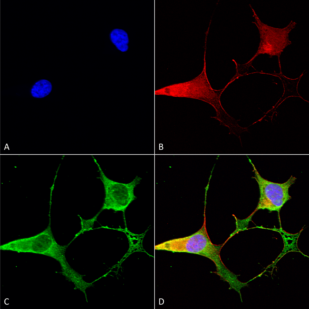
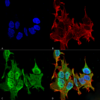
Description Immunocytochemistry/Immunofluorescence analysis using Mouse Anti-Ankyrin B Monoclonal Antibody, Clone S105-13 (56518). Tissue: Neuroblastoma cells (SH-SY5Y). Species: Human. Fixation: 4% PFA for 15 min. Primary Antibody: Mouse Anti-Ankyrin B Monoclonal Antibody (56518) at 1:50 for overnight at 4°C with slow rocking. Secondary Antibody: AlexaFluor 488 at 1:1000 for 1 hour at RT. Counterstain: Phalloidin-iFluor 647 (red) F-Actin stain; Hoechst (blue) nuclear stain at 1:800, 1.6mM for 20 min at RT. (A) Hoechst (blue) nuclear stain. (B) Phalloidin-iFluor 647 (red) F-Actin stain. (C) Ankyrin B Antibody (D) Composite.

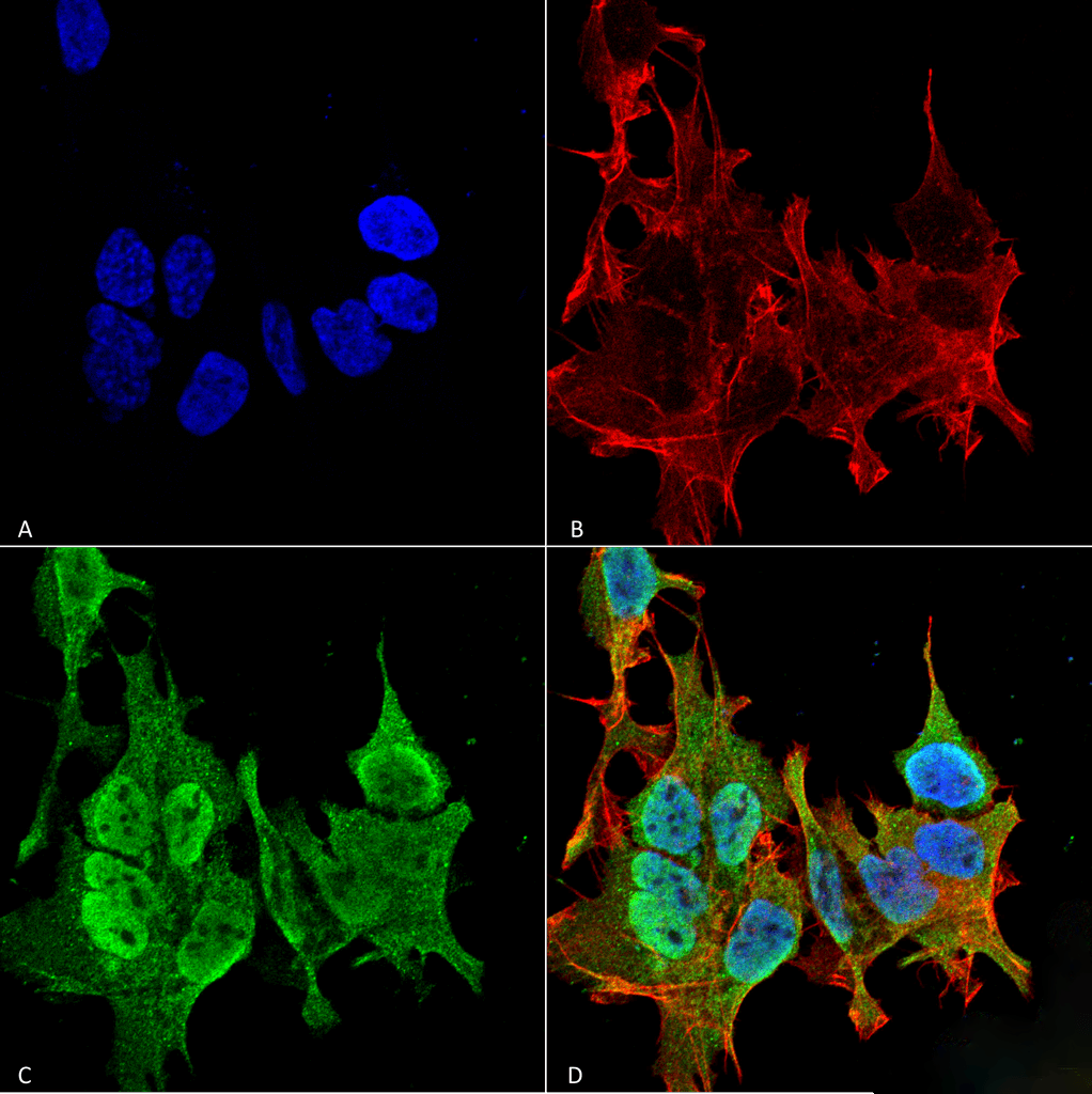
Description Nucleus. Magnification: 60X. (A) DAPI (blue) nuclear stain. (B) Phalloidin Texas Red F-Actin stain. (C) Ankyrin B Antibody. (D) Composite.

Description Immunocytochemistry/Immunofluorescence analysis using Mouse Anti-Ankyrin B Monoclonal Antibody, Clone S105-13 (56518). Tissue: Neuroblastoma cell line (SK-N-BE). Species: Human. Fixation: 4% Formaldehyde for 15 min at RT. Primary Antibody: Mouse Anti-Ankyrin B Monoclonal Antibody (56518) at 1:100 for 60 min at RT. Secondary Antibody: Goat Anti-Mouse ATTO 488 at 1:100 for 60 min at RT. Counterstain: Phalloidin Texas Red F-Actin stain; DAPI (blue) nuclear stain at 1:1000; 1:5000 for 60 min RT, 5 min RT. Localization: Cytoplasm
Handling
Storage This product is stable for at least 1 year at -20°C. Freeze in multiple aliquots to avoid repeated freeze-thaw cycles.
Dilution Instructions Dilute in PBS or medium that is identical to that used in the assay system.
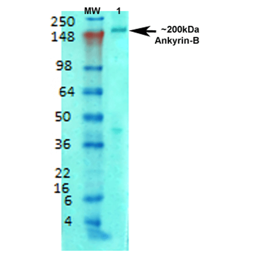
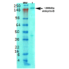
Application Instructions Immunoblotting: use at 1-2ug/mL. A band of >200kDa is detected.
Immunohistochemistry: use at 1-5ug/mL.
These are recommended concentrations.
Enduser should determine optimal concentrations for their applications.
Positive control: rat brain membranes.
Immunohistochemistry: use at 1-5ug/mL.
These are recommended concentrations.
Enduser should determine optimal concentrations for their applications.
Positive control: rat brain membranes.
References & Data Sheet
Data Sheet  Download PDF Data Sheet
 Download PDF Data Sheet

