Anti-AIF (IN) Antibody (2267)
$445.00
| Host | Quantity | Applications | Species Reactivity | Data Sheet | |
|---|---|---|---|---|---|
| Rabbit | 100ug | ELISA,WB,IHC-P | Human, Mouse, Rat |  |
SKU: 2267
Categories: Antibody Products, Apoptosis Antibodies, Products
Overview
Product Name Anti-AIF (IN) Antibody (2267)
Description Anti-AIF (IN) Rabbit Polyclonal Antibody
Target AIF (IN)
Species Reactivity Human, Mouse, Rat
Applications ELISA,WB,IHC-P
Host Rabbit
Clonality Polyclonal
Isotype IgG
Immunogen Peptide corresponding to aa 517- 531 of human AIF. This sequence is identical to those of mouse and rat AIF.
Properties
Form Liquid
Concentration Lot Specific
Formulation PBS, pH 7.4.
Buffer Formulation Phosphate Buffered Saline
Buffer pH pH 7.4
Format Purified
Purification Purified by peptide immuno-affinity chromatography
Specificity Information
Specificity This antibody recognizes human, mouse, and rat AIF (67 kDa).
Target Name Connector enhancer of kinase suppressor of ras 1
Target ID AIF (IN)
Uniprot ID O95381
Alternative Names CNK
Gene Name CNKSR1
Gene ID 9131
Accession Number NP_001124318.2
Research Areas Apoptosis
Background A novel protein that causes chromatin condensation and DNA fragmentation has been designated Apoptosis Inducing Factor (AIF). AIF localizes in mitochondria and translocates to the nucleus when apoptosis is induced. This event is followed by the release of chytochrome c and caspase-9 from mitochondria. AIF is highly conserved between human and mouse and widely expressed in different cell types and tissues.
Application Images


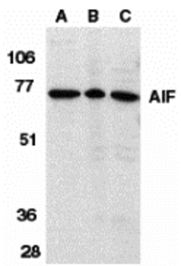
Description Western Blot Validation in Different Species
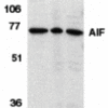
Loading: 15 ug of lysates per lane. Antibodies: AIF 2267, (1 µg/mL), 1h incubation at RT in 5% NFDM/TBST.Secondary: Goat anti-rabbit IgG HRP conjugate at 1:10000 dilution.Lane A: Human K562 cellsLane B: Rat heartLane C: Mouse heart
Loading: 15 ug of lysates per lane. Antibodies: AIF 2267, (1 µg/mL), 1h incubation at RT in 5% NFDM/TBST.Secondary: Goat anti-rabbit IgG HRP conjugate at 1:10000 dilution.Lane A: Human K562 cellsLane B: Rat heartLane C: Mouse heart

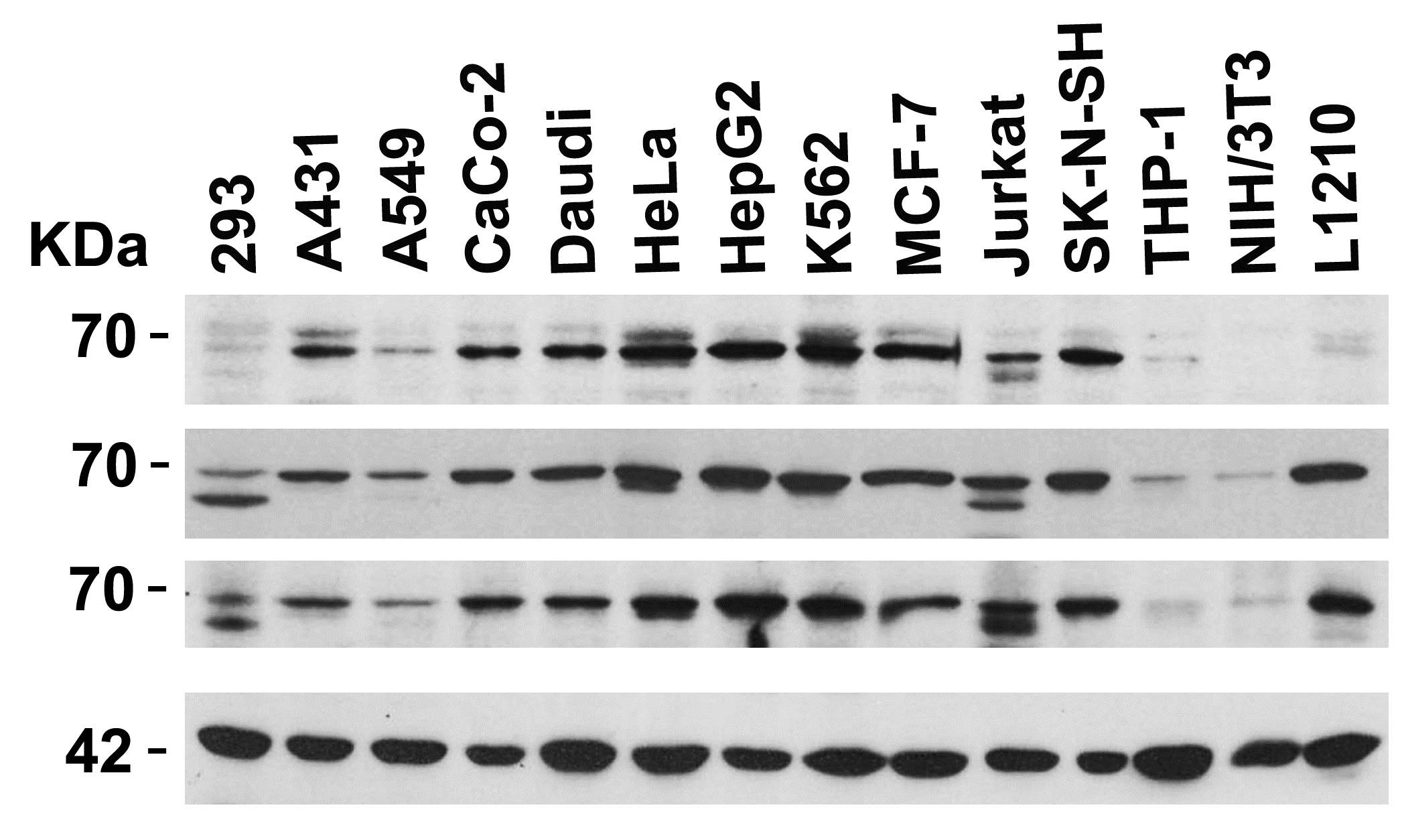
Description Independent Antibody Validation (IAV) via Protein Expression Profile in Cell Lines
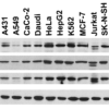
Loading: 15 µg of lysates per lane. Antibodies: AIF 2267, (1 ug/mL), AIF 2239, (1 ug/mL), AIF 2301, (2 ug/mL), and beta-actin (1 ug/mL), 1h incubation at RT in 5% NFDM/TBST.Secondary: Goat anti-rabbit IgG HRP conjugate at 1:10000 dilution.
Loading: 15 µg of lysates per lane. Antibodies: AIF 2267, (1 ug/mL), AIF 2239, (1 ug/mL), AIF 2301, (2 ug/mL), and beta-actin (1 ug/mL), 1h incubation at RT in 5% NFDM/TBST.Secondary: Goat anti-rabbit IgG HRP conjugate at 1:10000 dilution.

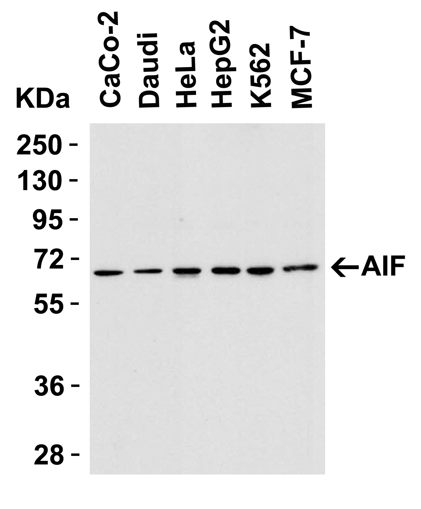
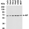
Description Western Blot Validation in Human Cell Lines
Loading: 15 ug of lysates per lane. Antibodies: AIF 2267, (1 ug/mL), 1h incubation at RT in 5% NFDM/TBST.Secondary: Goat anti-rabbit IgG HRP conjugate at 1:10000 dilution.
Loading: 15 ug of lysates per lane. Antibodies: AIF 2267, (1 ug/mL), 1h incubation at RT in 5% NFDM/TBST.Secondary: Goat anti-rabbit IgG HRP conjugate at 1:10000 dilution.

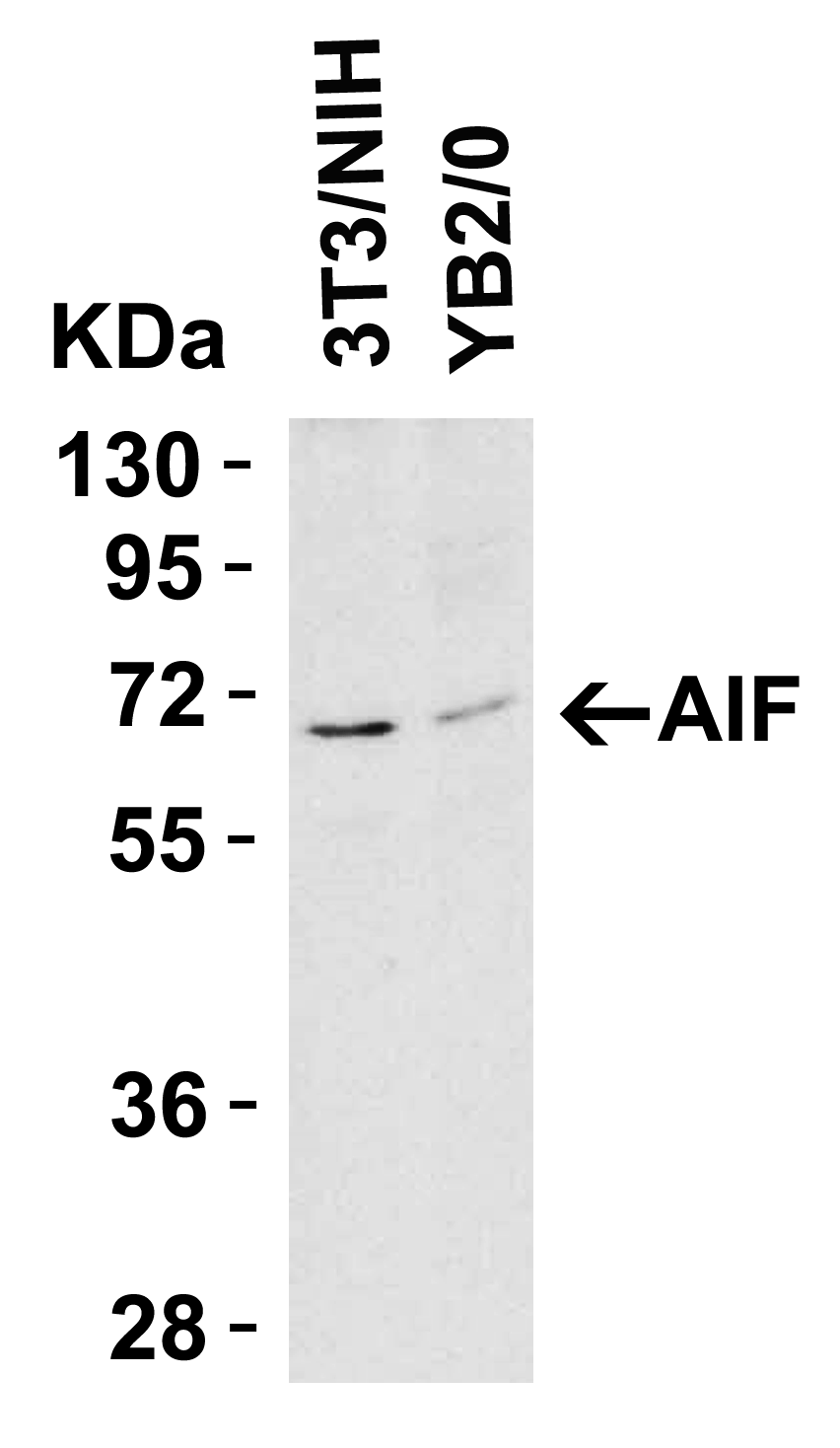
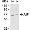
Description Western Blot Validation in Mouse and Rat Cell Lines
Loading: 15 ug of lysates per lane. Antibodies: AIF 2267, (1 ug/mL), 1h incubation at RT in 5% NFDM/TBST.Secondary: Goat anti-rabbit IgG HRP conjugate at 1:10000 dilution.
Loading: 15 ug of lysates per lane. Antibodies: AIF 2267, (1 ug/mL), 1h incubation at RT in 5% NFDM/TBST.Secondary: Goat anti-rabbit IgG HRP conjugate at 1:10000 dilution.

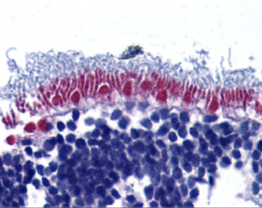
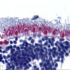
Description Immunohistochemistry Validation of AIF in Human Retina Tissue
Immunohistochemical analysis of paraffin-embedded human retina tissue using anti-AIF antibody (2267) at 10 ug/ml. Tissue was fixed with formaldehyde and blocked with 10% serum for 1 h at RT; antigen retrieval was by heat mediation with a citrate buffer (pH6). Samples were incubated with primary antibody overnight at 4 C. A goat anti-rabbit IgG H&L (HRP) at 1/250 was used as secondary. Counter stained with Hematoxylin.
Immunohistochemical analysis of paraffin-embedded human retina tissue using anti-AIF antibody (2267) at 10 ug/ml. Tissue was fixed with formaldehyde and blocked with 10% serum for 1 h at RT; antigen retrieval was by heat mediation with a citrate buffer (pH6). Samples were incubated with primary antibody overnight at 4 C. A goat anti-rabbit IgG H&L (HRP) at 1/250 was used as secondary. Counter stained with Hematoxylin.

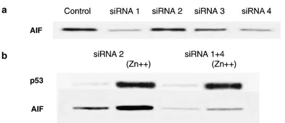
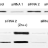
Description KD and Induced Validation of AIF in H1299 Cells(Stambolsky et al., 2006)
Western blot analysis of AIF knockdown with anti-AIF antibodies in H1299 cells. AIF expression was disrupted in AIF knockdown cells (siRNA1 and siRNA4). An increased expression of AIF was induced by ZnCl2 treatment, which was not observed in AIF knockdown cells.
Western blot analysis of AIF knockdown with anti-AIF antibodies in H1299 cells. AIF expression was disrupted in AIF knockdown cells (siRNA1 and siRNA4). An increased expression of AIF was induced by ZnCl2 treatment, which was not observed in AIF knockdown cells.

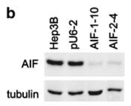
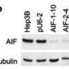
Description KD Validation of AIF in AIF Silenced Stable Cells(Apostolova et al., 2006)
AIF silencing is sustained in stable cell lines. Western blot analysis ofstable lines AIF-1-10, AIF-2-4 and pU6-2 using anti-AIF antibodies. AIF protein was disrupted after AIF silencing with AIF siRNA (AIF-1-10 and AIF-2-4) as compared to control (Hep3B and pU6-2).
AIF silencing is sustained in stable cell lines. Western blot analysis ofstable lines AIF-1-10, AIF-2-4 and pU6-2 using anti-AIF antibodies. AIF protein was disrupted after AIF silencing with AIF siRNA (AIF-1-10 and AIF-2-4) as compared to control (Hep3B and pU6-2).

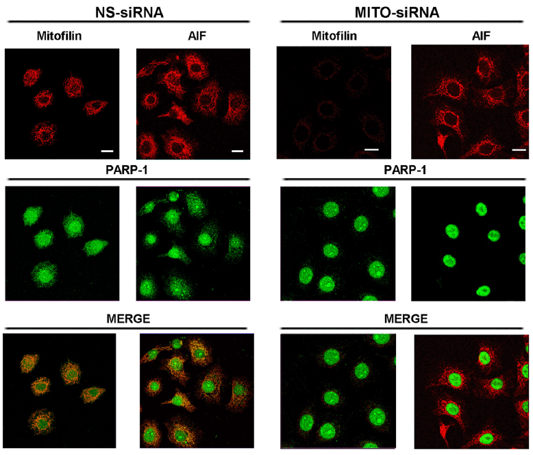
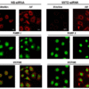
Description Immunofluorescence Validation of AIF in HeLa Cells (Rossi et al., 2009)
HeLa cells were transfected withMitofilin-specific (MITO-siRNA) or with nonspecific (NS-siRNA) siRNAs. AIF staining with anti-AIF antibodies was shown as a mitochondrial marker in the absence of Mitofilin.
HeLa cells were transfected withMitofilin-specific (MITO-siRNA) or with nonspecific (NS-siRNA) siRNAs. AIF staining with anti-AIF antibodies was shown as a mitochondrial marker in the absence of Mitofilin.

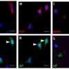
Description Immunofluorescence Validation of AIF in Rat Hippocampal Neurons (Hofer et al., 2011)
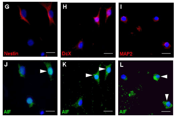
(G-L) After exposure to bacterial components, AIF colocalized in mature neurons (MAP2; I, L), immature neurons (DcX;H, K), and stem/progenitor cells (Nestin; G, J). AIF expression was detected by anti-AIF antibodies.
(G-L) After exposure to bacterial components, AIF colocalized in mature neurons (MAP2; I, L), immature neurons (DcX;H, K), and stem/progenitor cells (Nestin; G, J). AIF expression was detected by anti-AIF antibodies.

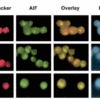
Description Subcellular Localization Validation of AIF in mononuclear cells (Gupta et al., 2424)
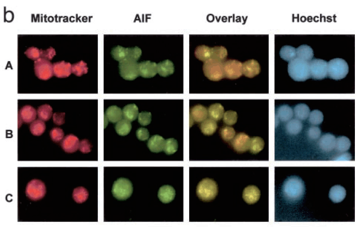
A shows mononuclear cells (MNCs) alone, B shows MNCs transfected with control plasmid, C shows MNCs transfected with Bcl-2 expression plasmid. Overlay is of Mitotracker (red) and AIF (green). Hoechst 33258 dye is used to examine chromatin fragmentation. The release of AIF form mitochondria is detected by anti-AIF antibodies.
A shows mononuclear cells (MNCs) alone, B shows MNCs transfected with control plasmid, C shows MNCs transfected with Bcl-2 expression plasmid. Overlay is of Mitotracker (red) and AIF (green). Hoechst 33258 dye is used to examine chromatin fragmentation. The release of AIF form mitochondria is detected by anti-AIF antibodies.

Description Induced Expression Validation of AIF in U937 Cells (Ikai et al., 2006)
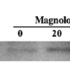
Release of AIF at 48 h after the treatment with 30 uM magnolol examined by Western blotAnalysis with anti-AIF antibodies. AIF release was markedly increased 48h after magnolol treatment.
Release of AIF at 48 h after the treatment with 30 uM magnolol examined by Western blotAnalysis with anti-AIF antibodies. AIF release was markedly increased 48h after magnolol treatment.
Handling
Storage This antibody is stable for at least one (1) year at -20°C. Avoid multiple freeze-thaw cycles.
Dilution Instructions Dilute in PBS or medium which is identical to that used in the assay system.
Application Instructions Immunoblotting: use at 1-10ug/mL
Immunohistochemistry: use at 10ug/mLImmunohistochemistry of AIF in human retina with AIF (IN) antibody at 10ug/mL.
These are recommended concentrations.
Endusers should determine optimal concentrations for their applications.
Positive control: Whole cell lysate from K562 cells.
Immunohistochemistry: use at 10ug/mLImmunohistochemistry of AIF in human retina with AIF (IN) antibody at 10ug/mL.
These are recommended concentrations.
Endusers should determine optimal concentrations for their applications.
Positive control: Whole cell lysate from K562 cells.
References & Data Sheet
Data Sheet  Download PDF Data Sheet
 Download PDF Data Sheet

