Anti-Id3 Antibody (56209)
$503.00
SKU: 56209
Categories: Antibody Products, Neuroscience and Signal Transduction Antibodies, Products
Overview
Product Name Anti-Id3 Antibody (56209)
Description Anti-Id3 Mouse Monoclonal Antibody
Target Id3
Species Reactivity Human, Mouse
Applications WB,ICC/IF,IP
Host Mouse
Clonality Monoclonal
Clone ID 2B11
Isotype IgG1
Immunogen Mouse Id3 gst-fusion protein (aa 1-119) expressed in E. coli.
Properties
Form Liquid
Concentration Lot Specific
Formulation 10 mM PBS, pH 7.4.
Buffer Formulation Phosphate Buffered Saline
Buffer pH pH 7.4
Format Purified
Purification Purified by Protein A affinity chromatography
Specificity Information
Specificity This antibody recognizes human and mouse Id3.
Target Name DNA-binding protein inhibitor ID-3
Target ID Id3
Uniprot ID Q02535
Alternative Names Class B basic helix-loop-helix protein 25, bHLHb25, Helix-loop-helix protein HEIR-1, ID-like protein inhibitor HLH 1R21, Inhibitor of DNA binding 3, Inhibitor of differentiation 3
Gene Name ID3
Sequence Location Nucleus.
Biological Function Transcriptional regulator (lacking a basic DNA binding domain) which negatively regulates the basic helix-loop-helix (bHLH) transcription factors by forming heterodimers and inhibiting their DNA binding and transcriptional activity. Implicated in regulating a variety of cellular processes, including cellular growth, senescence, differentiation, apoptosis, angiogenesis, and neoplastic transformation. Involved in myogenesis by inhibiting skeletal muscle and cardiac myocyte differentiation and promoting muscle precursor cells proliferation. Inhibits the binding of E2A-containing protein complexes to muscle creatine kinase E-box enhancer. Regulates the circadian clock by repressing the transcriptional activator activity of the CLOCK-ARNTL/BMAL1 heterodimer. {PubMed:8437843}.
Research Areas Neuroscience
Background Id3 is a 14 kD protein and a member of the Id family of helix-loop-helix (HLH) proteins. Id proteins are believed to be dominant negative regulators of HLH involved in cell differentiation and implicated in the control of cell growth, cellular differentiation, apoptosis, and carcinogenesis.
Application Images


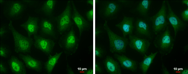
Description Id3 antibody [2B11] detects Id3 protein at cytoplasm and nucleus by immunofluorescent analysis.
Sample: HeLa cells were fixed in 4% paraformaldehyde at RT for 15 min.
Green: Id3 protein stained by Id3 antibody [2B11] (56209) diluted at 1:100.
Blue: Hoechst 33342 staining.
Scale bar = 10 um.
Sample: HeLa cells were fixed in 4% paraformaldehyde at RT for 15 min.
Green: Id3 protein stained by Id3 antibody [2B11] (56209) diluted at 1:100.
Blue: Hoechst 33342 staining.
Scale bar = 10 um.

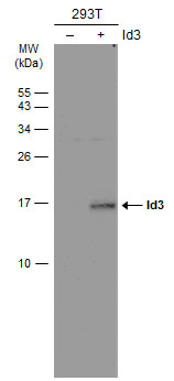
Description Non-transfected (–) and transfected (+) 293T whole cell extracts (30 ug) were separated by 15% SDS-PAGE, and the membrane was blotted with Id3 antibody [2B11] (56209) diluted at 1:1000. The HRP-conjugated anti-mouse IgG antibody was used to detect the primary antibody.
Handling
Storage This antibody is stable for at least one (1) year at -20°C. Avoid multiple freeze- thaw cycles.
Dilution Instructions Dilute in PBS or medium which is identical to that used in the assay system.
Application Instructions Immunoblotting and Immuno- precipitation: use at 1-5 ug/mL.
Positive controls: Id3 transfected Sol8 muscles cells and recombinant fusion protein.
Positive controls: Id3 transfected Sol8 muscles cells and recombinant fusion protein.
References & Data Sheet
Data Sheet  Download PDF Data Sheet
 Download PDF Data Sheet


