Anti-Hsp90 β Antibody (11106)
$419.00
| Host | Quantity | Applications | Species Reactivity | Data Sheet | |
|---|---|---|---|---|---|
| Rabbit | 100ul | WB,IHC,ICC/IF,ELISA | Human, Mouse, Rat |  |
SKU: 11106
Categories: Antibody Products, Heat Shock and Stress Protein Antibodies, Products
Overview
Product Name Anti-Hsp90 β Antibody (11106)
Description Anti-Hsp90 β Rabbit Polyclonal Antibody
Target Hsp90 β
Species Reactivity Human, Mouse, Rat
Applications WB,IHC,ICC/IF,ELISA
Host Rabbit
Clonality Polyclonal
Immunogen Purified recombinant human Hsp90beta
Properties
Form Liquid
Concentration Lot Specific
Formulation Whole antiserum
Buffer Formulation Whole Antiserum
Format Whole Antiserum
Purification Whole antiserum
Specificity Information
Specificity This antibody recognizes human, rat, and mouse Hsp90b. It does not cross- react with Hsp90alpha.
Target Name Heat shock protein HSP 90-β
Target ID Hsp90 β
Uniprot ID P08238
Alternative Names HSP 90, Heat shock 84 kDa, HSP 84, HSP84
Gene Name HSP90AB1
Gene ID 3326
Accession Number NP_031381.2
Sequence Location Cytoplasm, Melanosome, Nucleus, Secreted, Cell membrane, Dynein axonemal particle
Biological Function Molecular chaperone that promotes the maturation, structural maintenance and proper regulation of specific target proteins involved for instance in cell cycle control and signal transduction. Undergoes a functional cycle linked to its ATPase activity. This cycle probably induces conformational changes in the client proteins, thereby causing their activation. Interacts dynamically with various co-chaperones that modulate its substrate rPubMed:16478993, PubMed:18239673, PubMed:19696785, PubMed:20353823, PubMed:24613385, PubMed:32272059, PubMed:25973397, PubMed:26991466, PubMed:27295069}.
Research Areas Heat Shock& Stress Proteins
Background Hsp90 is a highly conserved and essential stress protein that is expressed in all eukaryotic cells. Hsp90 is involved in the folding, assembly, maturation, and stabilization of specific proteins as a member of a chaperone complex. It is also highly expressed in unstressed cells where it participates in controlling the activity, turnover, and trafficking of a variety of proteins. Most of the Hsp90-regulated proteins are involved in cell signaling and include the kinases, v-Src, Wee1, c-Raf, transcriptional regulators such as p53 and steroid receptors, and the polymerases of hepatitis B virus and telomerase.
Application Images



Description Immunocytochemistry/Immunofluorescence analysis using Rabbit Anti-Hsp90 beta Polyclonal Antibody (11106). Tissue: Cervical cancer cell line (HeLa). Species: Human. Fixation: 2% Formaldehyde for 20 min at RT. Primary Antibody: Rabbit Anti-Hsp90 beta Polyclonal Antibody (11106) at 1:120 for 12 hours at 4°C. Secondary Antibody: APC Goat Anti-Rabbit (red) at 1:200 for 2 hours at RT. Counterstain: DAPI (blue) nuclear stain at 1:40000 for 2 hours at RT. Localization: Cytoplasm. Melanosome. Magnification: 100x. (A) DAPI (blue) nuclear stain. (B) Anti-Hsp90 beta Antibody. (C) Composite.

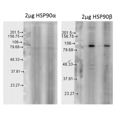
Description Western blot analysis of Human Cell line lysates showing detection of HSP90 beta protein using Rabbit Anti-HSP90 beta Polyclonal Antibody (11106). Load: 15 µgprotein. Block: 1.5% BSA for 30 minutes at RT. Primary Antibody: Rabbit Anti-HSP90 beta Polyclonal Antibody (11106) at 1:1000 for 2 hours at RT. Secondary Antibody: Donkey Anti-Rabbit IgG: HRP for 1 hour at RT. Left: 2 ug of Hsp90 Alpha, Right: 2 ug Hsp90 Beta.

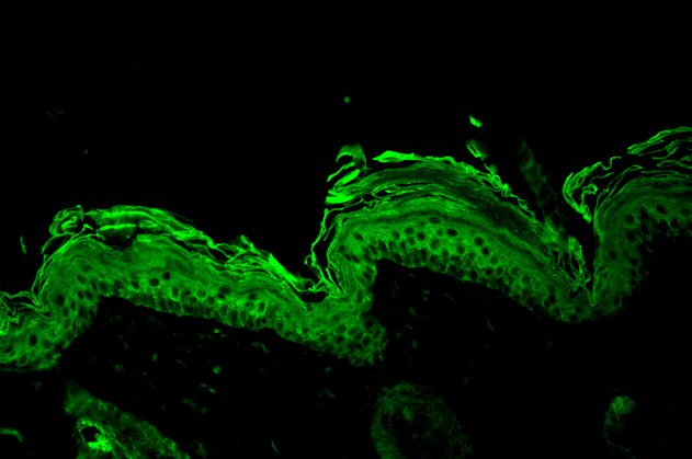
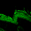
Description Immunohistochemistry analysis using Rabbit Anti-HSP90 beta Polyclonal Antibody (11106). Tissue: backskin. Species: Mouse. Fixation: Bouin's Fixative Solution. Primary Antibody: Rabbit Anti-HSP90 beta Polyclonal Antibody (11106) at 1:100 for 1 hour at RT. Secondary Antibody: FITC Goat Anti-Rabbit (green) at 1:50 for 1 hour at RT. Localization: Cytoplasm.

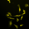
Description Immunocytochemistry/Immunofluorescence analysis using Rabbit Anti-Hsp90 beta Polyclonal Antibody (11106). Tissue: Cervical cancer cell line (HeLa). Species: Human. Fixation: 2% Formaldehyde for 20 min at RT. Primary Antibody: Rabbit Anti-Hsp90 beta Polyclonal Antibody (11106) at 1:120 for 12 hours at 4°C. Secondary Antibody: R-PE Goat Anti-Rabbit (yellow) at 1:200 for 2 hours at RT. Counterstain: DAPI (blue) nuclear stain at 1:40000 for 2 hours at RT. Localization: Cytoplasm. Melanosome. Magnification: 20x. (A) DAPI (blue) nuclear stain. (B) Anti-Hsp90 beta Antibody. (C) Composite.

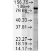
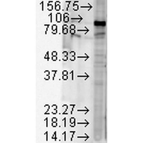
Description Western blot analysis of Human Cell line lysates showing detection of HSP90 beta protein using Rabbit Anti-HSP90 beta Polyclonal Antibody (11106). Load: 15 µgprotein. Block: 1.5% BSA for 30 minutes at RT. Primary Antibody: Rabbit Anti-HSP90 beta Polyclonal Antibody (11106) at 1:2000 for 2 hours at RT. Secondary Antibody: Donkey Anti-Rabbit IgG: HRP for 1 hour at RT.
Handling
Storage This antibody is stable for at least one (1) year at -20°C.
Dilution Instructions Dilute in PBS or medium which is identical to that used in the assay system.
Application Instructions Immunoblotting: use at 1:20,000- 1:40,000 (ECL). A band of ~90 kDa is detected.
Positive control: HeLa cell lysate
Positive control: HeLa cell lysate
References & Data Sheet
Data Sheet  Download PDF Data Sheet
 Download PDF Data Sheet


