Anti-Hsp90 Antibody (11098)
$338.00
| Host | Quantity | Applications | Species Reactivity | Data Sheet | |
|---|---|---|---|---|---|
| Mouse | 100ug | WB,IHC,ICC/IF,IP,ELISA,AM | Human, Mouse, Rabbit |  |
SKU: 11098
Categories: Antibody Products, Heat Shock and Stress Protein Antibodies, Products
Overview
Product Name Anti-Hsp90 Antibody (11098)
Description Anti-Hsp90 Mouse Monoclonal Antibody
Target Hsp90
Species Reactivity Human, Mouse, Rabbit
Applications WB,IHC,ICC/IF,IP,ELISA,AM
Host Mouse
Clonality Monoclonal
Clone ID H90-10
Isotype IgG2a
Immunogen Recombinant human Hsp90β
Properties
Form Liquid
Concentration Lot Specific
Formulation PBS, pH 7.4.
Buffer Formulation Phosphate Buffered Saline
Buffer pH pH 7.4
Format Purified
Purification Purified by Protein G affinity chromatography
Specificity Information
Specificity Detects 90kDa Hsp90b in all reactive species except chicken where it detects both alpha and beta isoforms.
Target Name Heat shock protein HSP 90-β
Target ID Hsp90
Uniprot ID P08238
Alternative Names HSP 90, Heat shock 84 kDa, HSP 84, HSP84
Gene Name HSP90AB1
Gene ID 3326
Accession Number NP_031381.2
Sequence Location Cytoplasm, Melanosome, Nucleus, Secreted, Cell membrane, Dynein axonemal particle
Biological Function Molecular chaperone that promotes the maturation, structural maintenance and proper regulation of specific target proteins involved for instance in cell cycle control and signal transduction. Undergoes a functional cycle linked to its ATPase activity. This cycle probably induces conformational changes in the client proteins, thereby causing their activation. Interacts dynamically with various co-chaperones that modulate its substrate rPubMed:16478993, PubMed:18239673, PubMed:19696785, PubMed:20353823, PubMed:24613385, PubMed:32272059, PubMed:25973397, PubMed:26991466, PubMed:27295069}.
Research Areas Heat Shock& Stress Proteins
Application Images


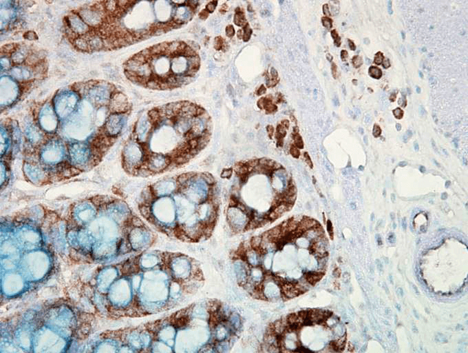
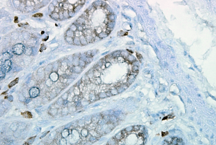
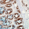
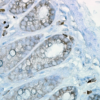
Description Immunohistochemistry analysis using Mouse Anti-Hsp90 Monoclonal Antibody, Clone H9010 (11098). Tissue: colon carcinoma. Species: Human. Fixation: Formalin. Primary Antibody: Mouse Anti-Hsp90 Monoclonal Antibody (11098) at 1:10000 for 12 hours at 4°C. Secondary Antibody: Biotin Goat Anti-Mouse at 1:2000 for 1 hour at RT. Counterstain: Mayer Hematoxylin (purple/blue) nuclear stain at 200 µl for 2 minutes at RT. Localization: Inflammatory cells. Magnification: 40x. This image was produced using an amplifying IHC wash buffer. The antibody has therefore been diluted more than is recommended for other applications.

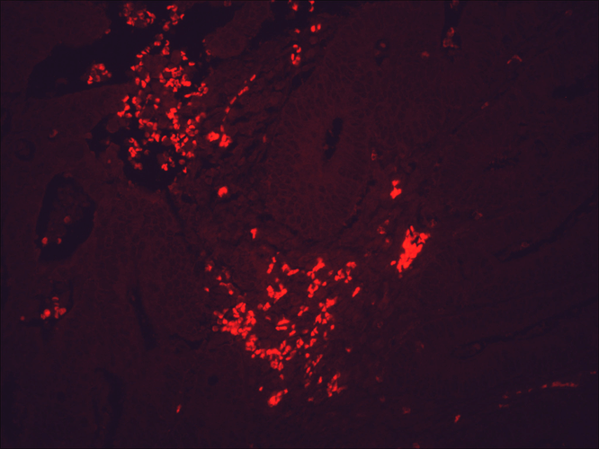
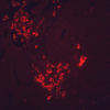
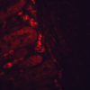
Description Immunohistochemistry analysis using Mouse Anti-Hsp90 Monoclonal Antibody, Clone H9010 (11098). Tissue: colon carcinoma. Species: Human. Fixation: Formalin. Primary Antibody: Mouse Anti-Hsp90 Monoclonal Antibody (11098) at 1:10000 for 12 hours at 4°C. Secondary Antibody: Alexa Fluor 555 Goat Anti-Mouse (red) at 1:5000 for 1 hour at RT. Magnification: 40x. This image was produced using an amplifying IHC wash buffer. The antibody has therefore been diluted more than is recommended for other applications.

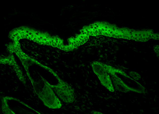
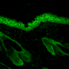
Description Immunohistochemistry analysis using Mouse Anti-Hsp90 Monoclonal Antibody, Clone H9010 (11098). Tissue: backskin. Species: Mouse. Fixation: Bouin's Fixative and paraffin-embedded. Primary Antibody: Mouse Anti-Hsp90 Monoclonal Antibody (11098) at 1:100 for 1 hour at RT. Secondary Antibody: FITC Goat Anti-Mouse (green) at 1:50 for 1 hour at RT. Localization: Epidermis.

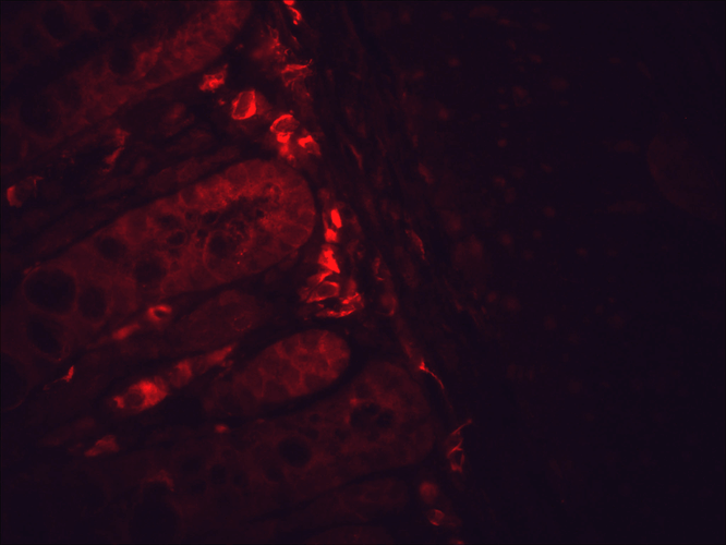
Description Immunohistochemistry analysis using Mouse Anti-Hsp90 Monoclonal Antibody, Clone H9010 (11098). Tissue: inflamed colon. Species: Mouse. Fixation: Formalin. Primary Antibody: Mouse Anti-Hsp90 Monoclonal Antibody (11098) at 1:10000 for 12 hours at 4°C. Secondary Antibody: Alexa Fluor 555 Goat Anti-Mouse (red) at 1:5000 for 1 hour at RT. Localization: Inflammatory and epithelial mucosa. Magnification: 40x. Inflammatory and epithelial mucosa. This image was produced using an amplifying IHC wash buffer. The antibody has therefore been diluted more than is recommended for other applications.

Description Immunohistochemistry analysis using Mouse Anti-Hsp90 Monoclonal Antibody, Clone H9010 (11098). Tissue: inflamed colon. Species: Mouse. Fixation: Formalin. Primary Antibody: Mouse Anti-Hsp90 Monoclonal Antibody (11098) at 1:10000 for 12 hours at 4°C. Secondary Antibody: Biotin Goat Anti-Mouse at 1:2000 for 1 hour at RT. Counterstain: Mayer Hematoxylin (purple/blue) nuclear stain at 200 µl for 2 minutes at RT. Localization: Inflammatory cells. Magnification: 40x. This image was produced using an amplifying IHC wash buffer. The antibody has therefore been diluted more than is recommended for other applications.

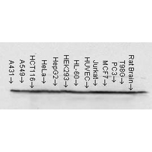
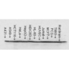
Description Western Blot analysis of Human cell lysates from various cell lines showing detection of Hsp90 protein using Mouse Anti-Hsp90 Monoclonal Antibody, Clone H9010 (11098). Load: 15 µg. Block: 1.5% BSA for 30 minutes at RT. Primary Antibody: Mouse Anti-Hsp90 Monoclonal Antibody (11098) at 1:1000 for 2 hours at RT. Secondary Antibody: Sheep Anti-Mouse IgG: HRP for 1 hour at RT.

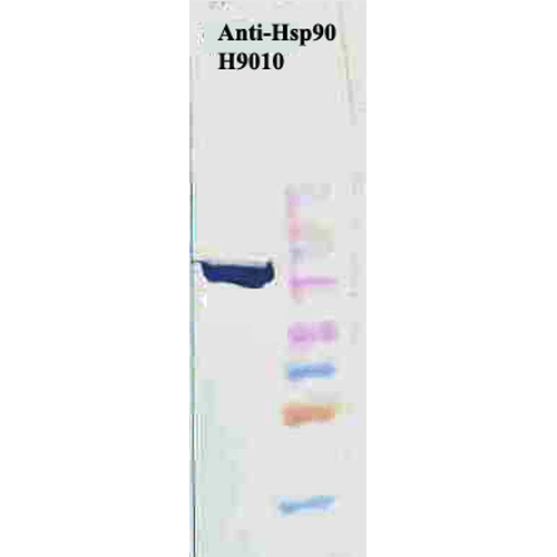
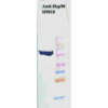
Description Western Blot analysis of Human Cervical cancer cell line (HeLa) lysate showing detection of Hsp90 protein using Mouse Anti-Hsp90 Monoclonal Antibody, Clone H9010 (11098). Primary Antibody: Mouse Anti-Hsp90 Monoclonal Antibody (11098) at 1:1000. Secondary Antibody: HRP Goat Anti-Mouse.

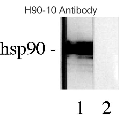
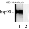
Description Western blot analysis of Human Lysates showing detection of Hsp90 protein using Mouse Anti-Hsp90 Monoclonal Antibody, Clone H9010 (11098). Primary Antibody: Mouse Anti-Hsp90 Monoclonal Antibody (11098) at 1:1000. Comparison of clone H9010 behavior with Hsp90 human beta (1) and Hsp90 human alpha (2). Courtesy of: David Toft, Mayo Clinic.

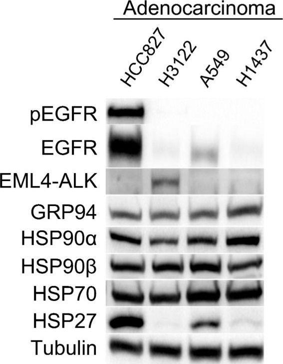
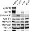
Description Characterization of a panel of adenocarcinoma cell lines. Western blot analysis of adenocarcinoma cell lines whose oncodrivers are either directly (epidermal growth factor receptor (EGFR) in HCC827 and EML4-ALK in H3122 cell lines) or indirectly Kirsten rat sarcoma (KRAS) viral oncogene homolog) in the A549 cell line) related to HSP90, as well as an EGFR, anaplastic lymphoma kinase (ALK) and KRAS wild-type cell line (H1437). This technique was employed to study protein expression levels of constitutive and inducible HSP90, other related heat shock proteins (HSPs) (GRP94, HSP70 and HSP27) as well as the HSP90 clients EGFR, its phosphorylated form pEGFR, and EML4-ALK. For Western blots, a representative image is shown. Densitometry analysis may be found in Figure S1.
Handling
Storage This antibody is stable for at least one (1) year at -20°C to -70°C. Store product in appropriate aliquots to avoid multiple freeze-thaw cycles.
Dilution Instructions Dilute in PBS or medium which is identical to that used in the assay system.
Application Instructions Immunoblotting: use at 1-5ug/mL. A band of ~90kDa is detected. Detection of Hsp90β in tumor cell lines and rat brain with #11098 at 1ug/mL. Detection of Hsp90β in HeLa cell lysate with #11098 at 1ug/mL.
Immunohistochemistry: use at 0.5-10ug/mL. Detection of Hsp90β in human colon carcinoma Detection of Hsp90β in mouse skin with #11098 with #11098 at 0.5ug/mL. at 10ug/mL.
Immunohistochemistry: use at 0.5-10ug/mL. Detection of Hsp90β in human colon carcinoma Detection of Hsp90β in mouse skin with #11098 with #11098 at 0.5ug/mL. at 10ug/mL.
References & Data Sheet
References Wang, Q., Li, X., et al.Stem Cell Res Ther . 2019 Nov 26
10(1):348
Lei, W., Duron, D. I., et al.Front Mol Neurosci. 2019 Nov 29
12:294
Impact of Heat Shock Protein 90 Inhibition on the Proteomic Profile of Lung Adenocarcinoma as Measured by Two-Dimensional Electrophoresis Coupled with Mass Spectrometry. Cells (2019) [31370342]
10(1):348
Lei, W., Duron, D. I., et al.Front Mol Neurosci. 2019 Nov 29
12:294
Impact of Heat Shock Protein 90 Inhibition on the Proteomic Profile of Lung Adenocarcinoma as Measured by Two-Dimensional Electrophoresis Coupled with Mass Spectrometry. Cells (2019) [31370342]
Data Sheet  Download PDF Data Sheet
 Download PDF Data Sheet

