Anti-HSF1 Antibody (12518)
$397.00
| Host | Quantity | Applications | Species Reactivity | Data Sheet | |
|---|---|---|---|---|---|
| Rat | 100ug | WB,IHC,ICC/IF,IP,ELISA,GS | Human, Mouse, Rat, Raabbit, Bovine, Guinea Pig, Hamster, Monkey |  |
SKU: 12518
Categories: Antibody Products, Heat Shock and Stress Protein Antibodies, Products
Overview
Product Name Anti-HSF1 Antibody (12518)
Description Anti-HSF1 Rat Monoclonal Antibody
Target HSF1
Species Reactivity Human, Mouse, Rat, Raabbit, Bovine, Guinea Pig, Hamster, Monkey
Applications WB,IHC,ICC/IF,IP,ELISA,GS
Host Rat
Clonality Monoclonal
Clone ID 10H8
Isotype IgG1
Immunogen Recombinant mouse HSF-1
Properties
Form Liquid
Concentration Lot Specific
Formulation PBS, pH 7.4.
Buffer Formulation Phosphate Buffered Saline
Buffer pH pH 7.4
Format Purified
Purification Purified by Protein G affinity chromatography
Specificity Information
Specificity This antibody recognizes an ~85 kDa protein (inactive form) in unstressed cell lysates and an ~95 kDa protein (active form) in heat-shocked cell lysates of human, mouse, rat, rabbit, bovine, guinea pig, hamster, and monkey.
Target Name Heat shock factor protein 1
Target ID HSF1
Uniprot ID Q00613
Alternative Names HSF 1, Heat shock transcription factor 1, HSTF 1
Gene Name HSF1
Accession Number NP_005517
Sequence Location Nucleus, Cytoplasm, Nucleus, nucleoplasm, Cytoplasm, perinuclear region, Cytoplasm, cytoskeleton, spindle pole, Cytoplasm, cytoskeleton, microtubule organizing center, centrosome, Chromosome, centromere, kinetochore
Biological Function Functions as a stress-inducible and DNA-binding transcription factor that plays a central role in the transcriptional activation of the heat shock response (HSR), leading to the expression of a large class of molecular chaperones heat shock proteins (HSPs) that protect cells from cellular insults' damage (PubMed:1871105, PubMed:11447121, PubMed:1986252, PubMed:7760831, PubMed:7623826, PubMed:8946918, PubMed:8940068, PubMed:9341107, PubMed:9121459, PubMed:9727490, PubMed:9499401, PubMed:9535852, PubMed:12659875, PubMed:12917326, PubMed:15016915, PubMed:25963659, PubMed:26754925, PubMed:18451878). In unstressed cells, is present in a HSP90-containing multichaperone complex that maintains it in a non-DNA-binding inactivated monomeric form (PubMed:9727490, PubMed:11583998, PubMed:16278218). Upon exposure to heat and other stress stimuli, undergoes homotrimerization and activates HSP gene transcription through binding to site-specific heat shock elements (HSEs) present in the promoter regions of HSP genes (PubMed:1871105, PubMed:1986252, PubMed:8455624, PubMed:7935471, PubMed:7623826, PubMed:8940068, PubMed:9727490, PubMed:9499401, PubMed:10359787, PubMed:11583998, PubMed:12659875, PubMed:16278218, PubMed:25963659, PubMed:26754925). Upon heat shock stress, forms a chromatin-associated complex with TTC5/STRAP and p300/EP300 to stimulate HSR transcription, therefore increasing cell survival (PubMed:18451878). Activation is reversible, and during the attenuation and rPubMed:10359787, PubMed:11447121, PubMed:11583998, PubMed:12659875, PubMed:12917326, PubMed:14707147, PubMed:15016915, PubMed:16278218, PubMed:17897941, PubMed:18451878, PubMed:1871105, PubMed:18794143, PubMed:1986252, PubMed:25963659, PubMed:26359349, PubMed:26727489, PubMed:26754925, PubMed:7623826, PubMed:7760831, PubMed:7935471, PubMed:8455624, PubMed:8940068, PubMed:8946918, PubMed:9121459, PubMed:9341107, PubMed:9499401, PubMed:9535852, PubMed:9727490}.; (Microbial infection) Plays a role in latent human immunodeficiency virus (HIV-1) transcriptional reactivation. Binds to the HIV-1 long terminal repeat promoter (LTR) to reactivate viral transcription by recruiting cellular transcriptional elongation factors, such as CDK9, CCNT1 and EP300. {PubMed:27189267}.
Research Areas Heat Shock& Stress Proteins
Background Heat shock factor 1 (HSF1) is a heat shock transcription factor that activates the transcription of genes encoding products required for protein folding, processing, targeting, degradation, and function. Up- regulation of expression of heat shock proteins in response to stress occurs at the level of transcription through a heat shock element and a HSF transcription factor. Amino acid sequences for most HSFs are highly conserved. A DNA binding domain is at the N-terminus, hydrophobic repeats (essential to the formation of active trimers) are adjacent to this binding domain, and another short hydrophobic repeat (necessary for suppression of trimerization) occurs toward the C-terminus. In higher eukaryotes, HSF1 is distributed diffusely in the cell cytoplasm and nucleus in unstressed cells. On exposure to heat shock or other stresses, HSF1 localizes to discrete nuclear granules; on recovery from stress, it returns to a diffuse nuclear- cytoplasmic distribution.
Application Images


Description Immunocytochemistry/Immunofluorescence analysis using Rat Anti-HSF1 Monoclonal Antibody, Clone 10H8 (12518). Tissue: Heat Shocked cervical cancer cells (HeLa). Species: Human. Fixation: 2% Formaldehyde for 20 min at RT. Primary Antibody: Rat Anti-HSF1 Monoclonal Antibody (12518) at 1:100 for 12 hours at 4°C. Secondary Antibody: FITC Goat Anti-Rat (green) at 1:200 for 2 hours at RT. Counterstain: DAPI (blue) nuclear stain at 1:40000 for 2 hours at RT. Localization: Diffuse nuclear and cytoplasmic staining. Magnification: 100x. (A) DAPI (blue) nuclear stain. (B) Anti-HSF1 Antibody. (C) Composite. Heat Shocked at 42°C for 1h.

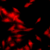
Description Immunocytochemistry/Immunofluorescence analysis using Rat Anti-HSF1 Monoclonal Antibody, Clone 10H8 (12518). Tissue: Heat Shocked cervical cancer cells (HeLa). Species: Human. Fixation: 2% Formaldehyde for 20 min at RT. Primary Antibody: Rat Anti-HSF1 Monoclonal Antibody (12518) at 1:100 for 12 hours at 4°C. Secondary Antibody: APC Goat Anti-Rat (red) at 1:200 for 2 hours at RT. Counterstain: DAPI (blue) nuclear stain at 1:40000 for 2 hours at RT. Localization: Diffuse nuclear and cytoplasmic staining. Magnification: 20x. (A) DAPI (blue) nuclear stain. (B) Anti-HSF1 Antibody. (C) Composite. Heat Shocked at 42°C for 1h.

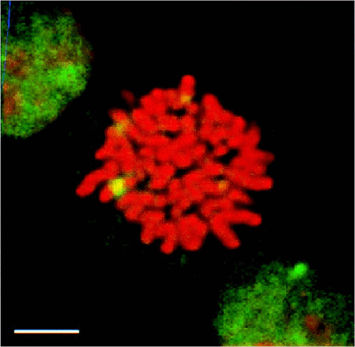
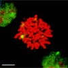
Description Immunocytochemistry/Immunofluorescence analysis using Rat Anti-HSF1 Monoclonal Antibody, Clone 10H8 (12518). Tissue: Heat Shocked mitotic HeLa cells. Species: Human. Primary Antibody: Rat Anti-HSF1 Monoclonal Antibody (12518) at 1:1000. HSF1 stained green. Courtesy of: Morimoto Lab, Northwestern University, USA.

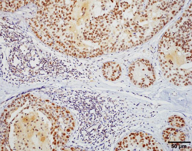
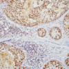
Description Immunohistochemistry analysis using Rat Anti-HSF1 Monoclonal Antibody, Clone 10H8 (12518). Tissue: Breast carcinoma. Species: Human. Fixation: 10% Formalin Solution for 20 hours at RT. Primary Antibody: Rat Anti-HSF1 Monoclonal Antibody (12518) at 1:1000 for 40 min. Secondary Antibody: Dako labeled Polymer HRP Anti-rat IgG, DAB Chromogen (brown) (Dako Envision+ System) for 30 min at RT. Counterstain: Mayer's Hematoxylin (purple/blue) nuclear stain for 1 minute at RT. Localization: Nuclear. Magnification: 100X. Courtesy of: Dr. Sandro Santagata, Harvard Medical School.

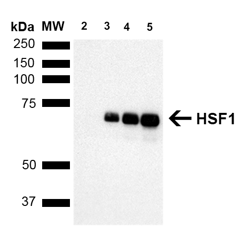
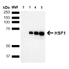
Description Western Blot analysis of Human Breast adenocarcinoma cell line (MCF7) showing detection of ~65 kDa HSF1 protein using Rat Anti-HSF1 Monoclonal Antibody, Clone 10H8 (12518). Lane 1: MW ladder. Lane 2: HSF1 null lysate prepared from mouse embryonic fibroblasts. Lane 3: MCF7 lysate (5 µg). Lane 4: MCF7 lysate (10 µg). Lane 5: MCF7 lysate (20 µg). Block: 1.5% BSA for 30 minutes at RT. Primary Antibody: Rat Anti-HSF1 Monoclonal Antibody (12518) at 1:1000 for 2 hours at RT. Secondary Antibody: Goat Anti-Rat IgG: HRP for 1 hour at RT. Predicted/Observed Size: ~65 kDa. Courtesy of: Dr. Sandro Santagata, Harvard Medical School.
Handling
Storage This antibody is stable for at least one (1) year at -20°C.
Dilution Instructions Dilute in PBS or medium which is identical to that used in the assay system.
Application Instructions Immunoblotting: use at 1-2ug/mL. A band of ~85 and/or ~95 kDa is detected.
Immunocytochemistry: use at 10ug/mL.
Immunoprecipitation: use at 12.5ug/mL
These are recommended concentrations. User should determine optimal concentrations for their application.
Positive control: HeLa cell lysate
Immunocytochemistry: use at 10ug/mL.
Immunoprecipitation: use at 12.5ug/mL
These are recommended concentrations. User should determine optimal concentrations for their application.
Positive control: HeLa cell lysate
References & Data Sheet
References Cotto, J et al. 1997 J Cell Science 110: 2925-2934.
PMID 9359875
Data Sheet  Download PDF Data Sheet
 Download PDF Data Sheet


